ventilation in fish and insects
1/36
There's no tags or description
Looks like no tags are added yet.
Name | Mastery | Learn | Test | Matching | Spaced |
|---|
No study sessions yet.
37 Terms
how many pairs of gills do most bony fish have
5 pairs
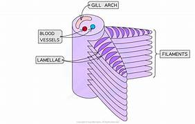
gill structure components
gill arch
gill filaments (primary lamellae)
gill plate (secondary lamellae)
operculum
bony flap that covers the gills
how many rows of primary lamellae does each gill have
2 rows attached to a gill arch
secondary lamellae
folded surface of primary lamellae to increase surface area
ventilation in bony fish process
mouth opens (operculum closed)
buccal cavity floor is lowered
increases the volume and decreases the pressure of the buccal cavity below outside
water rushes into the buccal cavity down a pressure gradient
mouth closes which raises the floor of the buccal cavity
volume decreases and pressure increases inside the buccal cavity higher that opercular cavity
increased pressure forces open operculum
water is forced over the gills and out of the operculum
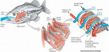
countercurrent flow process
blood flows along the gill arch and out along the filaments to the secondary lamellae
the blood flows in the opposite direction to the water flow across the gills
this helps the fish absorb as much oxygen as possible as there is always a concentration gradient

what is countercurrent flow
when the blood flows along the lamellae in the opposite direction to the water flowing over the gills to maintain a concentration gradient of oxygen
why is countercurrent flow efficient
water and blood concentration gradient of oxygen is maintained over the whole lamellae
so oxygen can diffuse down the concentration gradient from the water to the blood
why is countercurrent flow more efficient than concurrent flow
in concurrent flow the concentration of oxygen in the blood and in the water will eventually equalise so there would be no concentration gradient and no oxygen exchange would take place
in countercurrent flow the concentration of oxygen in the water is always higher than in the blood so as much oxygen as possible diffuses into the blood
how to dissect fish gills
wear apron and gloves
place fish on dissection tray
push back operculum and use scissors to remove gills
cut each gill arch trough the bone at the top and bottom
draw and annotate it along with a scale bar
how do fish ensure a large surface area for gas exchange
primary and secondary lamellae
how do insects ensure a large surface area for gas exhange
tracheoles and tracheal fluid
how do fish ensure a steep concentration gradient for gas exchange
countercurrent flow
how do insects ensure a steep concentration gradient for gas exchange
cells are always respiring so always producing co2
some insects can ‘pump’ air in and out of their bodies to replace air saturated with co2
how do fish ensure a short diffusion distance for gas exchange
very thin lamellae
blood in close contact with water
walls of capillaries are 1 cell thick
how do insects ensure a short diffusion distance for gas exchange
tracheoles walls are very thin
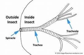
tracheal system features
cuticle (exoskeleton)
spiracle
trachea
tracheole
cuticle (exoskeleton) of an insect
provides protection and structure
spiracle strucure
tiny holes in an insects body with that can open and close to prevent water loss and have sensory hairs
spiracle function
where gases enter and where water can evaporate from an insect
trachea of an insect structure
network of internal tubes with o rings of chitin to prevent collapsing
trachea of an insect function
air filled pipes in an insect for gas exchange
tracheole structure
fine respiratory tube of the trachea of an insect with thin permeable walls
tracheole function
extend throughout the whole insect and go directly into respiring cells to provide o2
similarities between an insect and mammals respiratory system
have trachea
have structures with thin permeable walls
differences between an insect and mammals respiratory system
mammals have lungs and insects dont
insect trachea has o chitin but mammal trachea has c cartilage
insect trachea splits into tracheoles but mammal trachea splits into bronchi
what affects rate of ventilation in large insects
rhythmic abdominal movements
wing movements when larger insects are flying
flexible trachea wall
how do insects limit water loss
spiracles have a small surface area to volume ratio
spiracles cal open and close
waterproof exoskeleton contains lipid layer
water can only evaporate through spiracles
how does the flexible trachea wall affect ventilation in large insects
can ventilate the tracheal system by expanding and acting as air sacs which can be squeezed by the action on the flight muscles
how can movements of the wings affect ventilation in large insects
alter the volume of the thorax
if thorax volume decreases air pressure increases and air is pushed out
opposite if volume increases
how can rhythmic abdominal movements affect ventilation in large insects
locusts
as the abdomen expands the spiracles at the front end of their body open and air enters
as the abdomen reduces in volume the spiracles at the rear end of the body cavity open and air leaves
what are the examples of large insects
locusts
crickets
how the tracheal system works in insects
oxygen travels down the concentration gradient to the cells
carbon dioxide moves down its own concentration gradient to the spiracles
the trachea branches off into tracheoles
tracheoles contain tracheal fluid which oxygen dissolves in
oxygen diffuses from tracheal fluid into body cells
carbon dioxide diffuses in the opposite direction
the body cavity is filled with haemolymph
tracheal fluid
increases surface area and the rate of diffusion of oxygen from the tracheoles to the cells of an insect
haemolymph
transports nutrients and waste around the insect body
not contained within vessels
doesnt transport o2 or co2
transports sugars and proteins (hormones)
how to dissect an insect
grasshoppers or cockroaches
fix insect to dissecting board with dissecting pins through legs
cut and remove piece of the exoskeleton from along the length of its abdomen to examine the tracheae
fill the abdomen with saline solution with a syringe
network of very think grey tubes are the tracheae
can examine under a light microscope with a wet mount slide