Laboratories 12: Cardiovascular System
1/102
There's no tags or description
Looks like no tags are added yet.
Name | Mastery | Learn | Test | Matching | Spaced | Call with Kai |
|---|
No analytics yet
Send a link to your students to track their progress
103 Terms
Heart
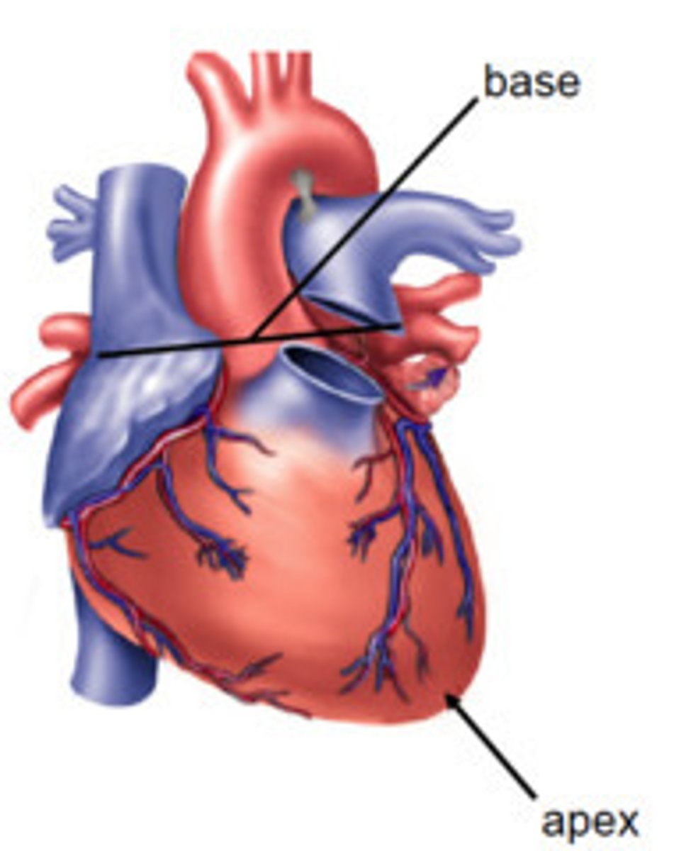
base and apex

base of heart
posterior surface of the heart
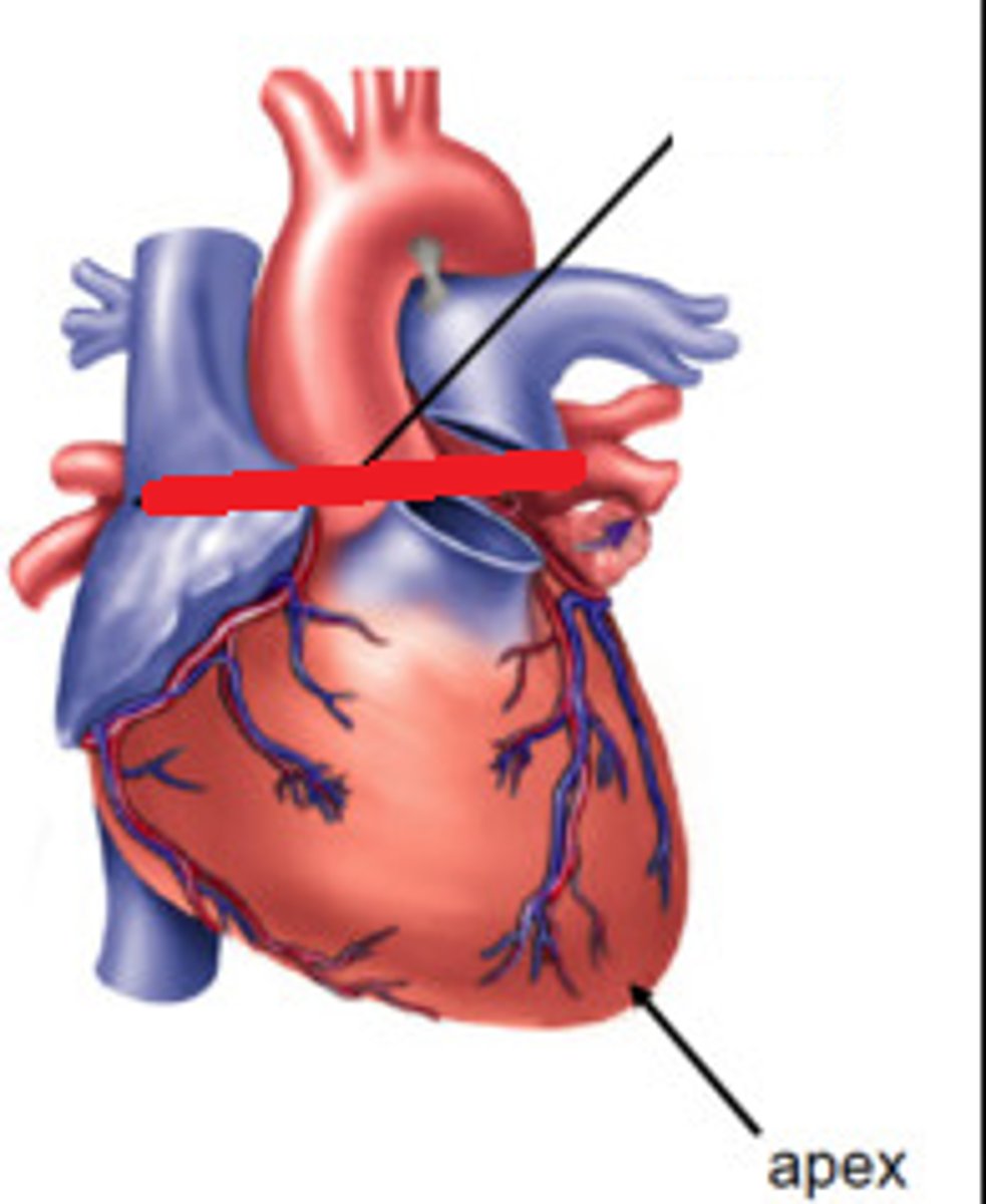
apex of the heart
lower tip of the heart
Septa and Surface Landmarks
yay!
interatrial septum
divides the right and left atrium
interatrial sulcus
separates the left and right atria
atrioventricular septum
divides right atrium and right ventricle
divides left atrium and left ventricle
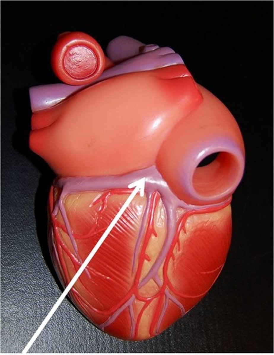
coronary sulcus
Groove separating atria from ventricles. AKA Atrioventricular sulcus

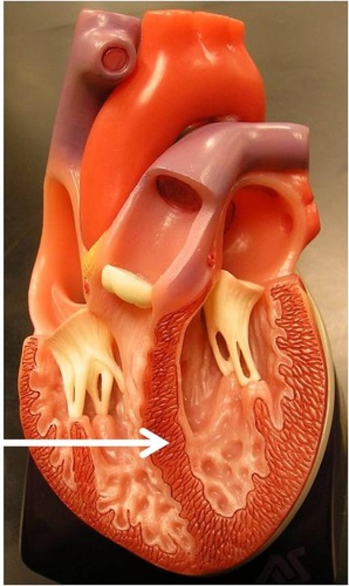
interventricular septum
divides right and left septum
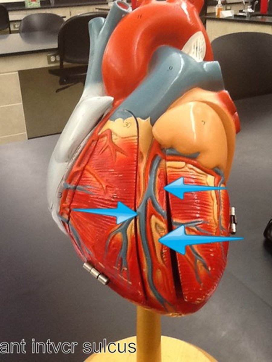
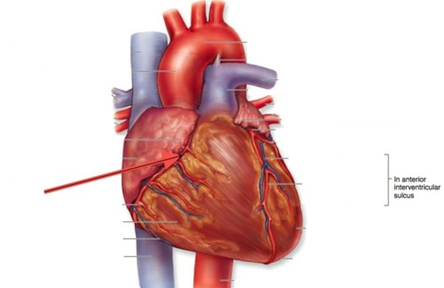
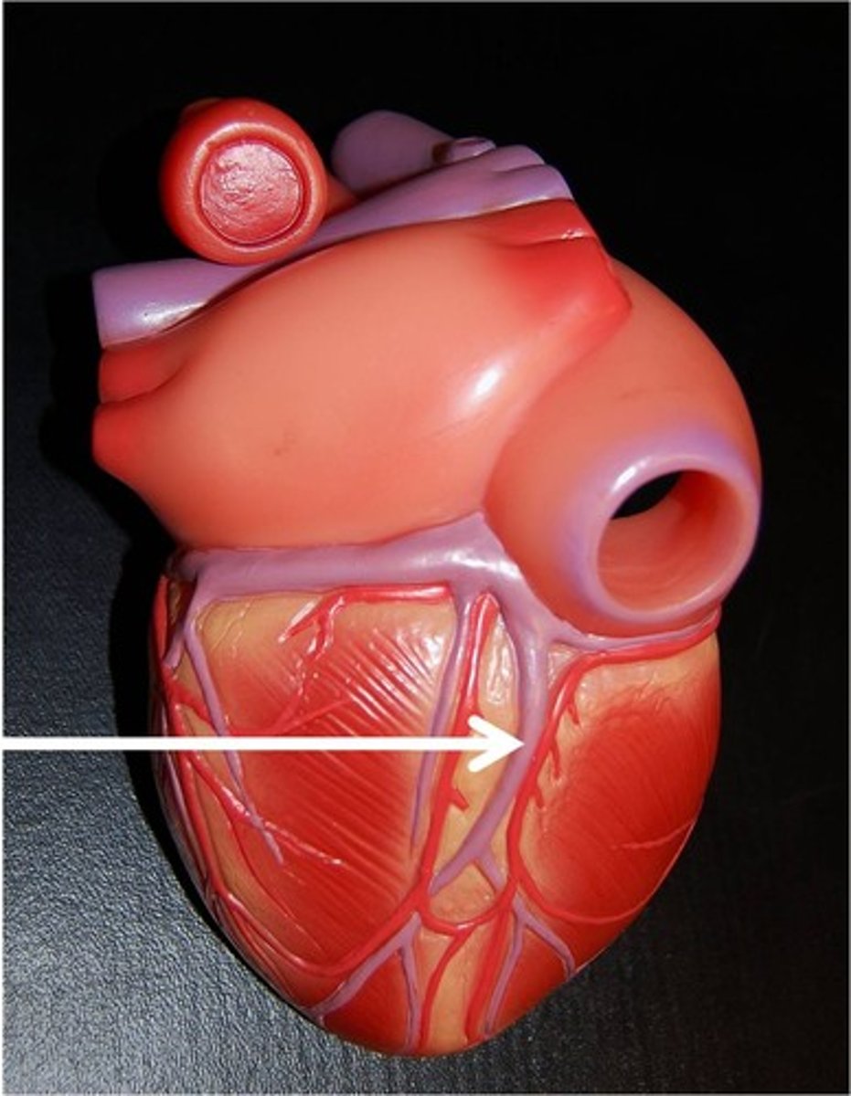
anterior interventricular sulcus,
marks the boundary between the ventricles anteriorly
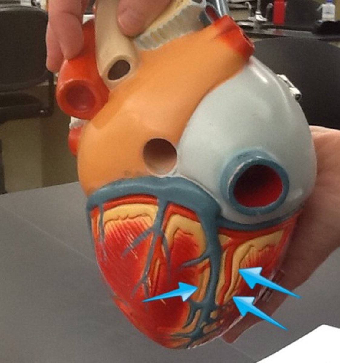
posterior interventricular sulcus
marks the boundary between the ventricles posteriorly
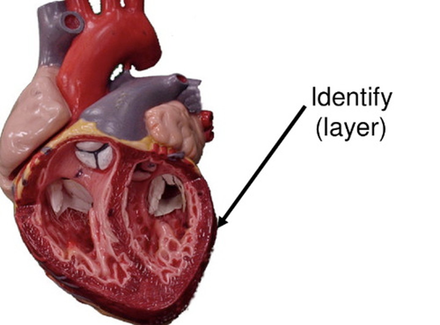
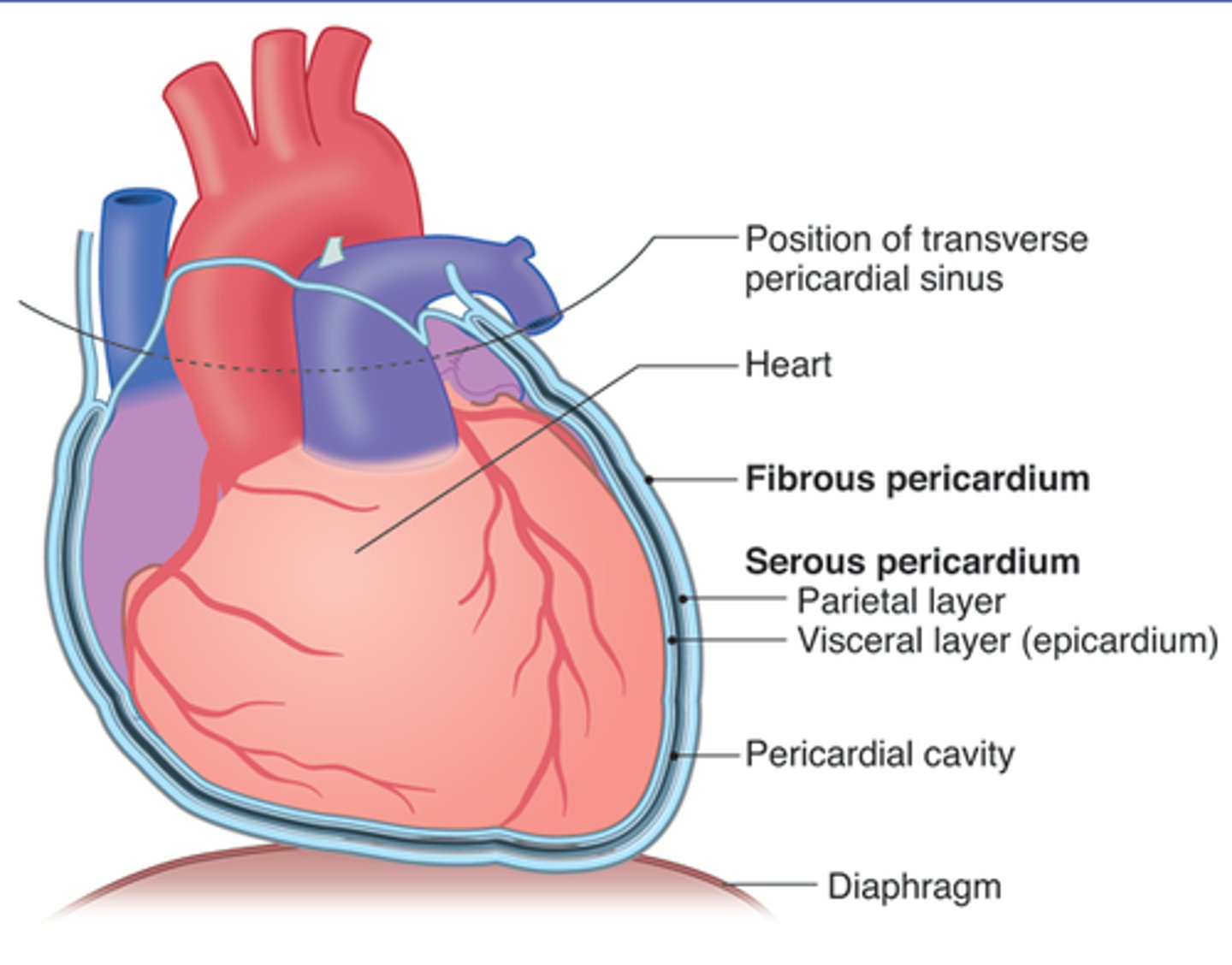
Layers of the Heart Wall
yay
endocardium
inner lining of the heart, simple squamous epithelium
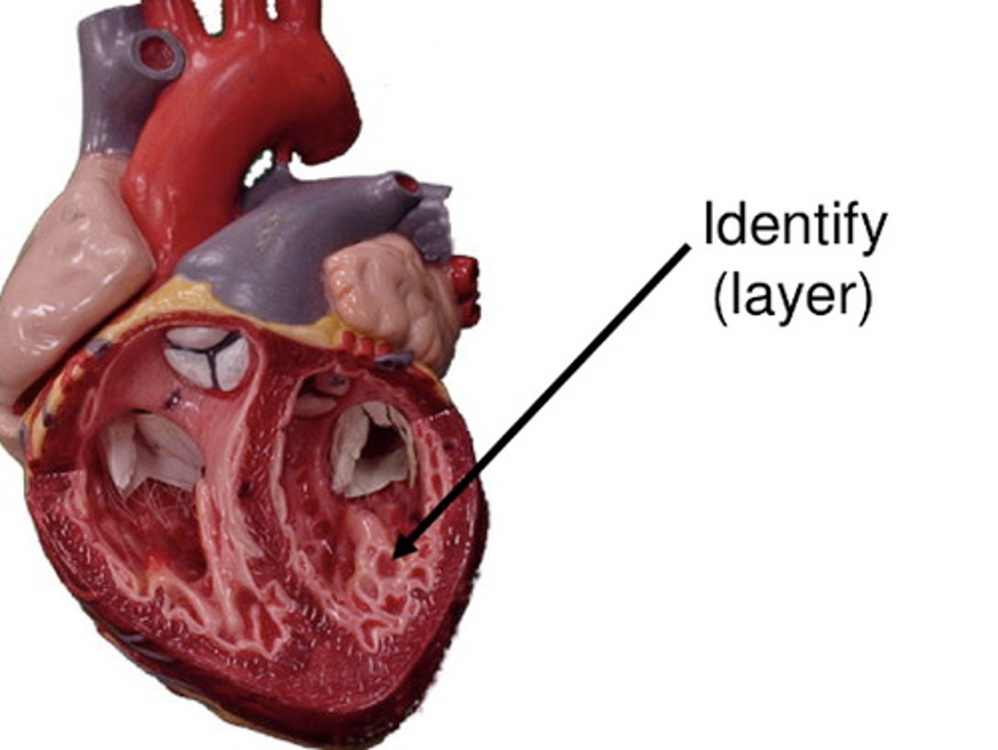
myocardium
middle layer, cardiac muscle tissue
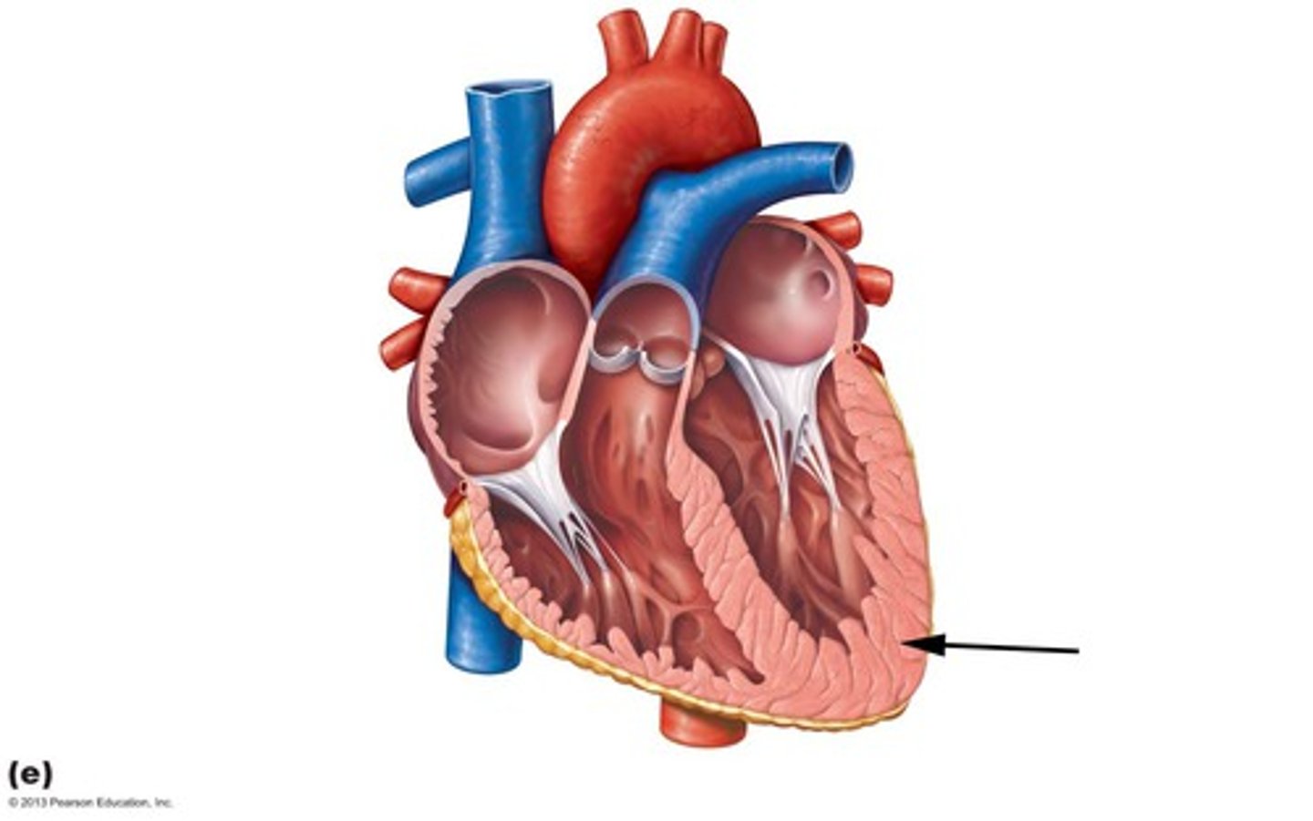
epicardium/visceral pericardium
outer layer of the heart. composed of simple squamous epithelium, areolar connective tissue, and adipose connective tissue
Coverings of the Heart
yay
visceral pericardium
layer closest to the heart

pericardial cavity
contains serous fluid
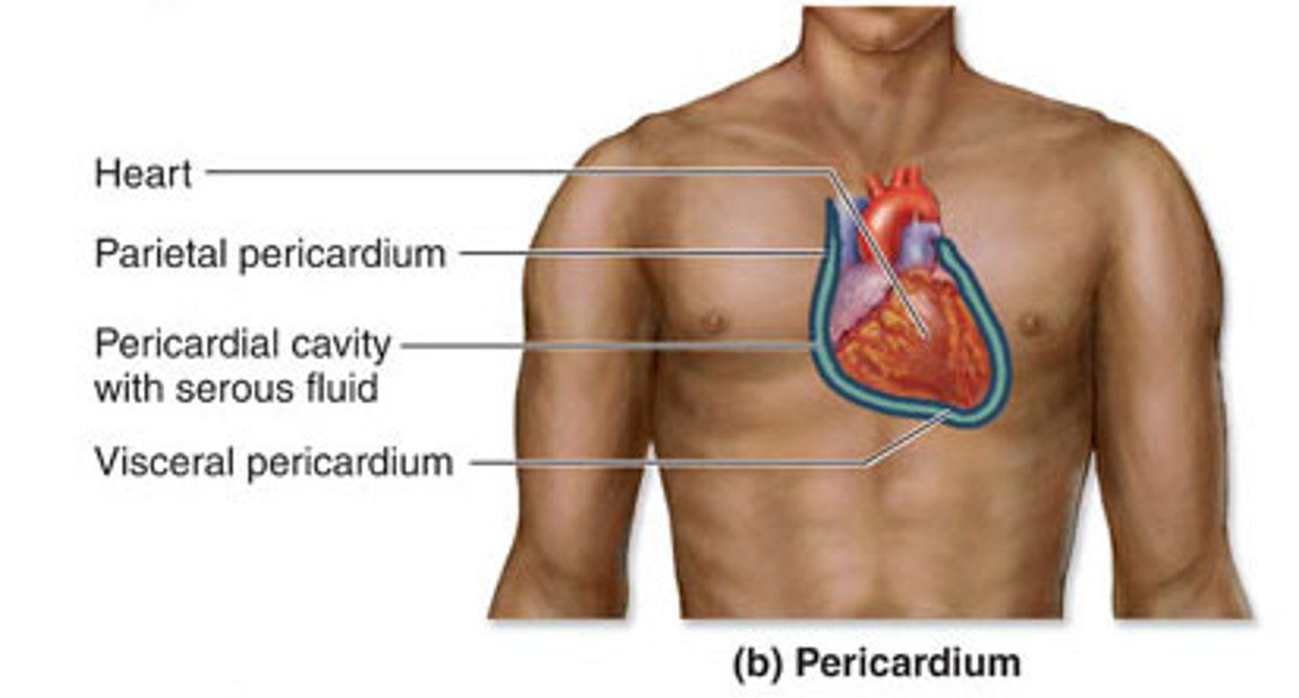
pericardial sac
surrounds the heart. parietal pericardium and fibrous pericardium
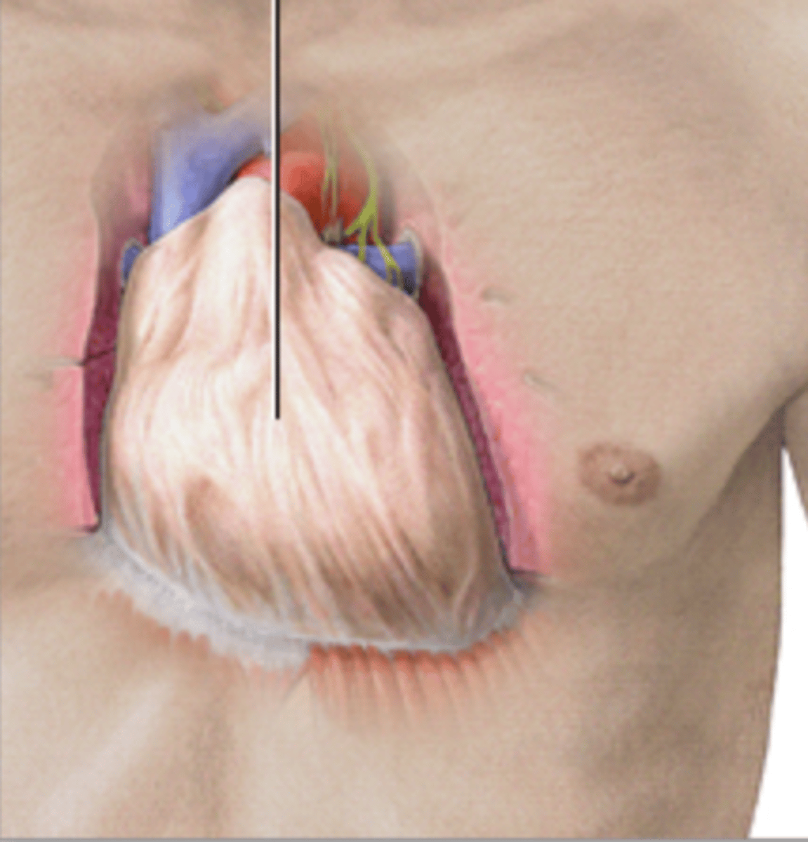
parietal pericardium
outer layer of the pericardium
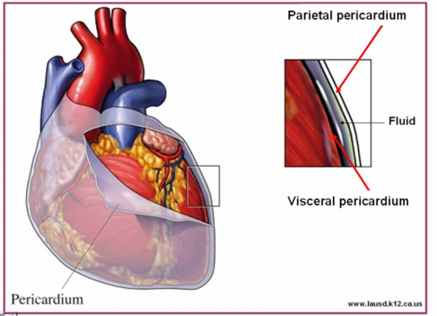
fibrous pericardium
dense connective tissue
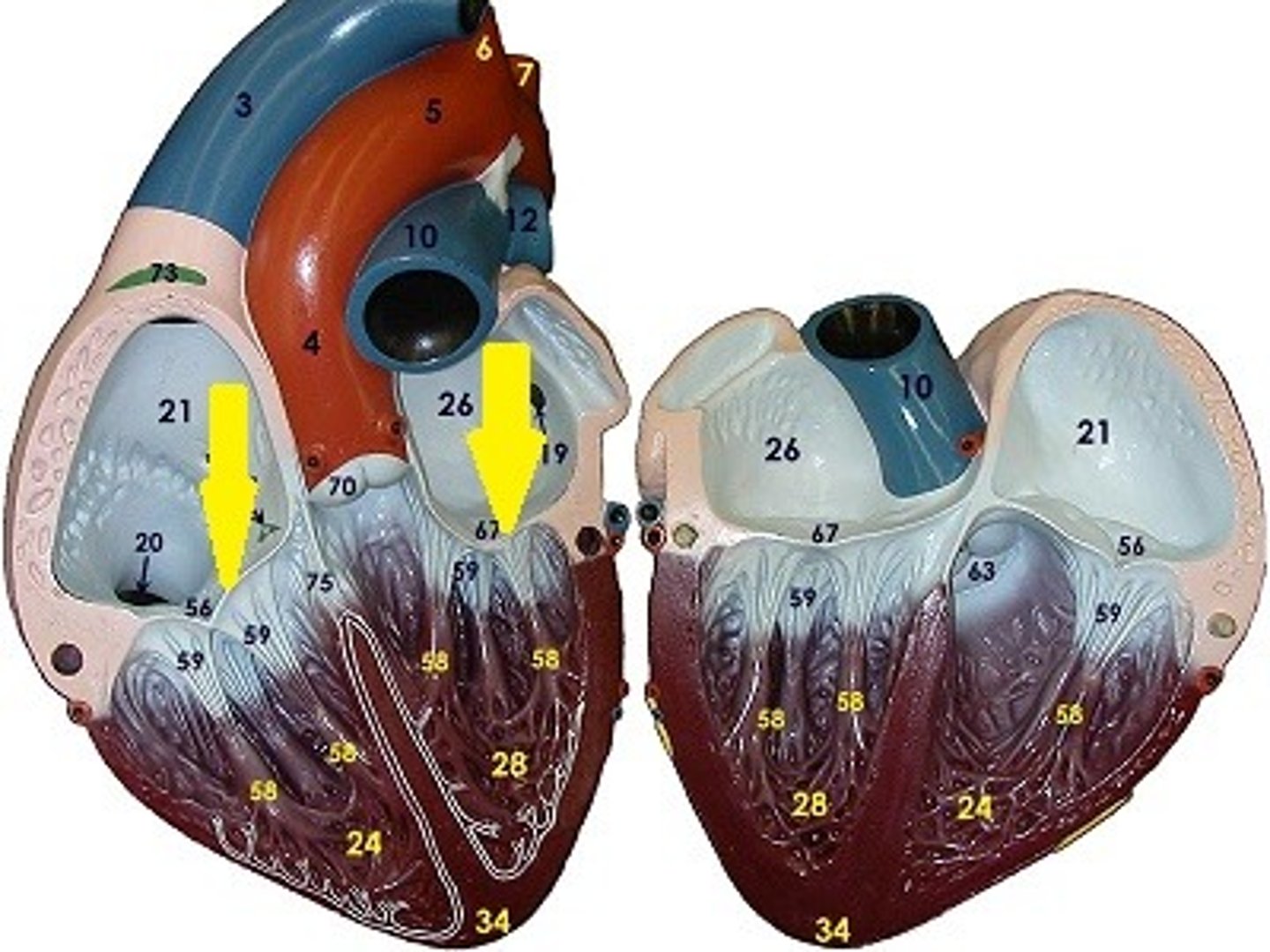
Heart Chambers
right atrium, right ventricle, left atrium, left ventricle
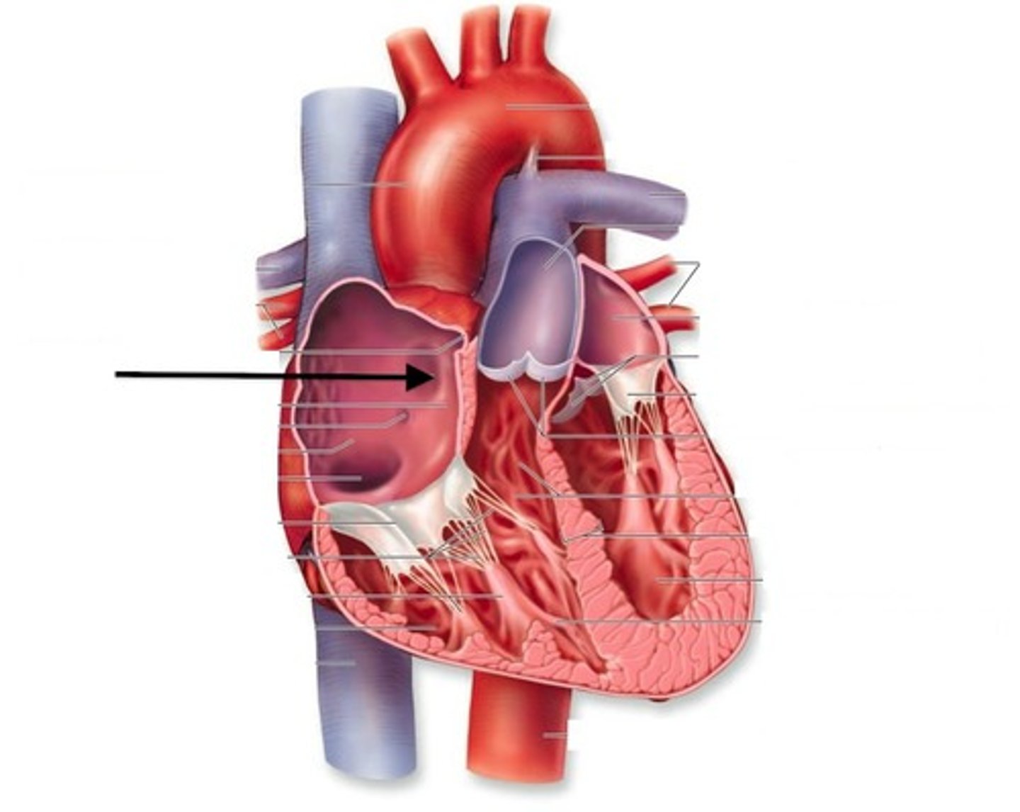
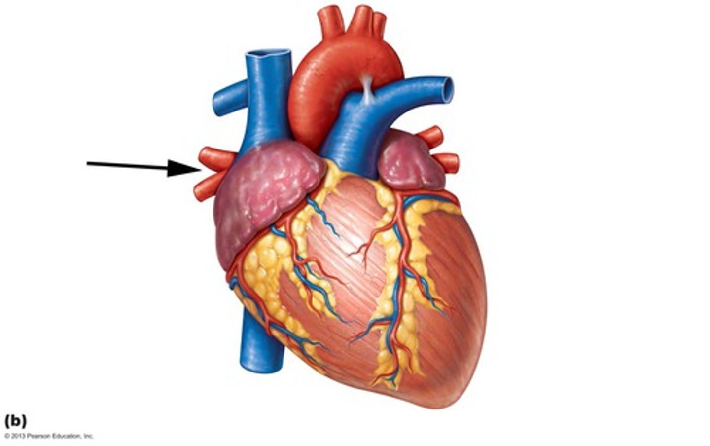
right atrium
yay
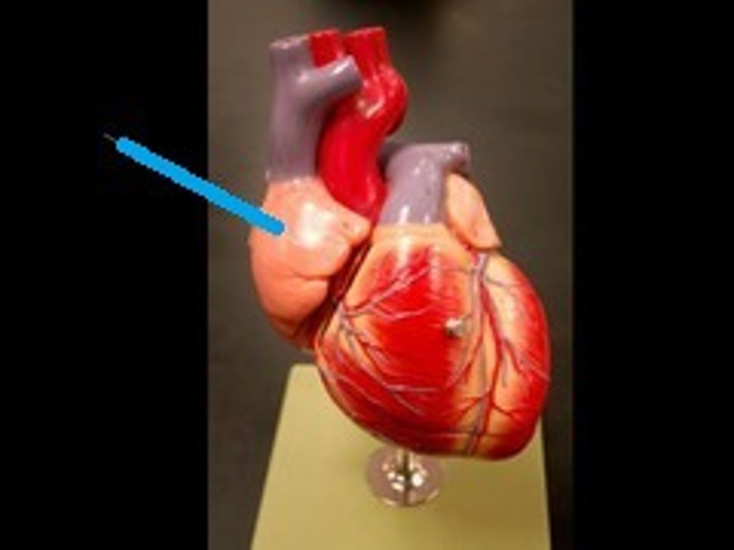
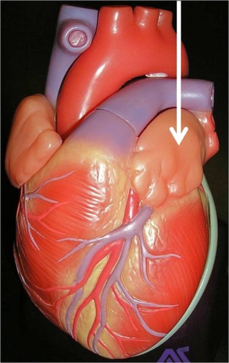
Right auricle
AKA right atrial appendage. Little flap
pectinate muscles
has ridges that allow blood to swirl and prevent clotting
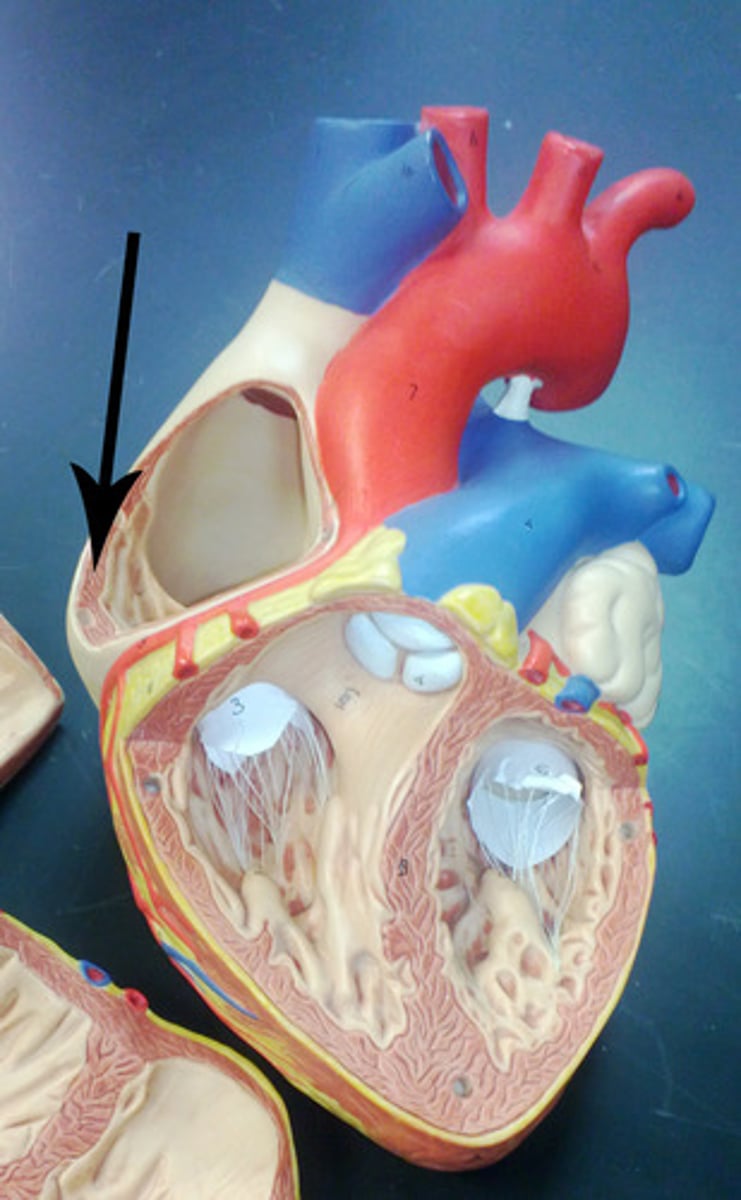
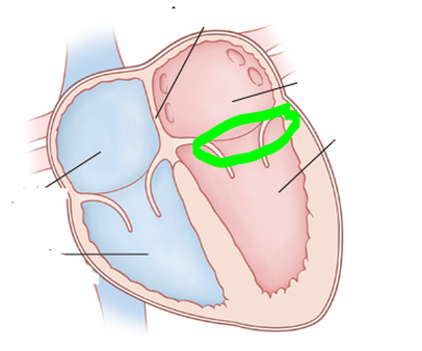
fossa ovalis
depression, fetal reminent of the foramen ovale allowing right and left atrial blood transfer
superior vena cava
returns dexoygenated blood from above diaphragm to right atrium
inferior vena cava
Returns deoxygenated blood from below diaphragm to right atrium
coronary sinus
Returns deoxygenated blood from below heart back to right atrium

right atrioventricular orifice
between atrium and ventricle
tricuspid valve (right atrioventricular valve)
valve between the right atrium and the right ventricle. 3 cusps
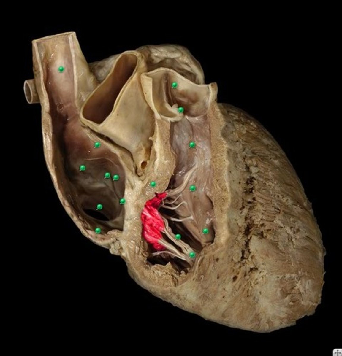
Right Ventricle
yay
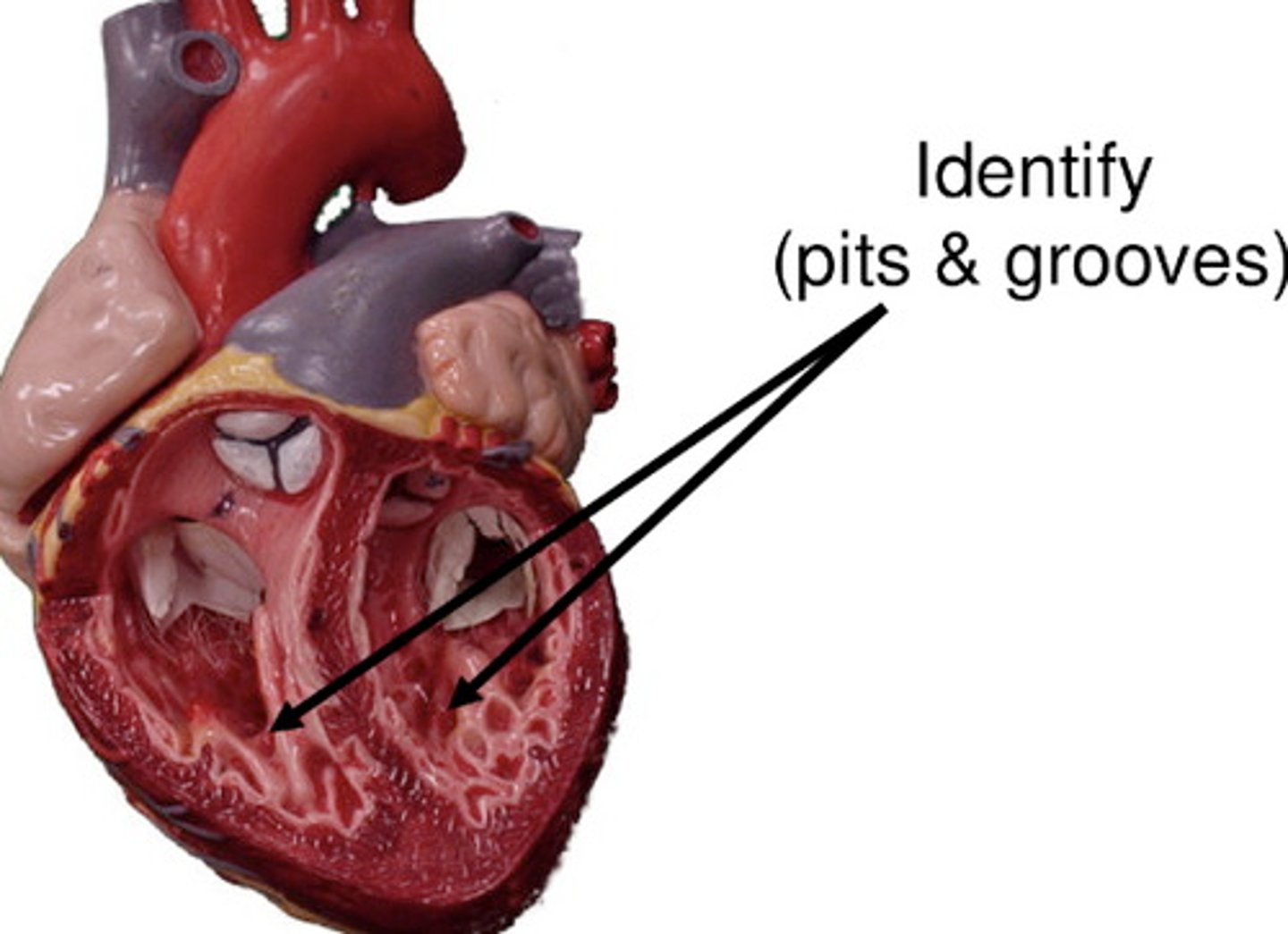
trabeculae carneae
allows blood to twirl and prevent clotting

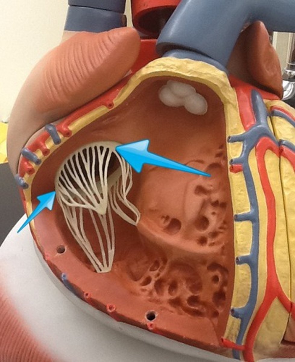
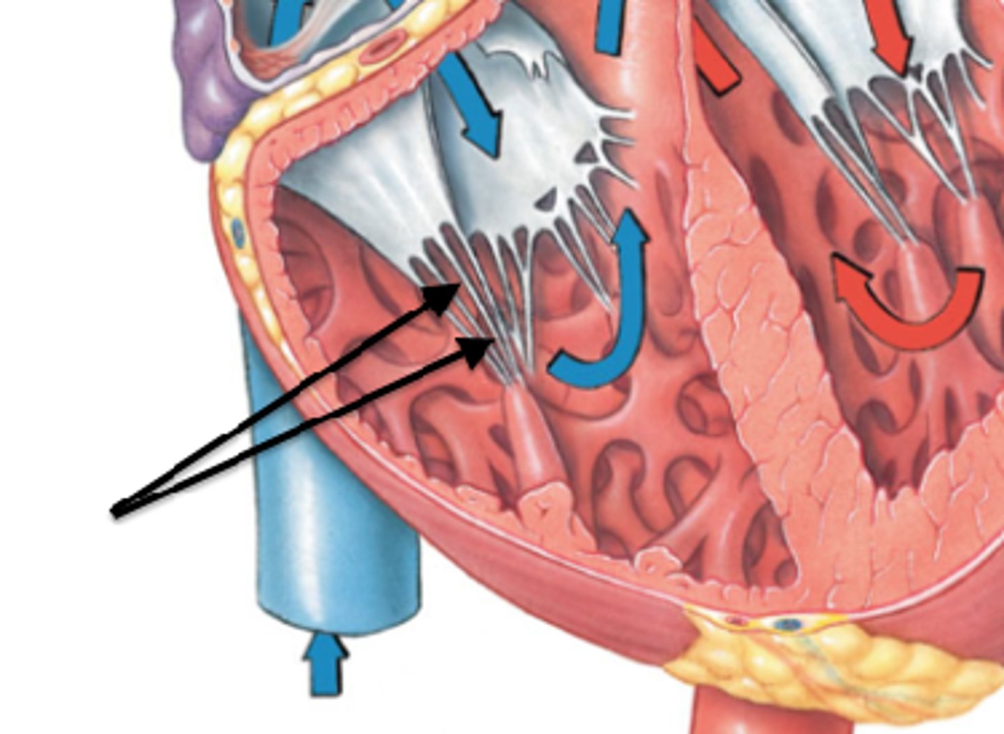
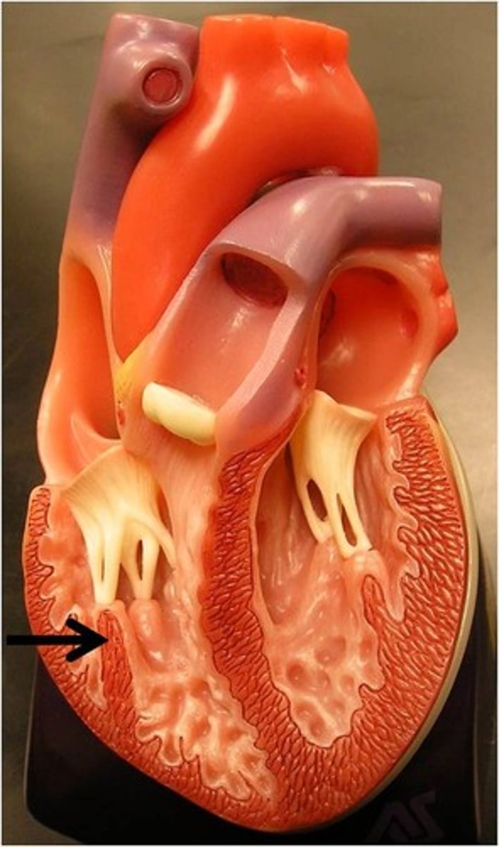
papillary muscles
anchor chordae tendineae

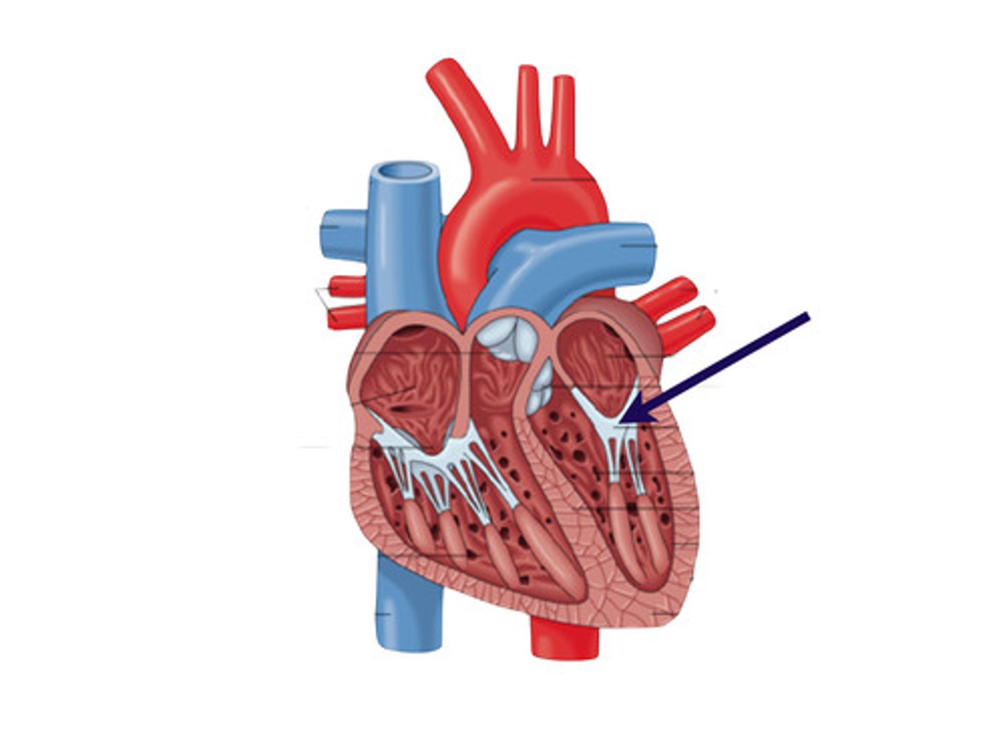
chordae tendineae
prevents right and left atrioventricular valves from everting back into atrium
pulmonary semilunar valve
prevents back flow of blood into right ventricle
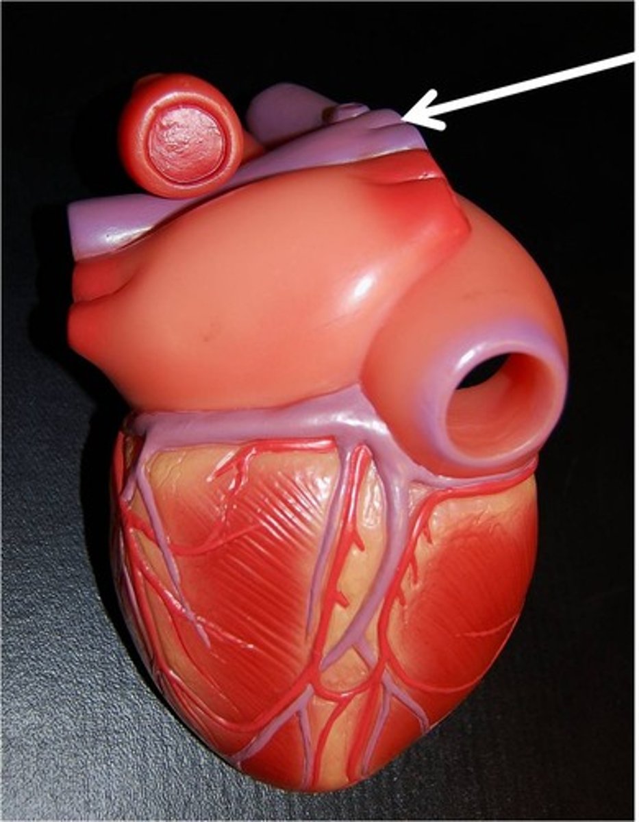
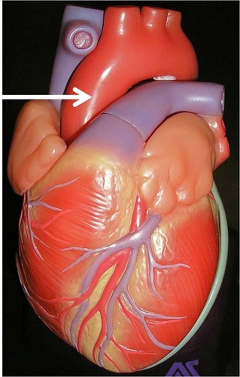
pulmonary trunk
deoxygenated blood leaves here
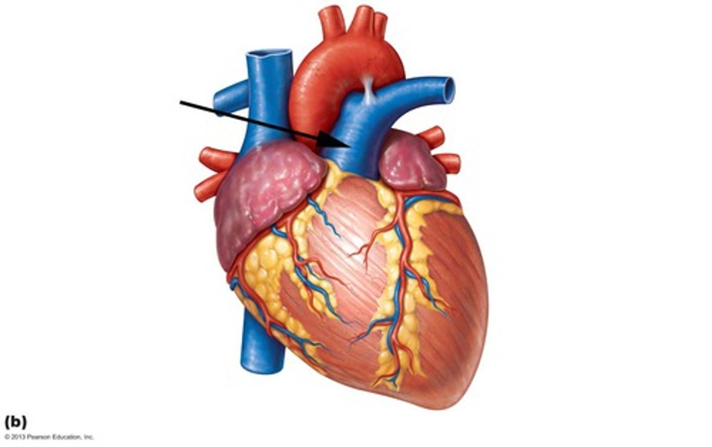
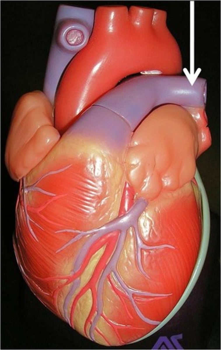
right pulmonary artery
takes blood to lungs to get oxygenated. Can see from posterior side
1 rt and lt artery
left pulmonary artery
takes blood to lungs to get oxygenated. Can see from anterior side.
1 rt and lt artery
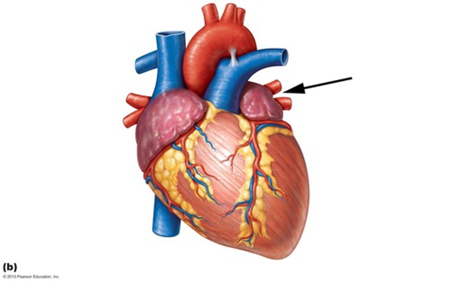
left atrium
yay
Left auricle
AKA left atrial appendage. Identify the flap.
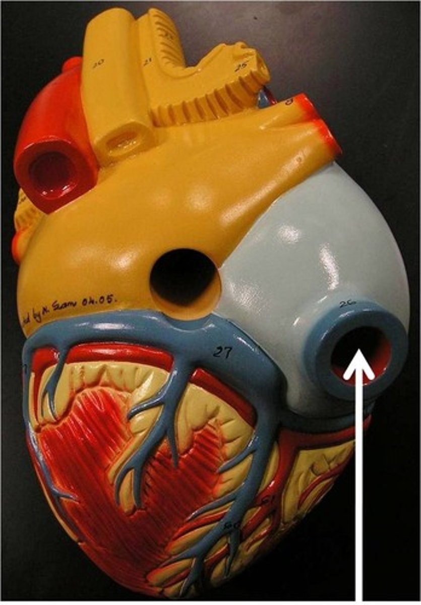
right pulmonary veins
bring oxygen-rich blood from the right lung to the left atrium
Left pulmonary veins
bring oxygen-rich blood from the left lung to the left atrium
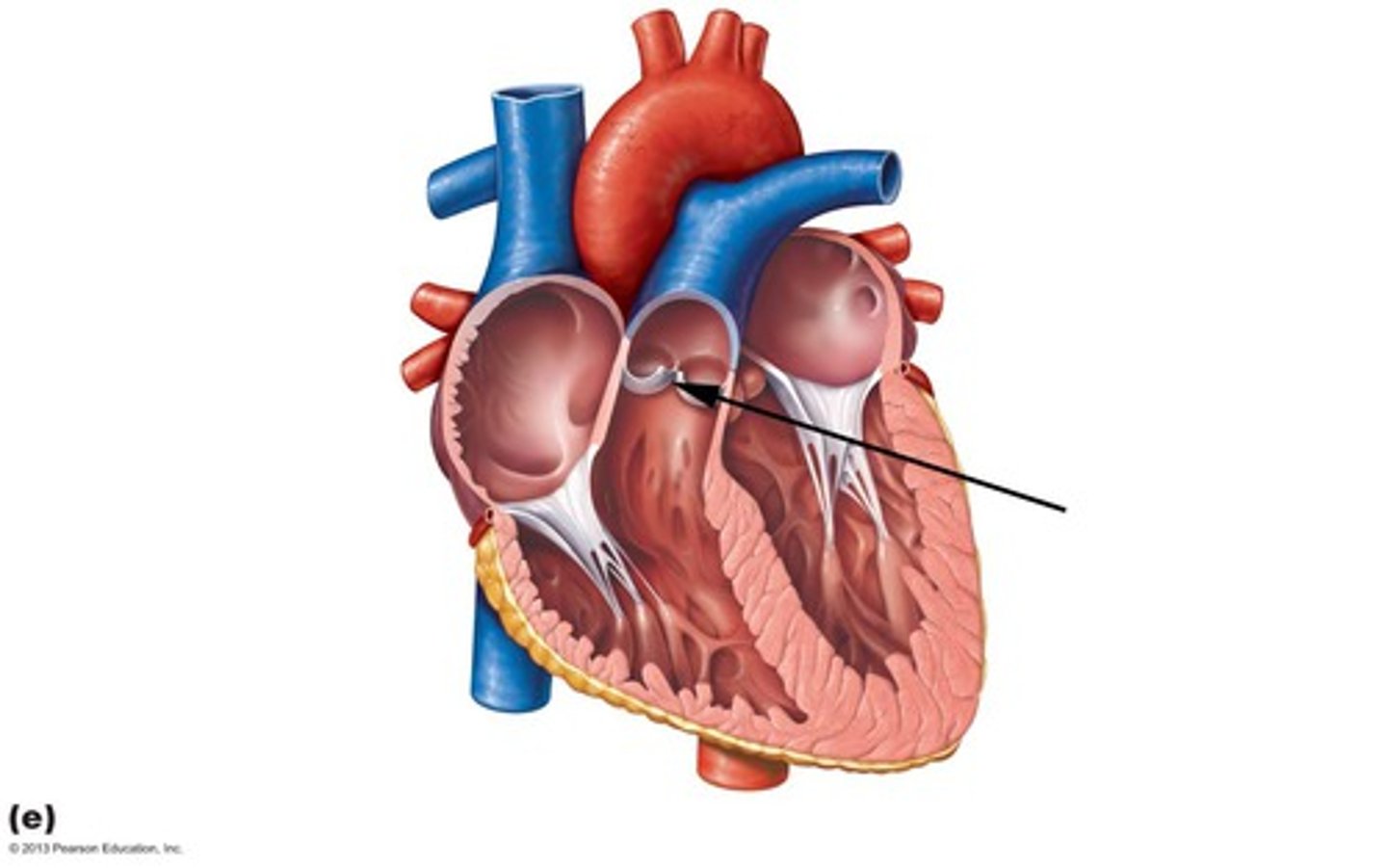
Left atrioventricular orifice
opening between left atrium and left ventricle. 2 cusps.
bicuspid, mitral, or lt. atrioventricular valve
prevents back flow of blood into left atrium
Left ventricle
yay
trabeculae carneae
muscular ridges on the internal surface of the ventricles
papillary muscles
anchor chordae tendineae
chordae tendineae
prevents right and left atrioventricular valves from everting back into atrium

aortic semilunar valve
prevents back flow of blood into the left ventricle

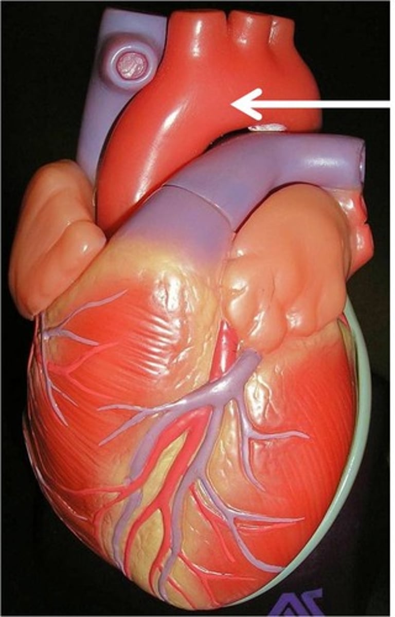
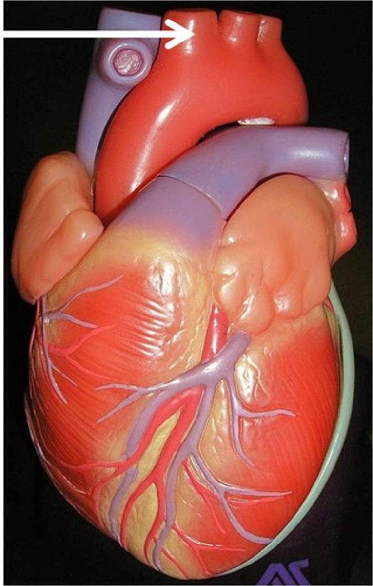
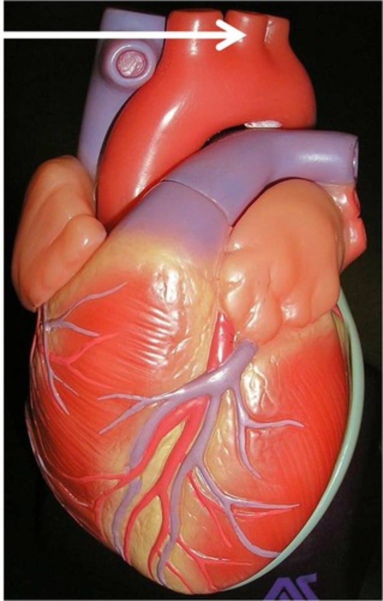
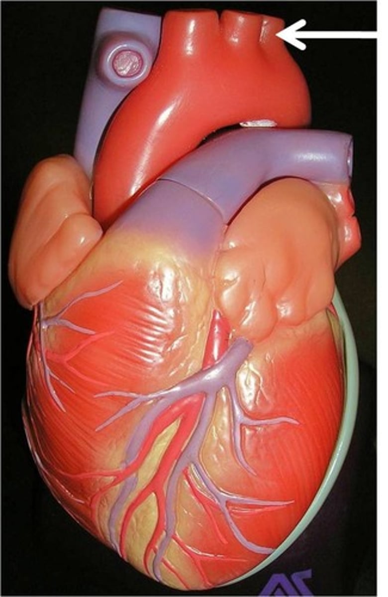
aorta
Largest artery in the body
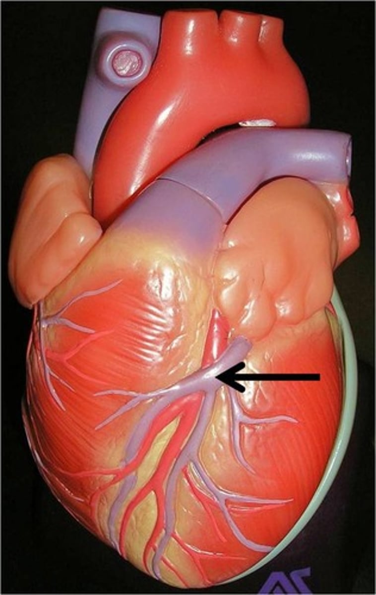
coronary arteries
yay
aorta
yay!

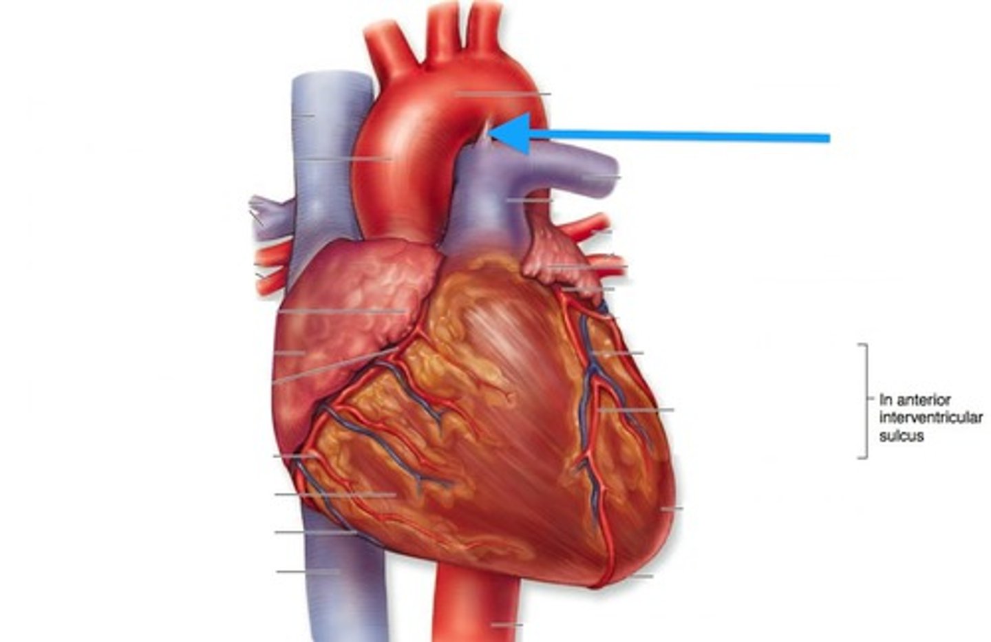
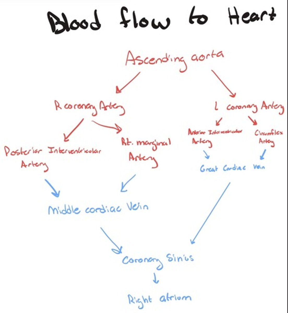
left coronary artery
Supplies blood to left heart structures.
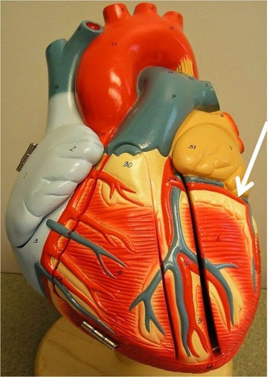
anterior interventricular artery/left anterior descending artery
...

circumflex artery
...
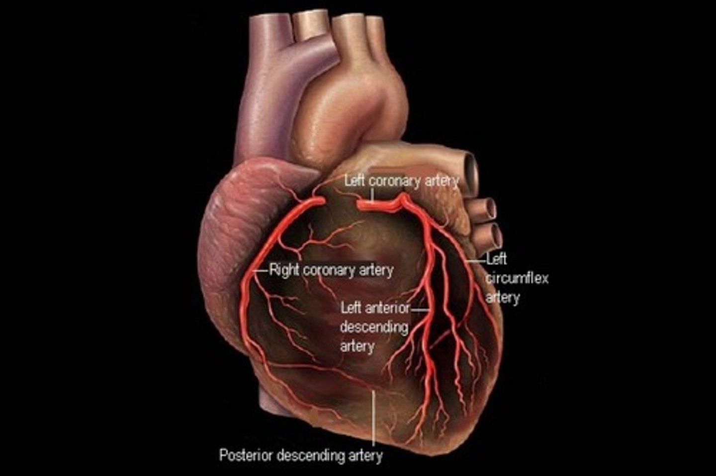
right coronary artery
supplies heart on right side, rungs along interventricular sulcus, can also see on posterior side
right marginal artery
...
posterior interventricular artery
...
cardiac veins
yay
great cardiac vein
...
middle cardiac vein
...
coronary sinus
...
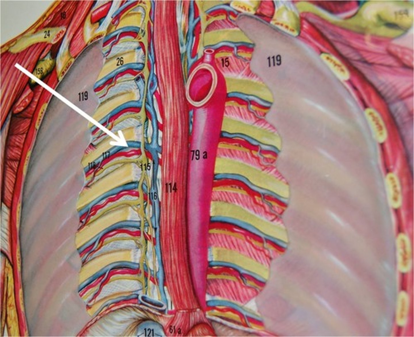
arteries within the thorax
yay
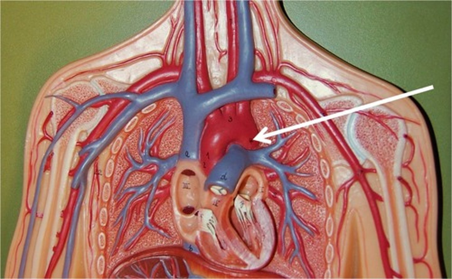
aorta
...

ascending aorta
has coronary arteries

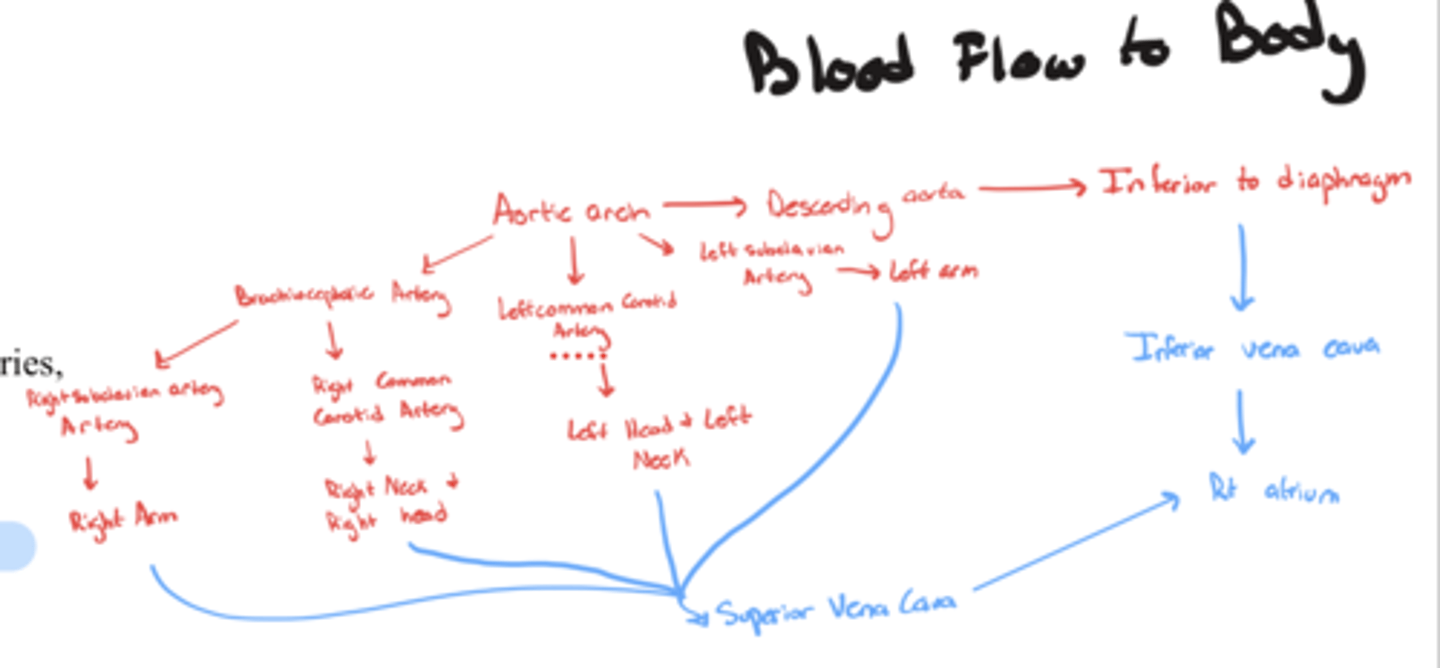
aortic arch
brachiocephalic artery: rt common carotid artery, rt subclavian artery
lt. common carotid artery
lt. subclavian artery
brachiocephalic artery
...
right common carotid artery
supplies blood to neck and head on right side
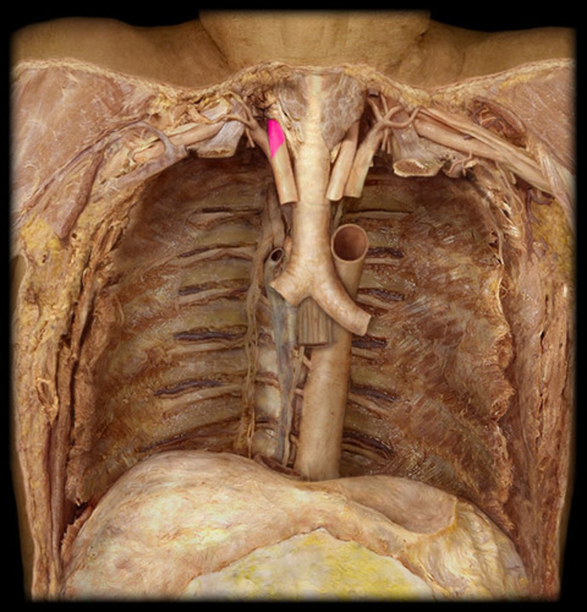
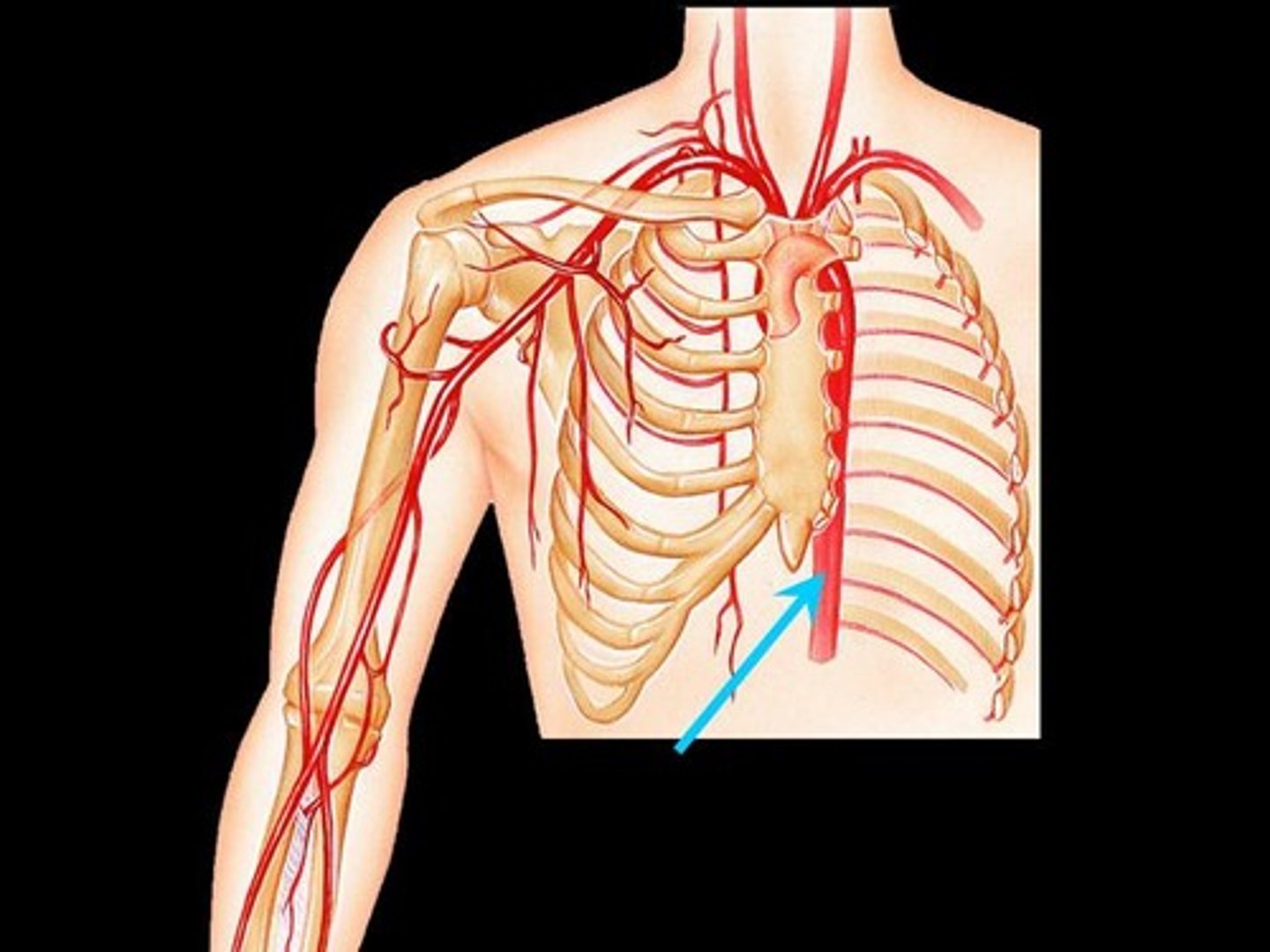
right subclavian artery
supplies blood to the right arm
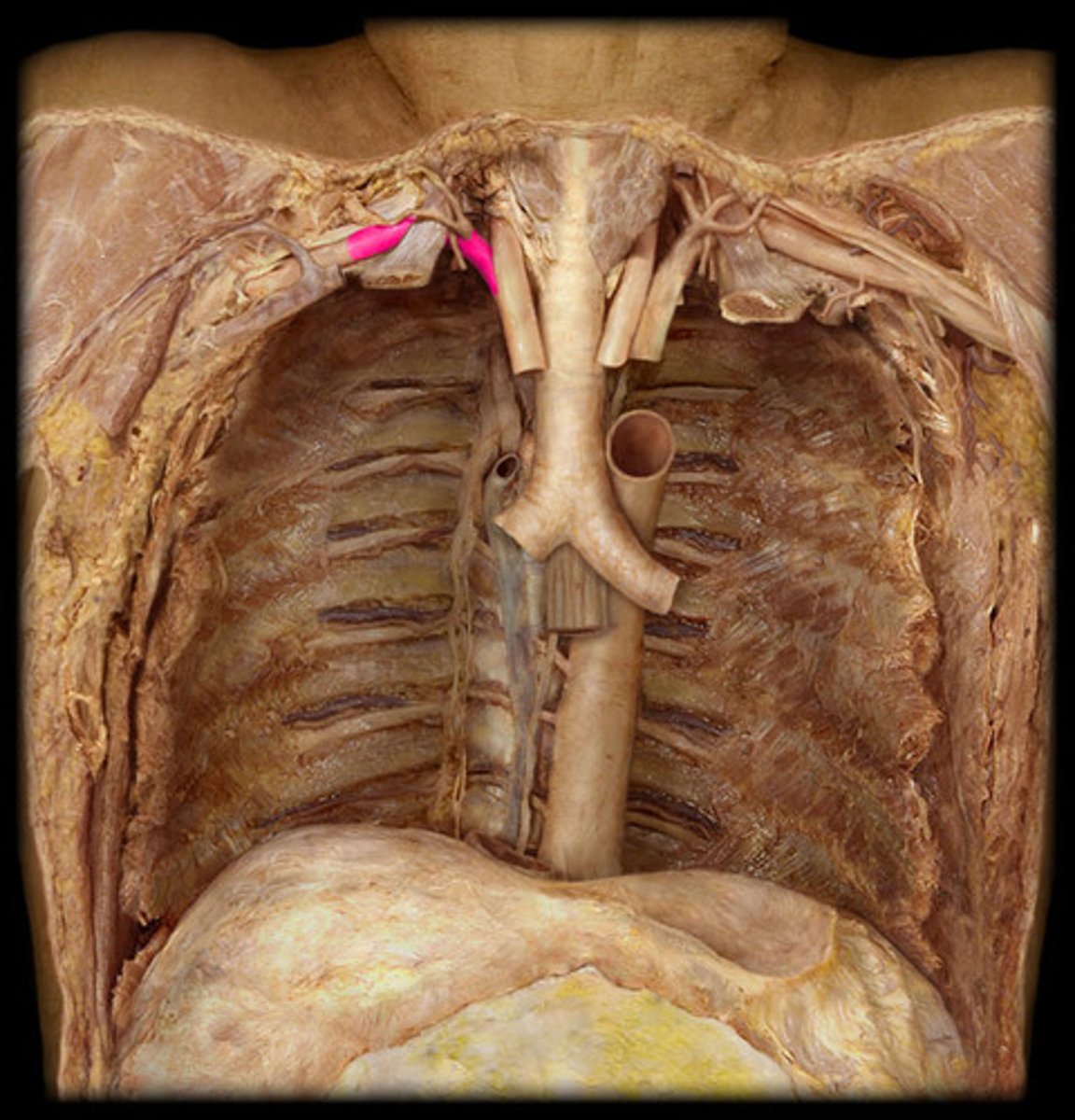
left common carotid artery
supplies blood to neck and head on left side
left subclavian artery
supplies blood to the left arm
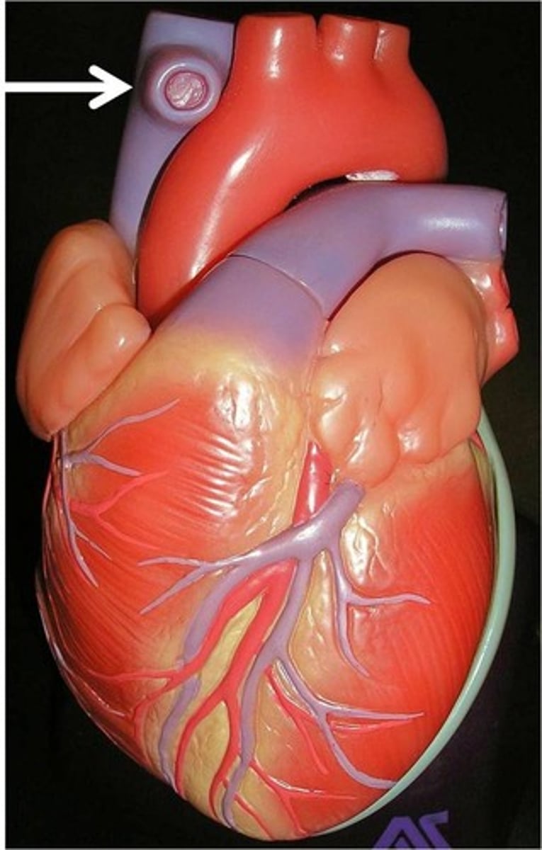
ligamentum arteriosum
remnant of ductus arteriosus. connects pulmonary trunk to aortic arch.
descending aorta
...
thoracic aorta
contains posterior intercostal arteries
posterior intercostal arteries
supply the vertebrae, spinal cord, intercostal muscles, and deep muscles of the back
Blood Flow Through Heart
1-Superior & Inferior Vena Cava
2-Right Atrium
3-Tricuspid Valve
4- Right Ventricle
5-Pulmonary Semilunar Valve
6-Pulmonary Trunk
7- Right and Left Pulmonary Artery
8- Right and Left Lungs
9- Right and Left Pulmonary Veins
10- Left Atrium
12- Mitral Valve (Bicuspid)
12-Left Ventricle
13- Aortic Semilunar Valve
14- Ascending Aorta
15- Aortic Arch
16- Descending Aorta
Blood Flow To Heart
...
Blood Flow to Body
...
Electrocardiogram (ECG/EKG)
record of the electrical activity of the heart
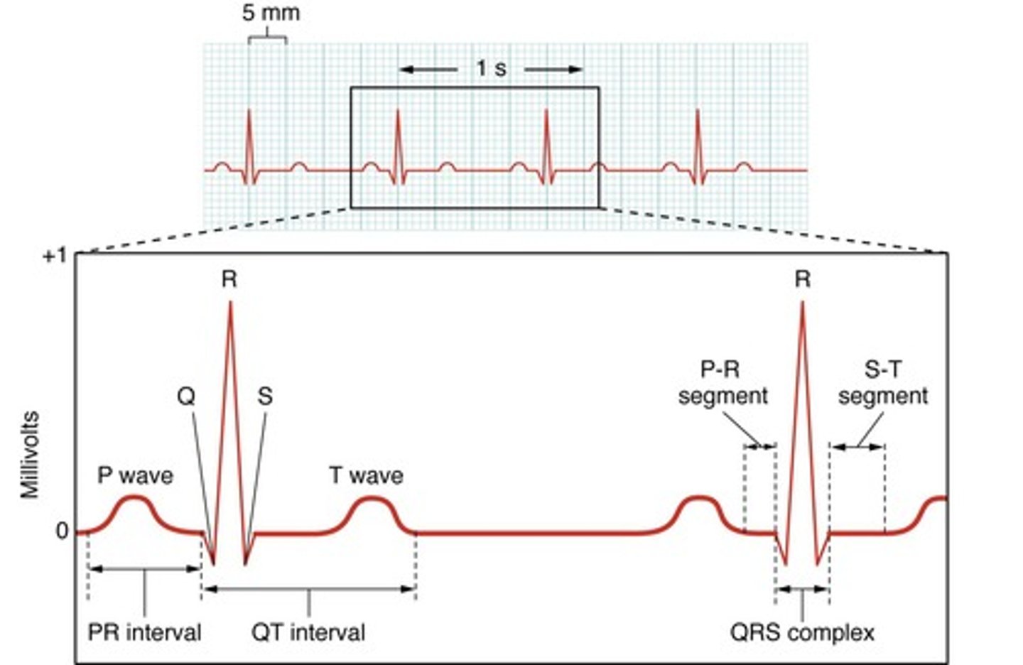
p wave
atrial depolarization
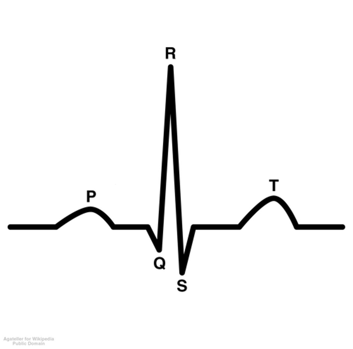
QRS Complex
Shows ventricular depolarization. However what actually occurs is ventricular depolarization and atrial repolarization (gets masked)

T wave
ventricular repolarization

Lub
First heart sound. closing of right and left atrioventricular valves
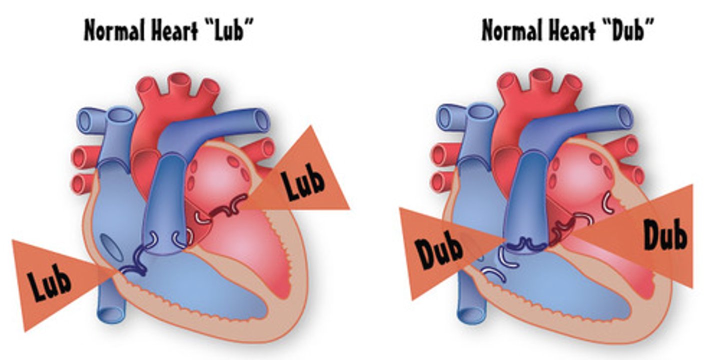
Dub
Second heart sound. closing of pulmonary and aortic semilunar valves

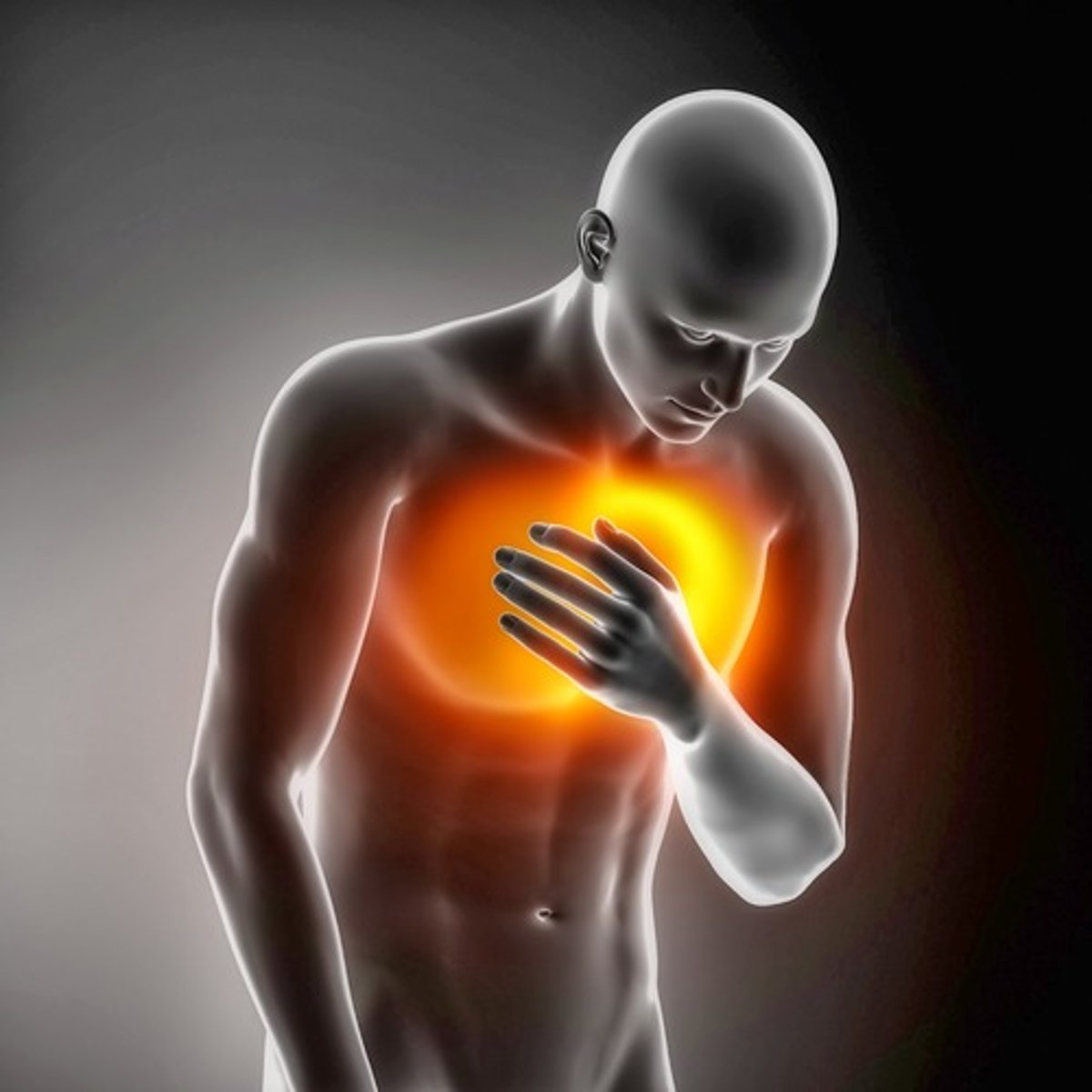
Angina pectoris
chest pain that results when the heart does not get enough oxygen
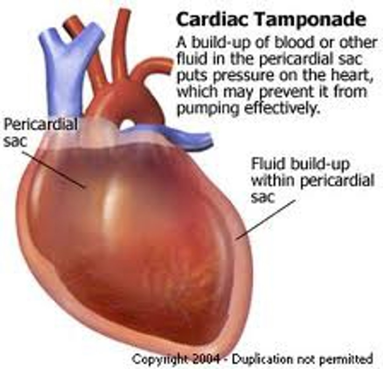
Cardiac Tamponade
acute compression of the heart caused by fluid accumulation in the pericardial cavity
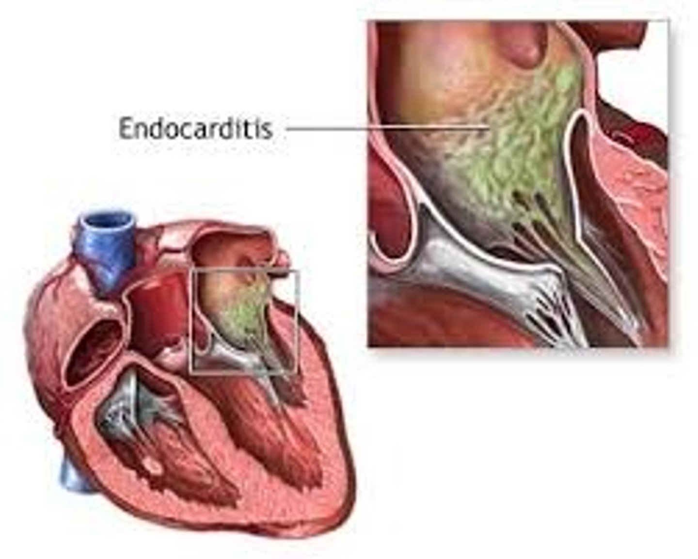
Endocarditis
inflammation of the endocardium
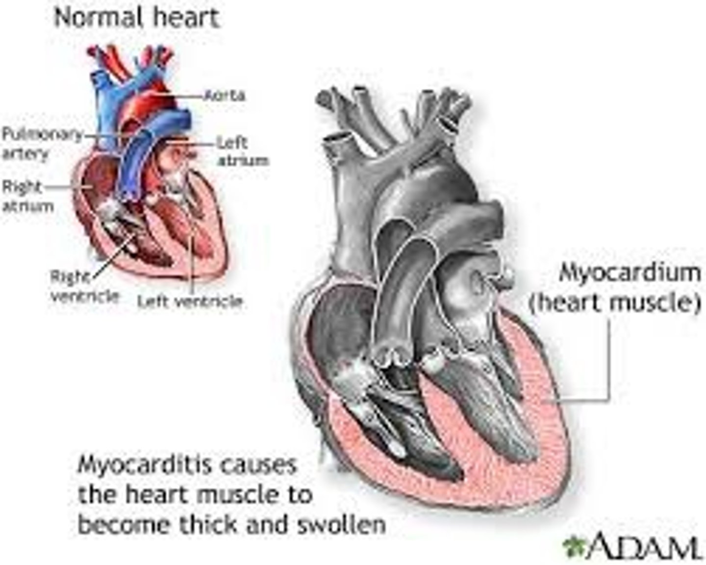
Myocarditis
inflammation of the myocardium
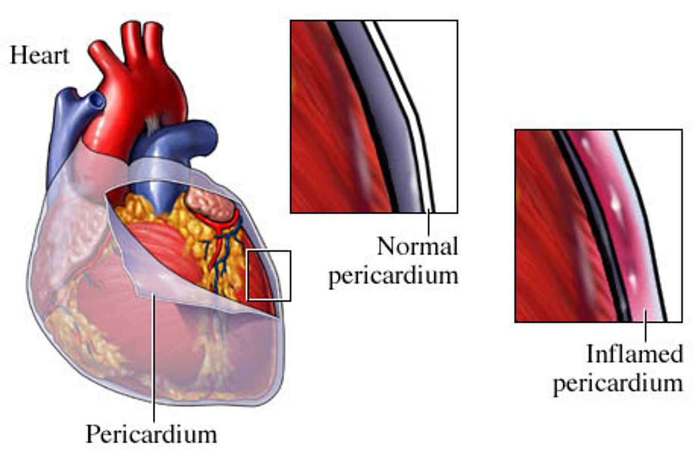
Pericarditis
inflammation of the pericardium
Congenital birth defects
Abnormalities present at birth, caused by genetic or environmental factors
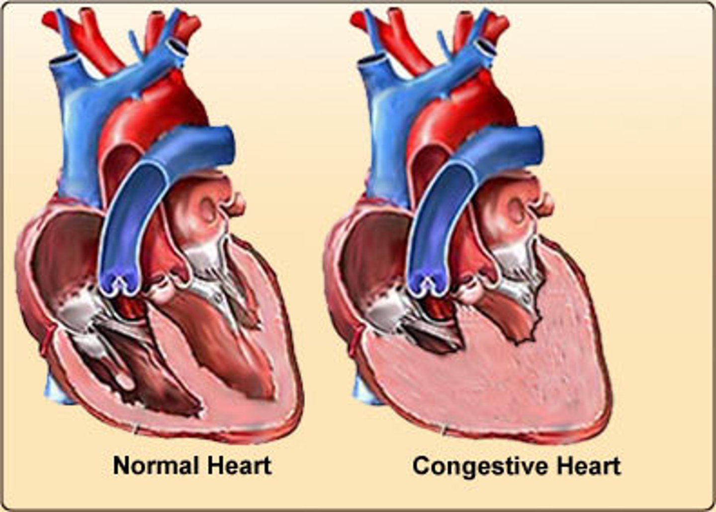
Congestive Heart Failure
heart is unable to pump its required amount of blood
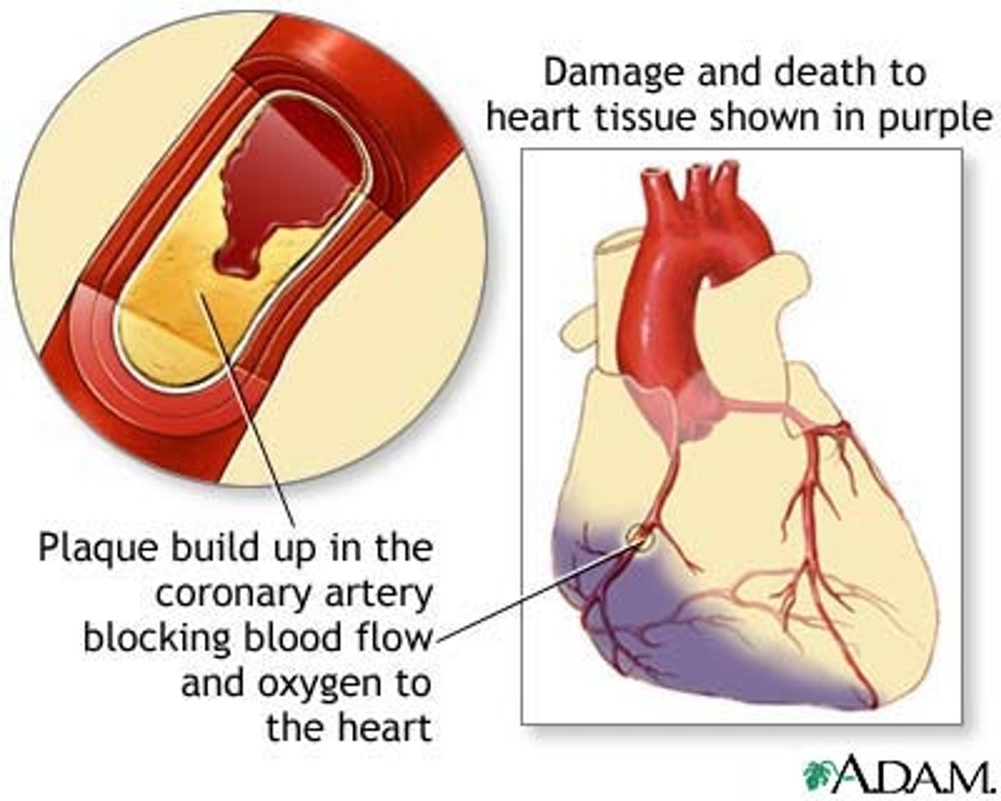
Coronary Artery disease (CAD)
disease of the arteries surrounding the heart
Heart Valve Disorders
one of the four heart valves does not open well or does not close tightly
Heart Block
interference with normal conduction of electrical impulses that control activity of the heart muscle
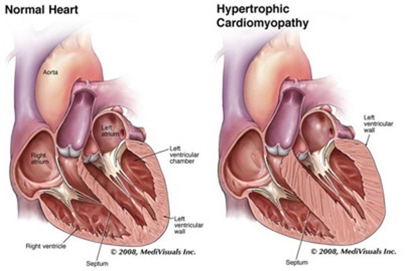
Hypertrophic Cardiomyopathy
heart muscle becomes enlarged and blocks blood flow
Myocardial infarction
the occlusion of one or more coronary arteries caused by plaque buildup (heart attack)
Pulmonary arterial hypertesion
Pulmonary arterial hypertension is a syndrome in which the blood pressure in the pulmonary arteries and pulmonary arterioles is elevated.
Tachycardia
fast heart rate
Bradycardia
slow heart rate