e
1/340
There's no tags or description
Looks like no tags are added yet.
Name | Mastery | Learn | Test | Matching | Spaced | Call with Kai |
|---|
No analytics yet
Send a link to your students to track their progress
341 Terms
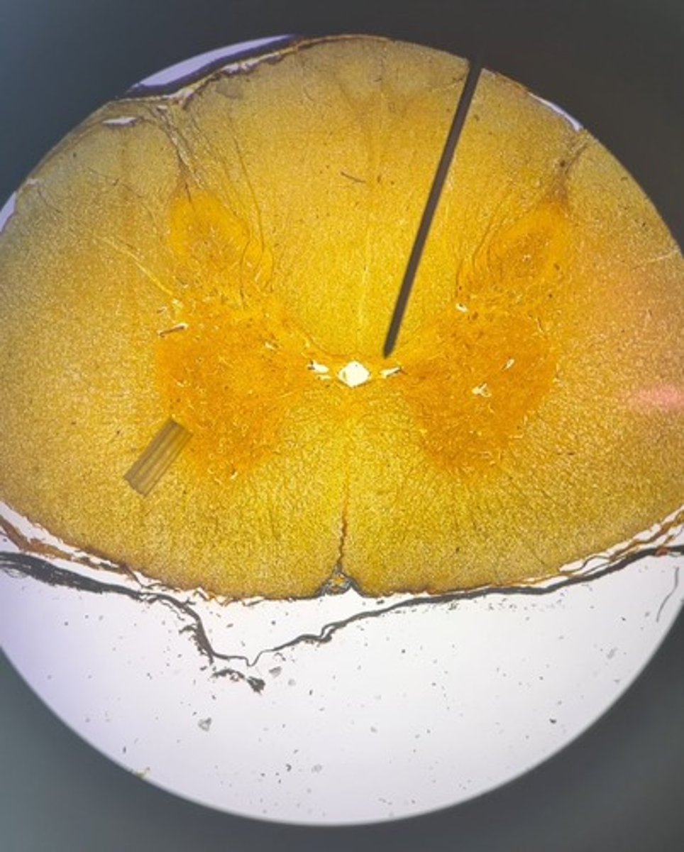
1. Gray commissure
2. Central canal
3. Posterior/ dorsal funiculus/ white column
4. Anterior/ ventral funiculus/ white column
1. What is the structure at the pointer?
2. What is the hole in the center near the pointer?
3. What is the lighter area of white matter above the pointer?
4. What is the lighter area of white matter below the pointer?

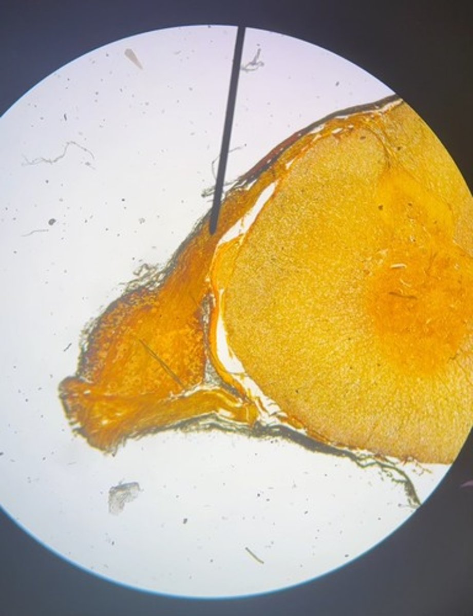
1. Dorsal root
2. Dorsal root ganglion
1. What is the structure at the pointer?
2. What is the large round structure to the lower left of the pointer?

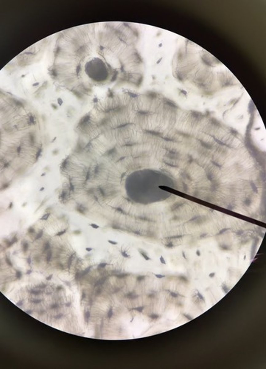
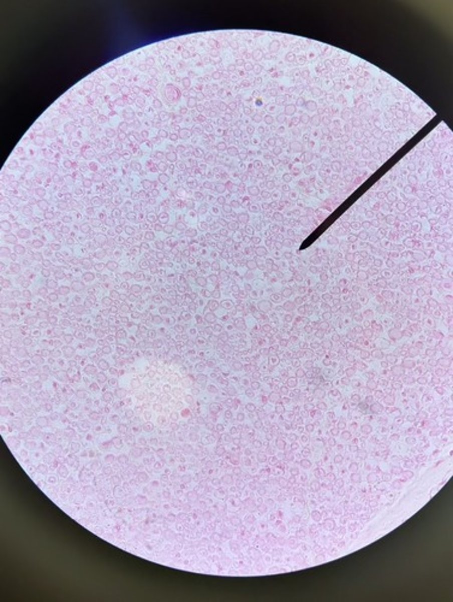
1. Osteon
2. Central canal
3. Lacunae
4. Canaliculi
1. What is one entire circular unit of compact bone seen here?
2. What structure is the pointer on?
3. What are the numerous smaller black "holes" seen in the slide?
4. what are the black lines or "cracks" seen in this slide?

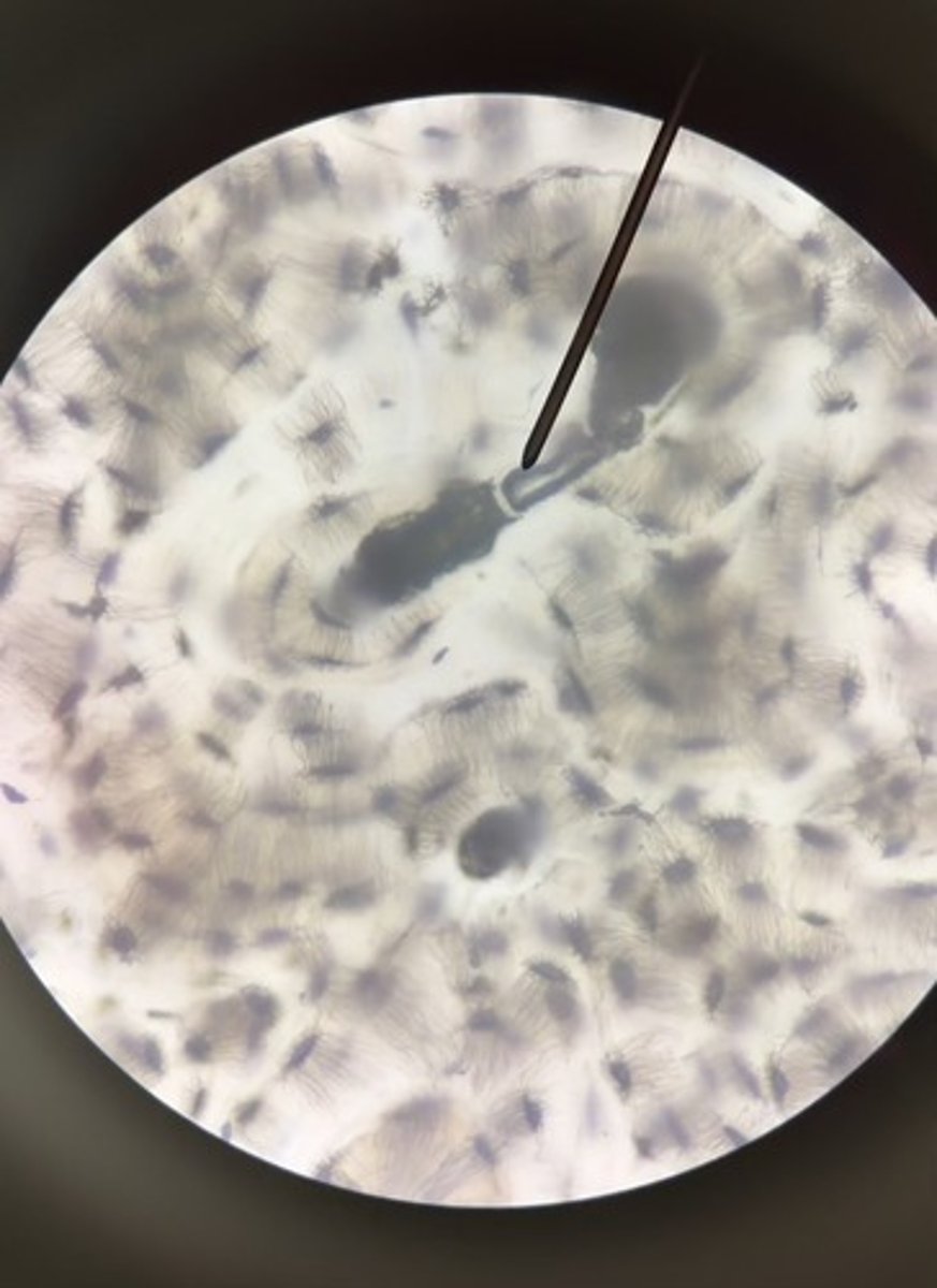
1. Perforating canal
2. Lamellae
1. What is the structure at the pointer?
2. What are the areas of hard bone matrix called?

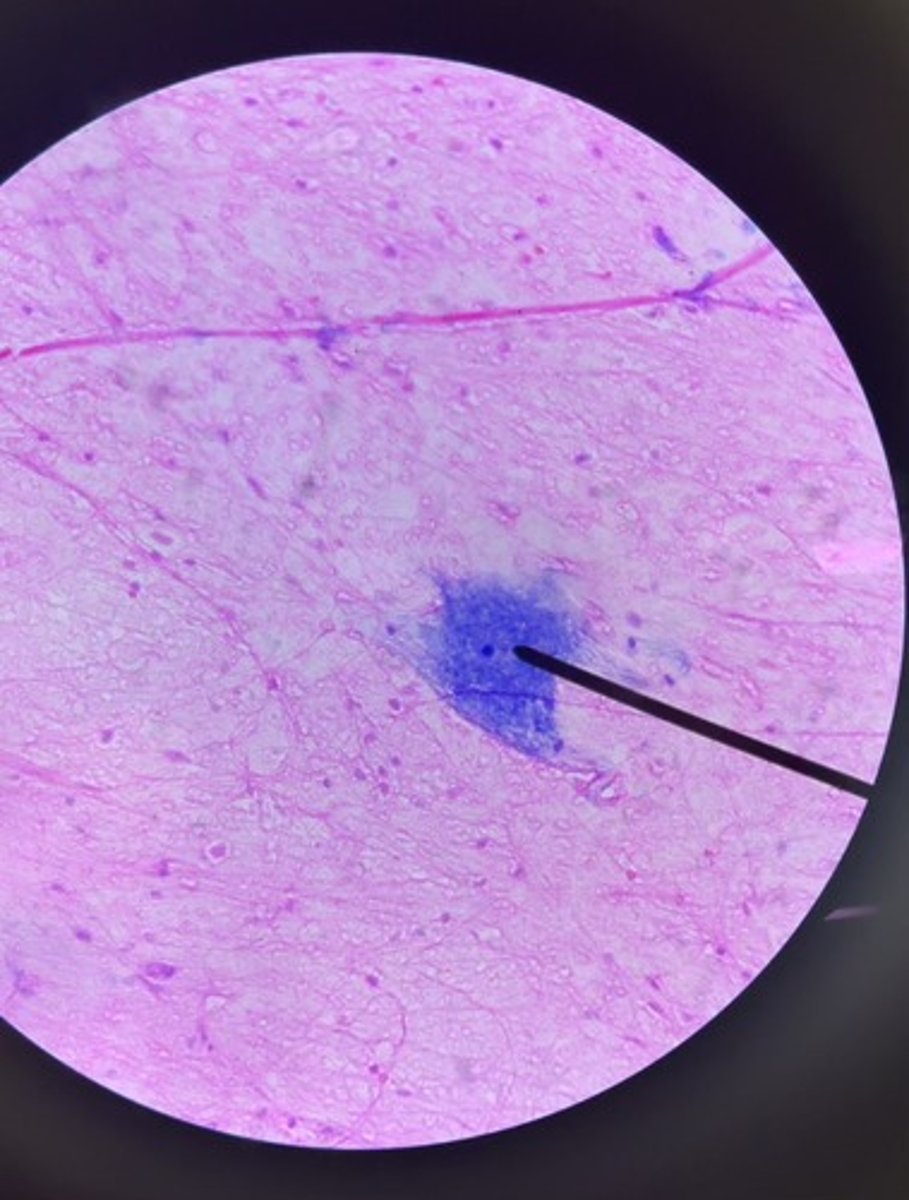
1. Multipolar neuron
2. Neuroglial cells or glial cells
1. What type of cell is seen at the pointer?
2. What type of cell is seen surrounding the cell at the pointer (smaller blue dots)?
1. Cross section of a nerve
2. Perineurium
3. Nerve fascicles
1. What are you looking at in this slide?
2. What connective tissue layer is the pointer on?
3. What are the large round structures called?
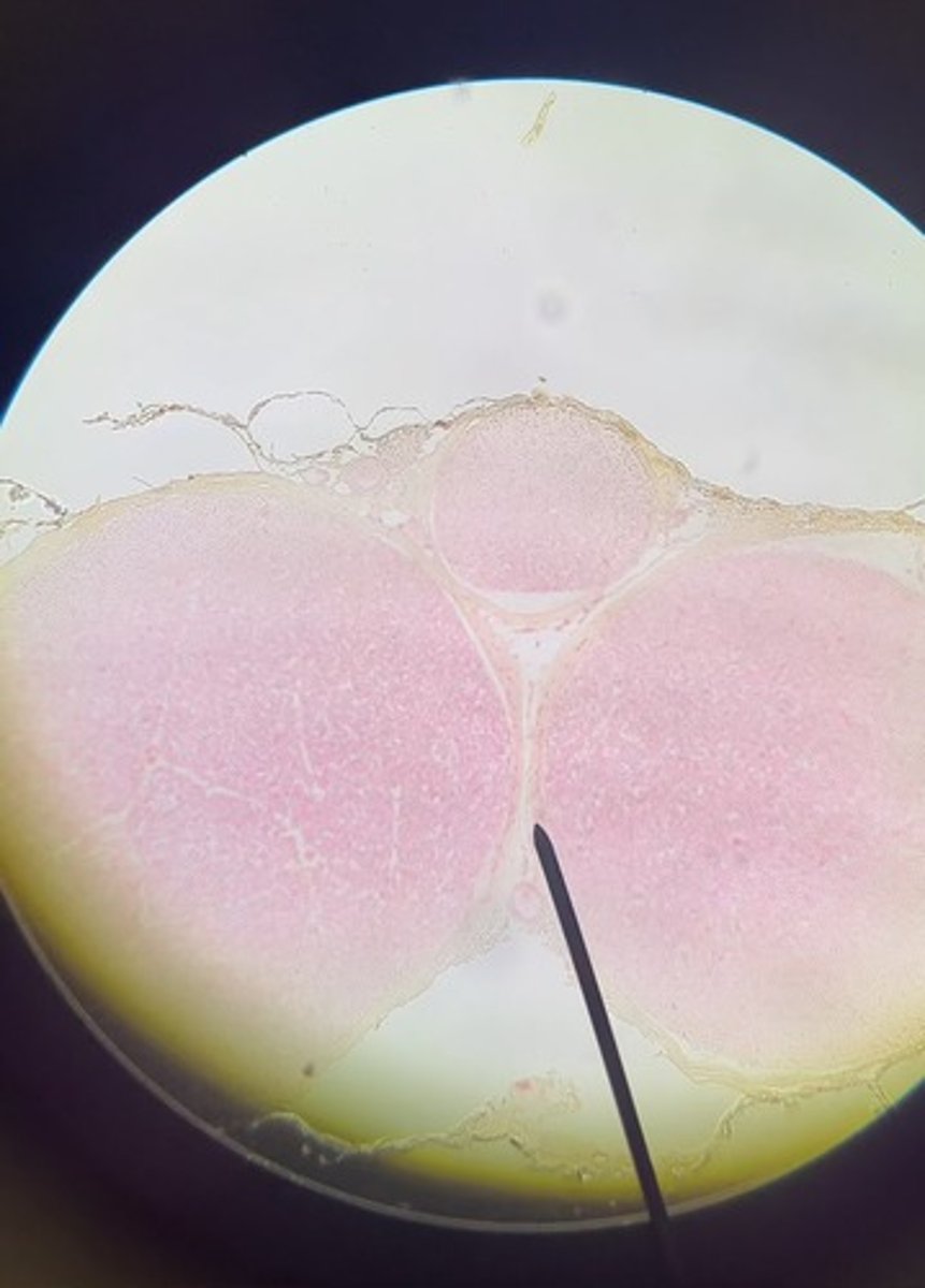
1. Axon
2. Myelin sheath
3. Endoneurium
1. What is the structure at the pointer (pink dot)?
2. What is the white area surrounding the pink dot?
3. What connective tissue layer would be found surrounding #1 and #2?

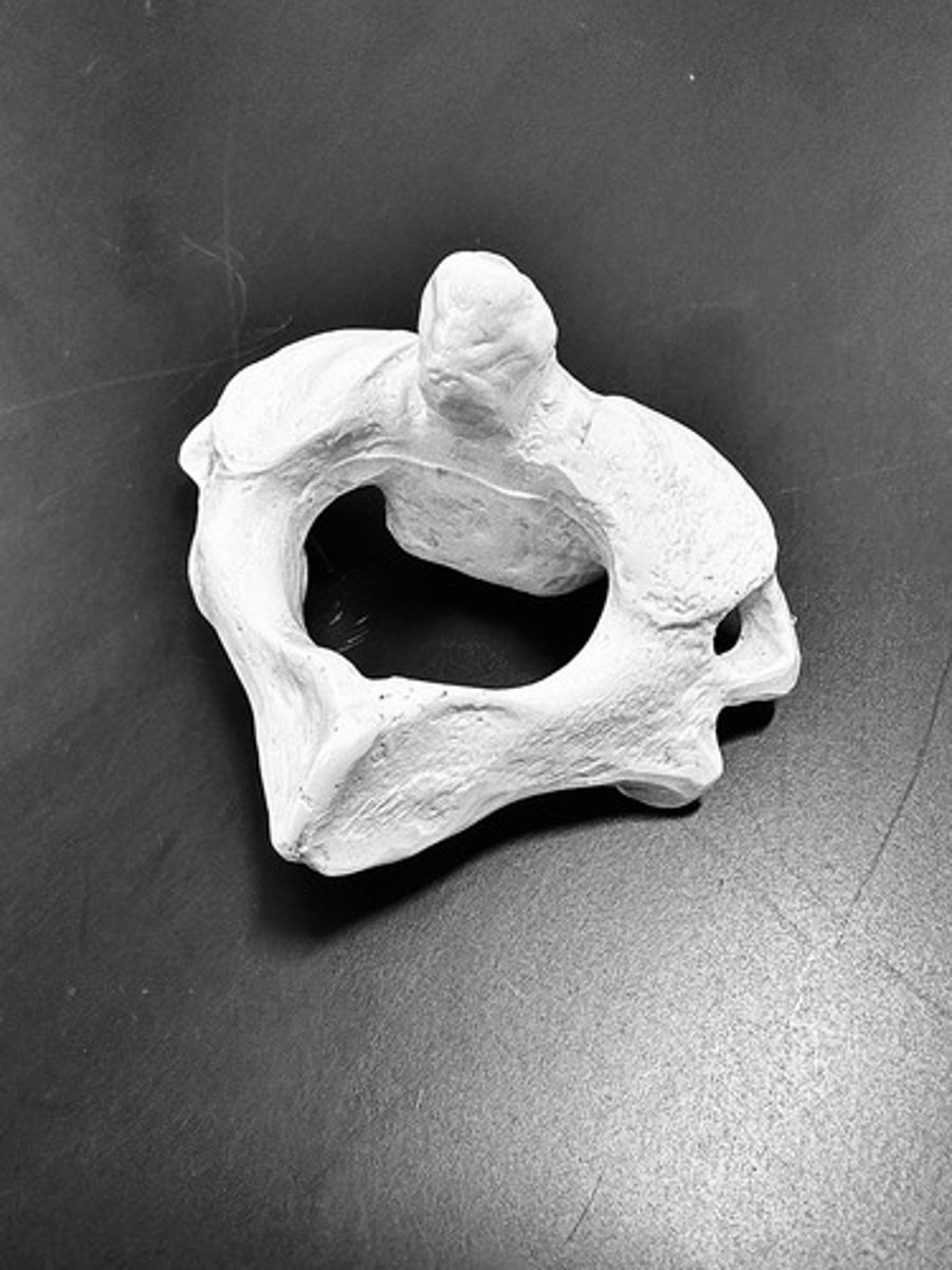
Atlas
The name for the first cervical vertebrae (C1)
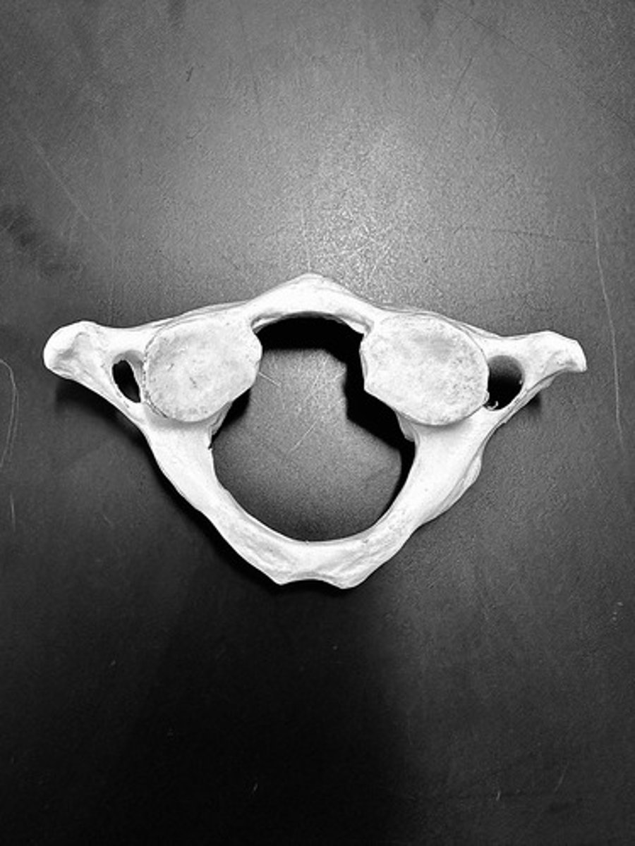
Axis
The name for the second cervical vertebrae (C2)
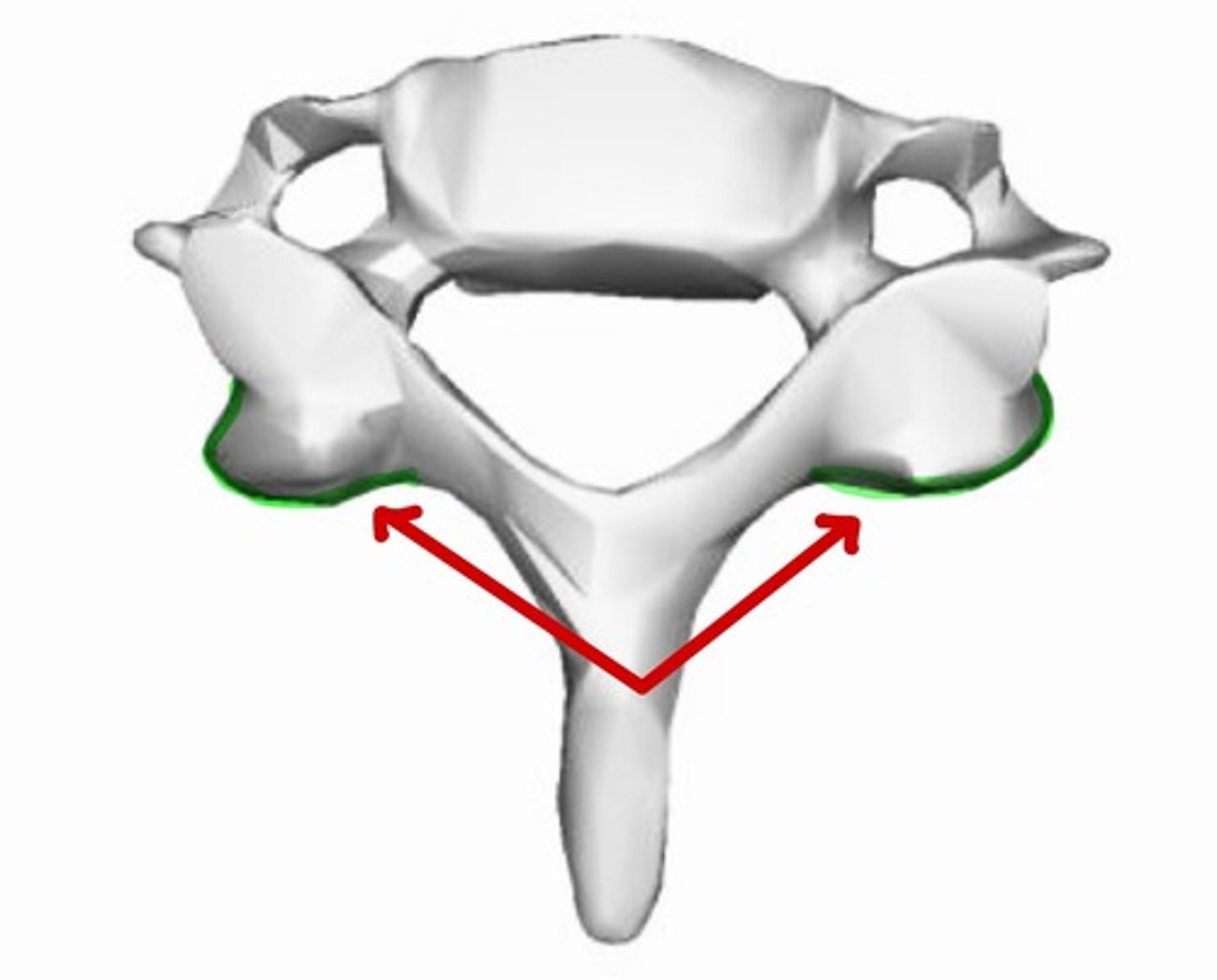
Lateral masses
anterior tubercle
posterior tubercle
Dens
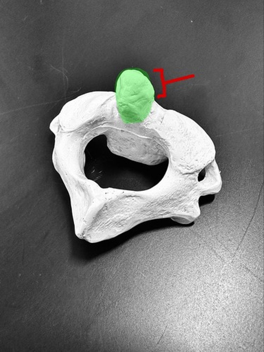
Body
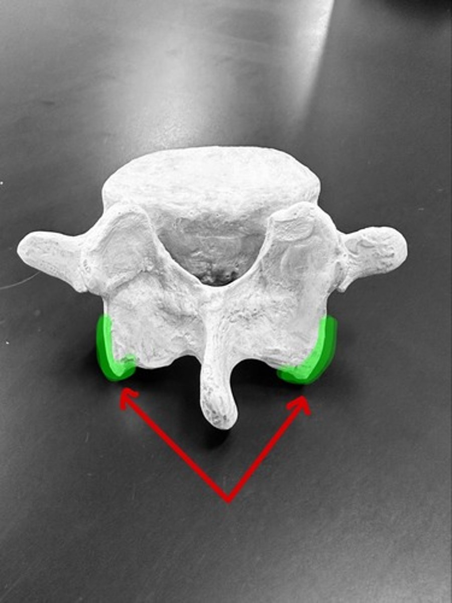
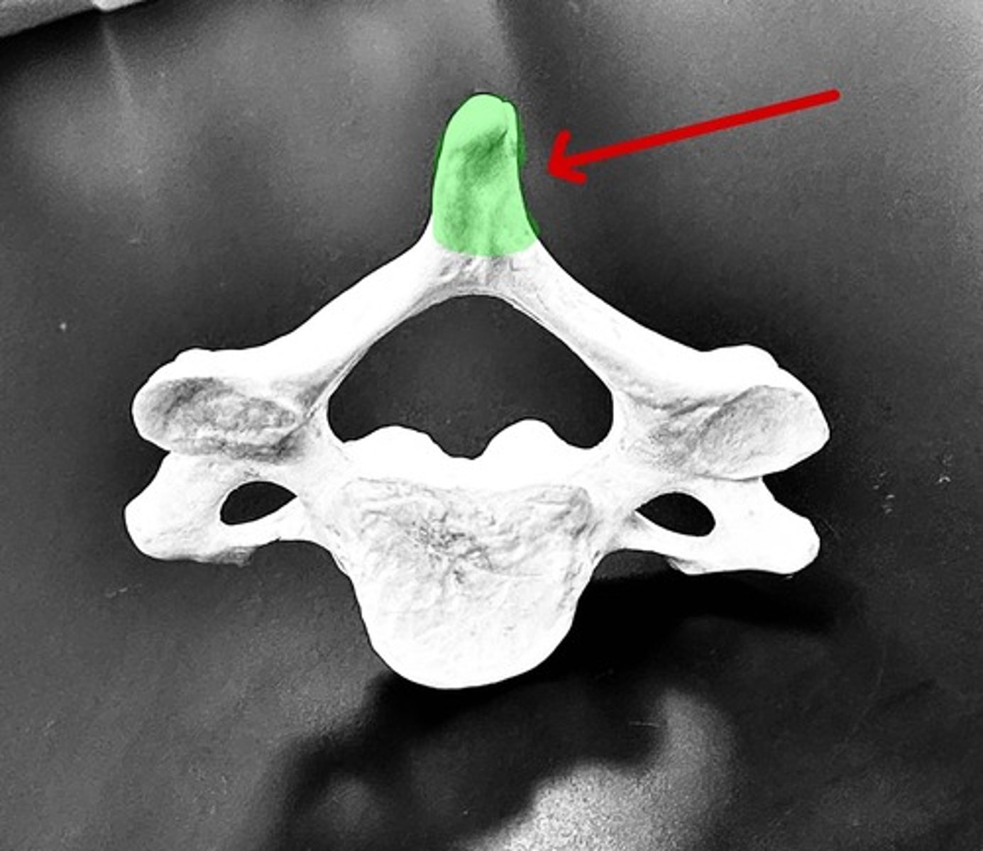
spinous process
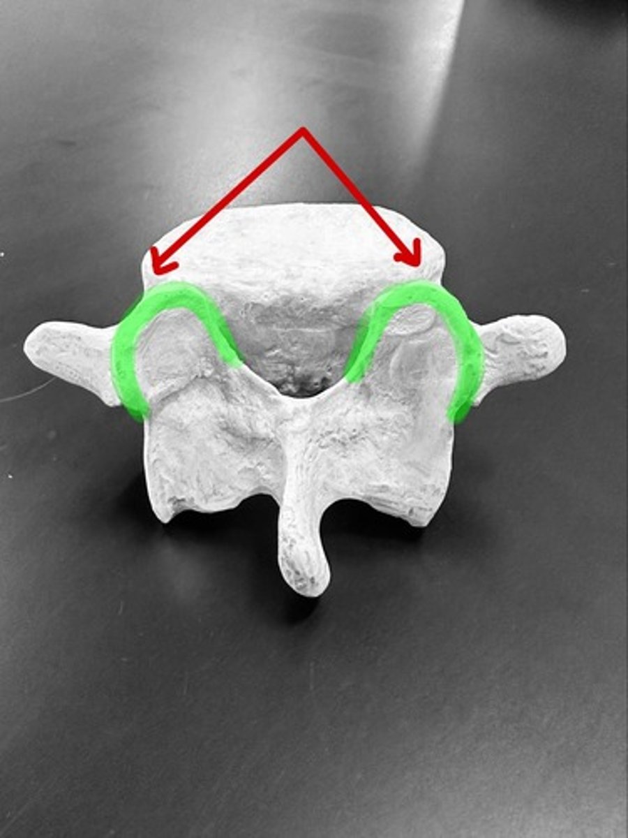
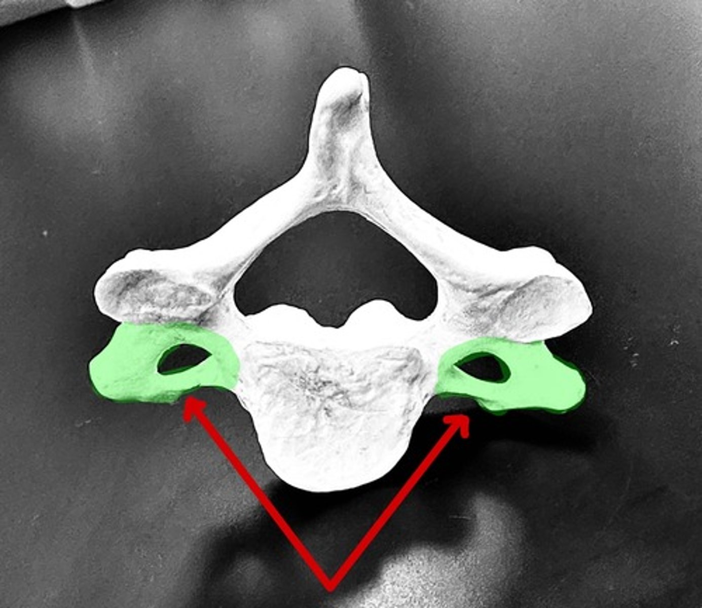
transverse process
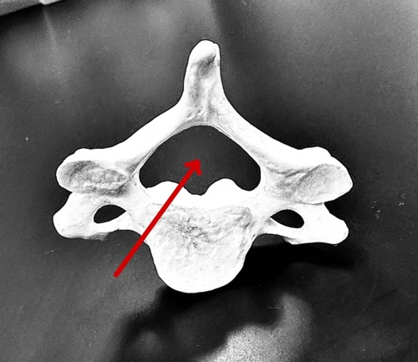
vertebral foramen
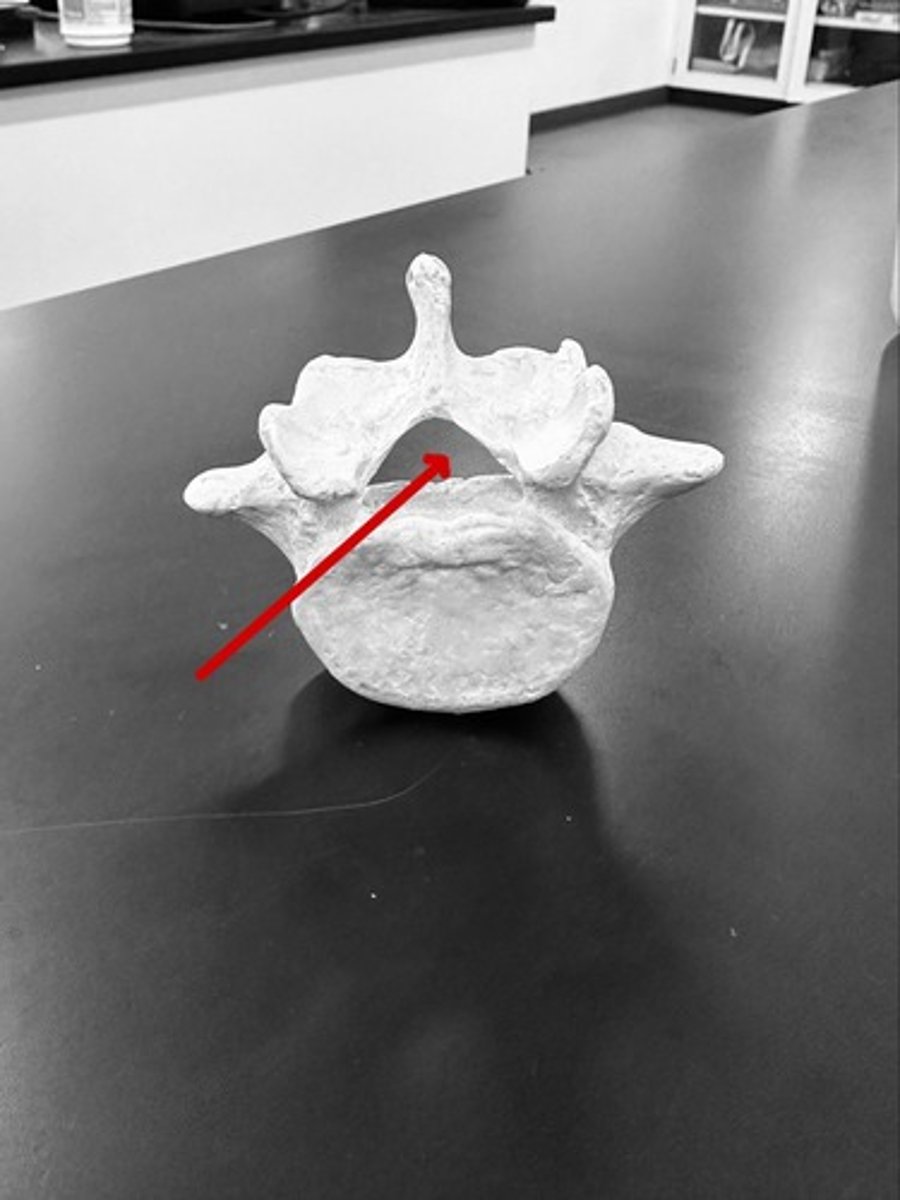
superior articular process
inferior articular process
pubic body
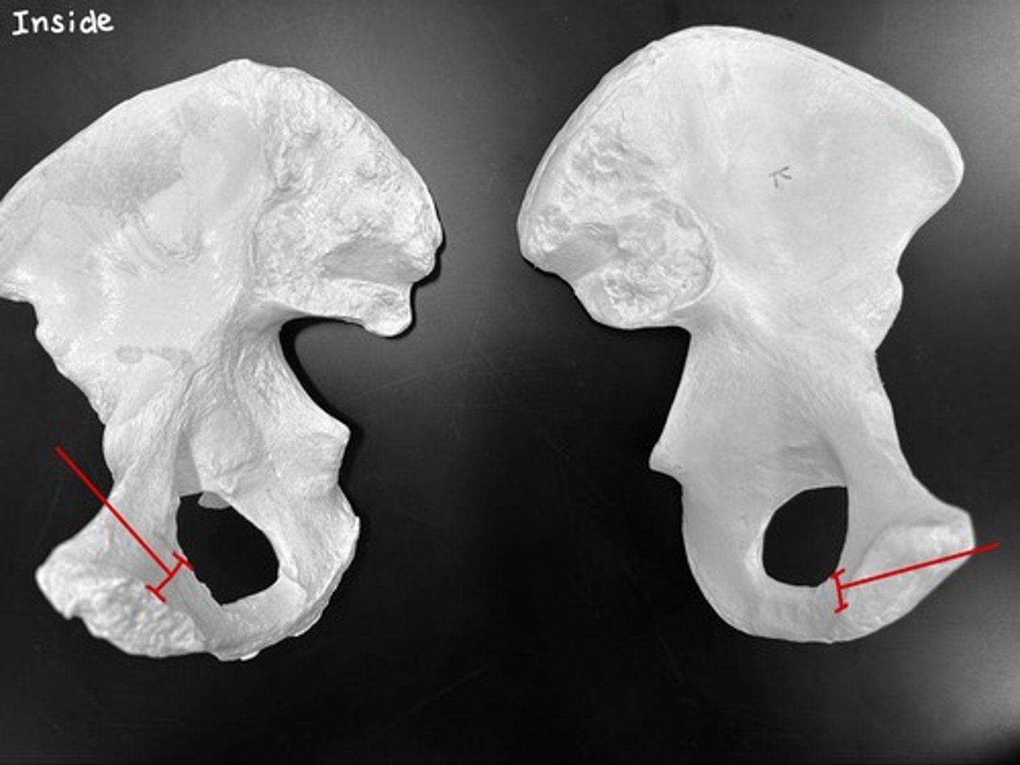
inferior pubic ramus
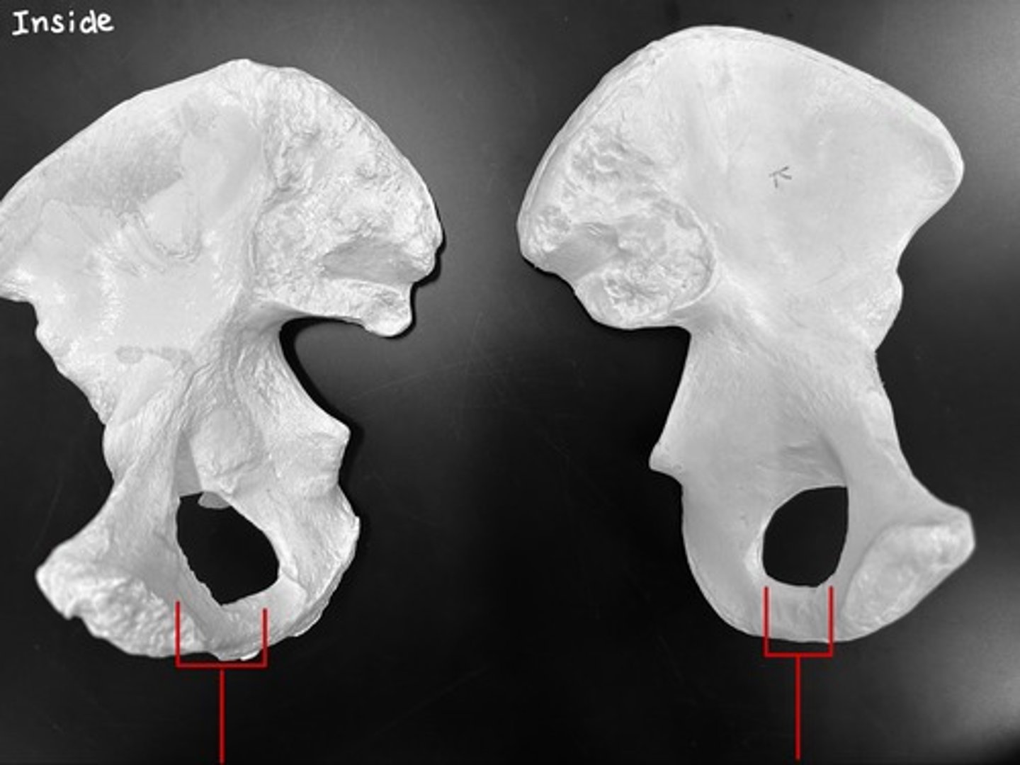
superior pubic ramus
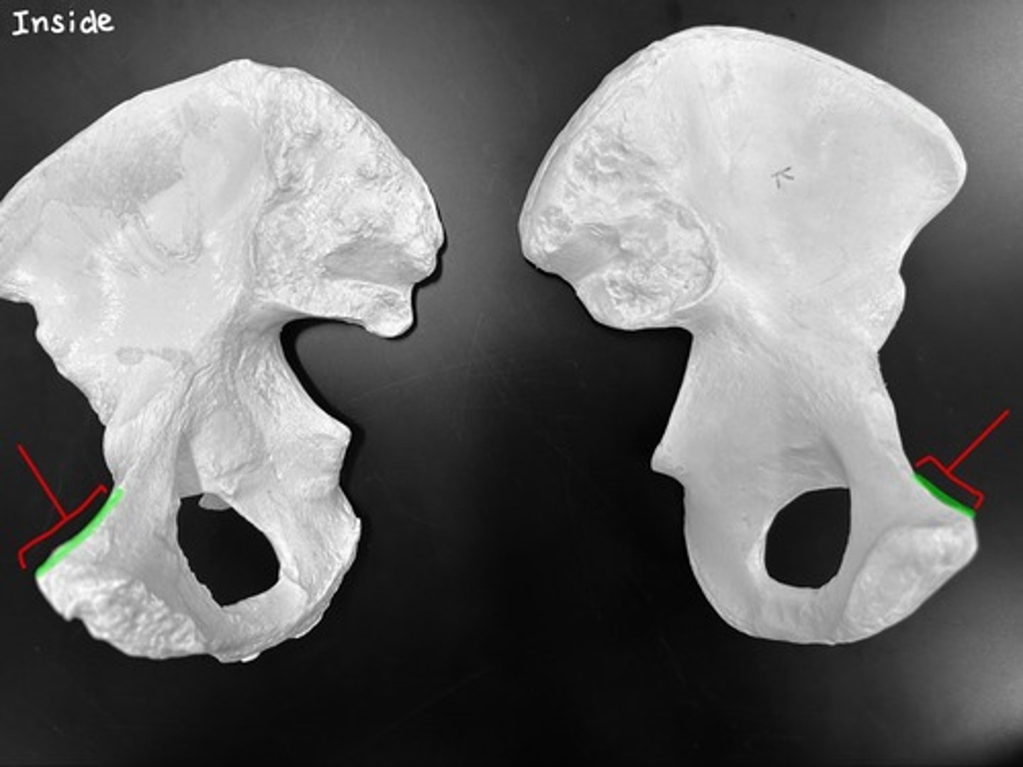
symphyseal/articular surface of pubis
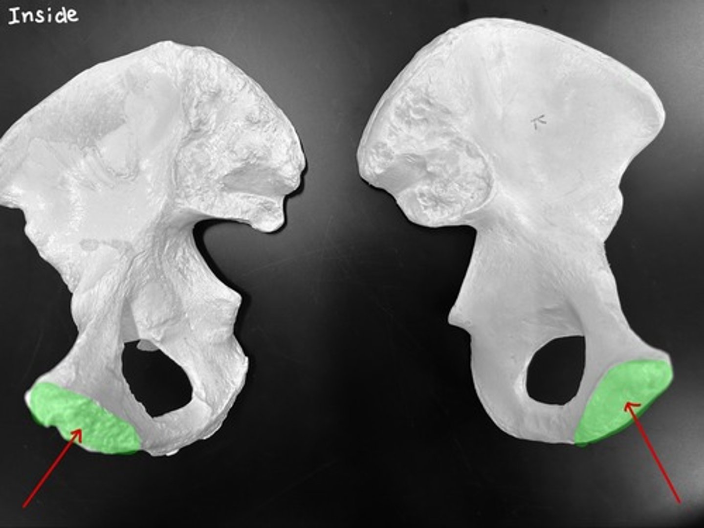
- symphyseal/articular surface of pubis is to the front
- acetabulum faces outward
How do you know which is left and which is right?
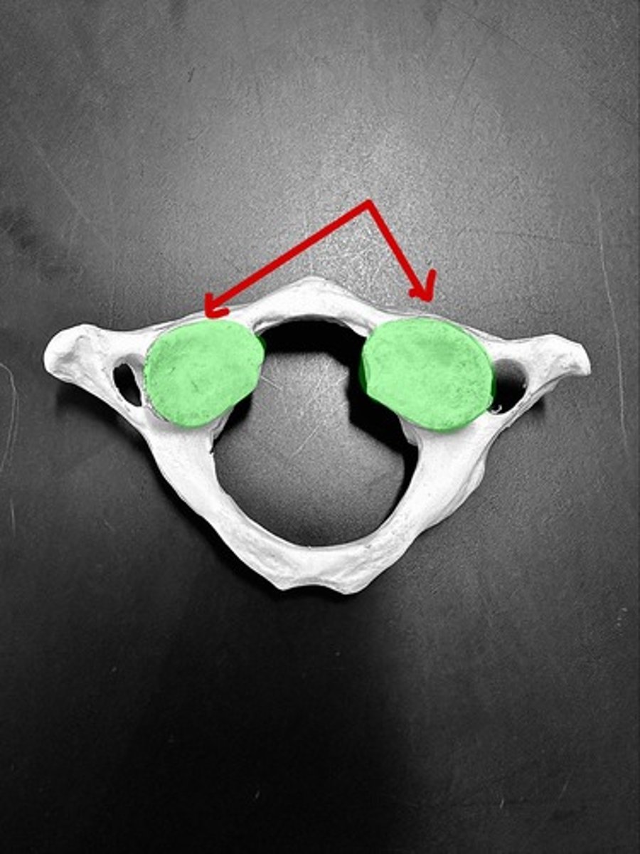
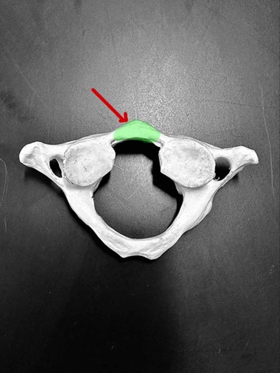
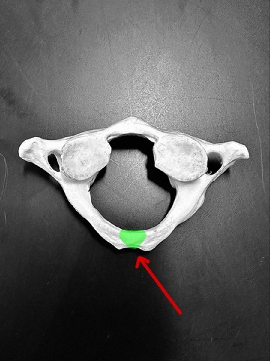
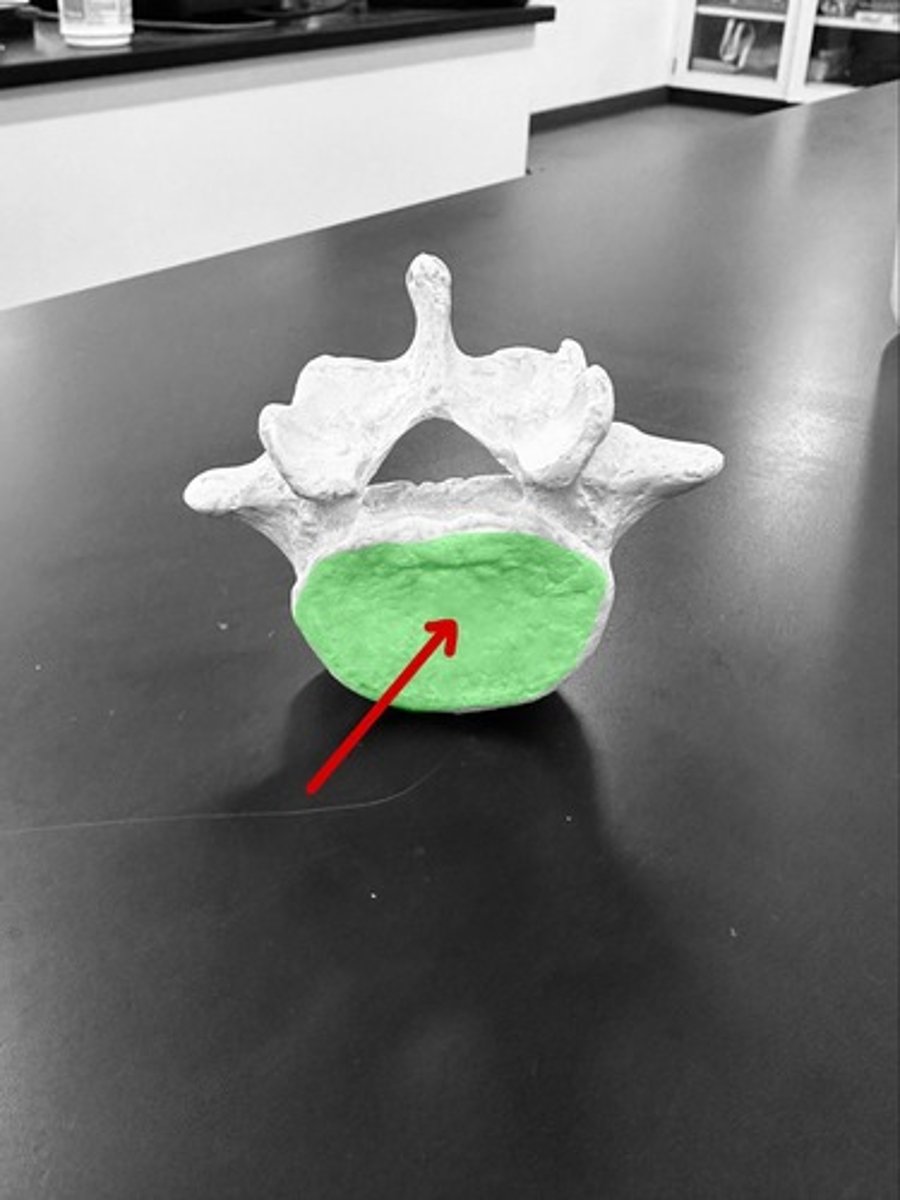
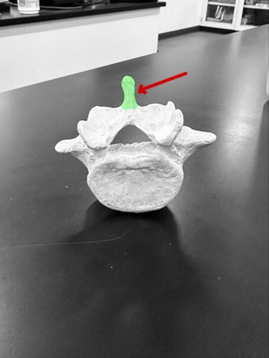
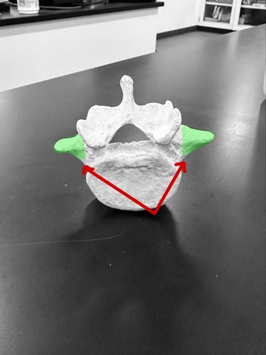
Body
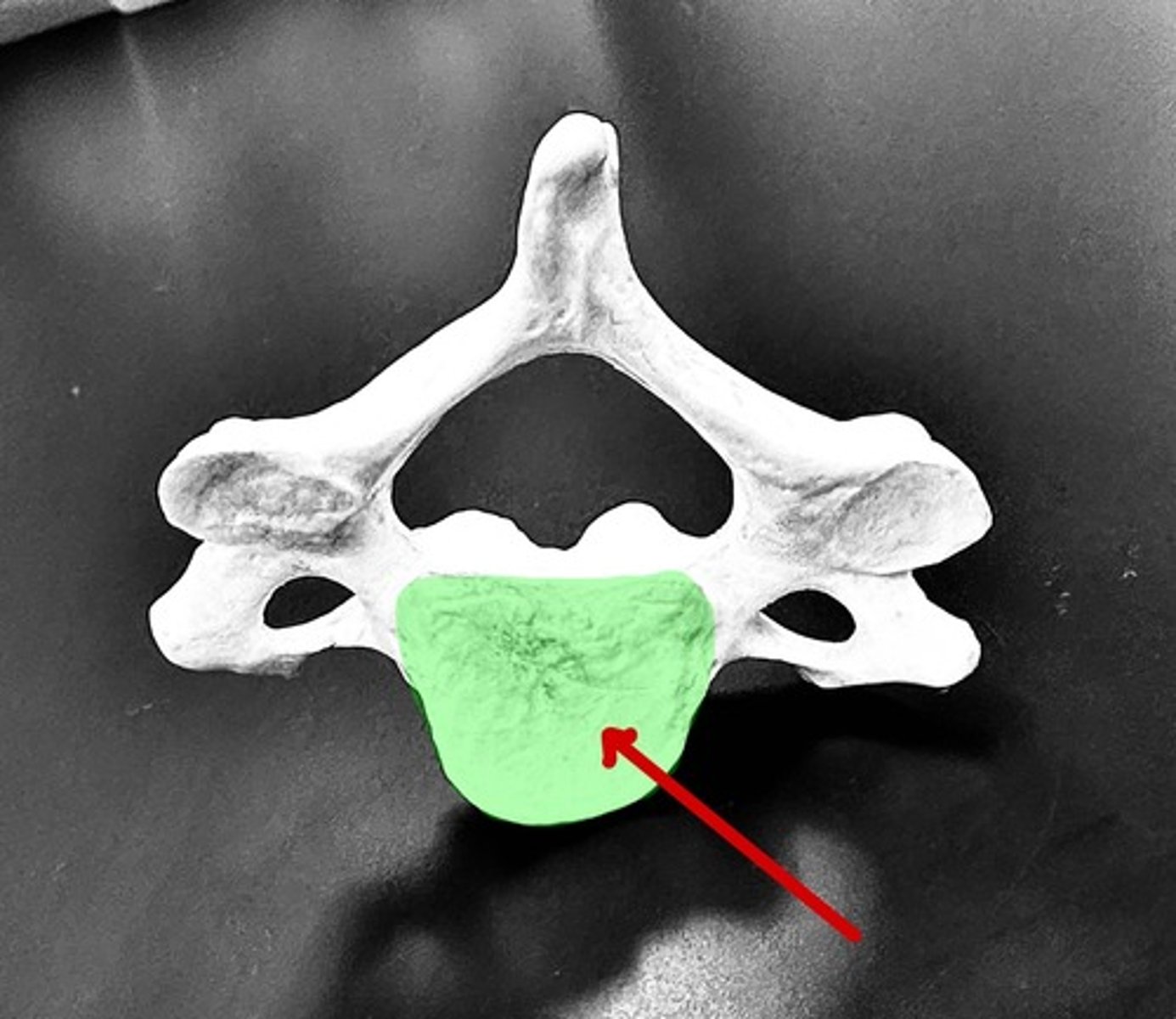
Spinous process
transverse process
vertebral foramen
superior articular process
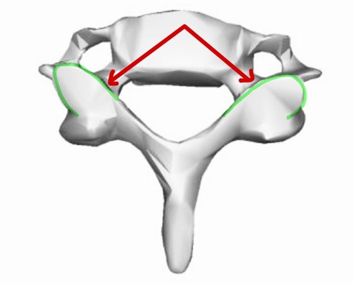
inferior articular process
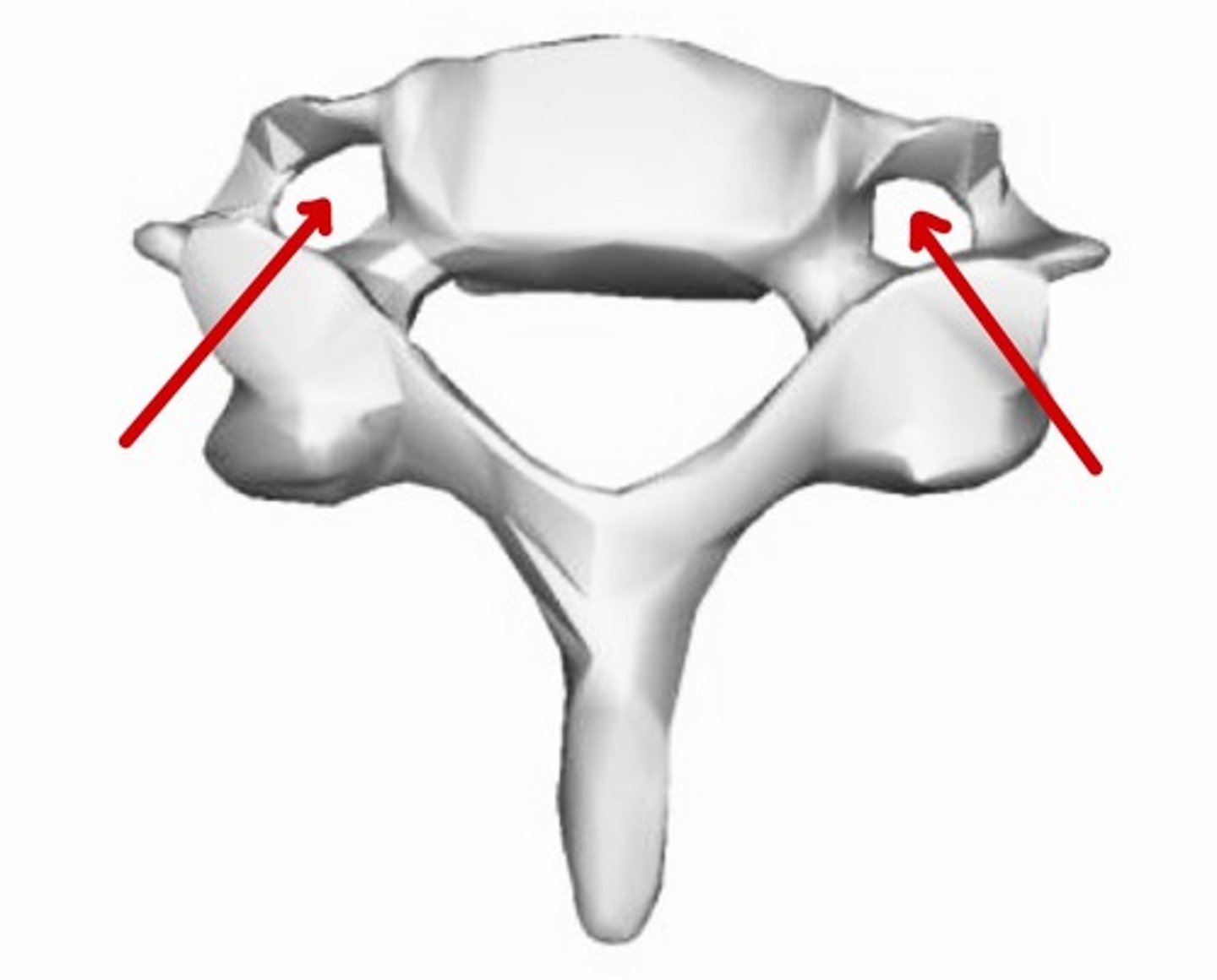
transverse foramen
Cerebrum
Processes conscious thoughts and sensation.
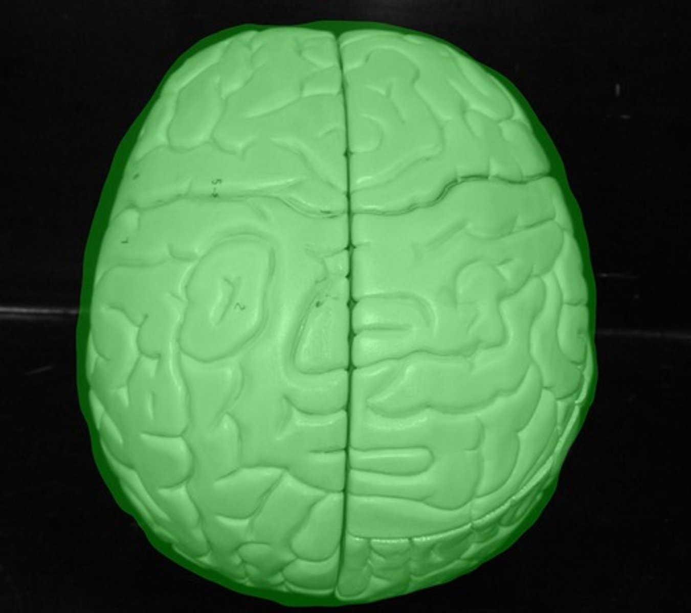
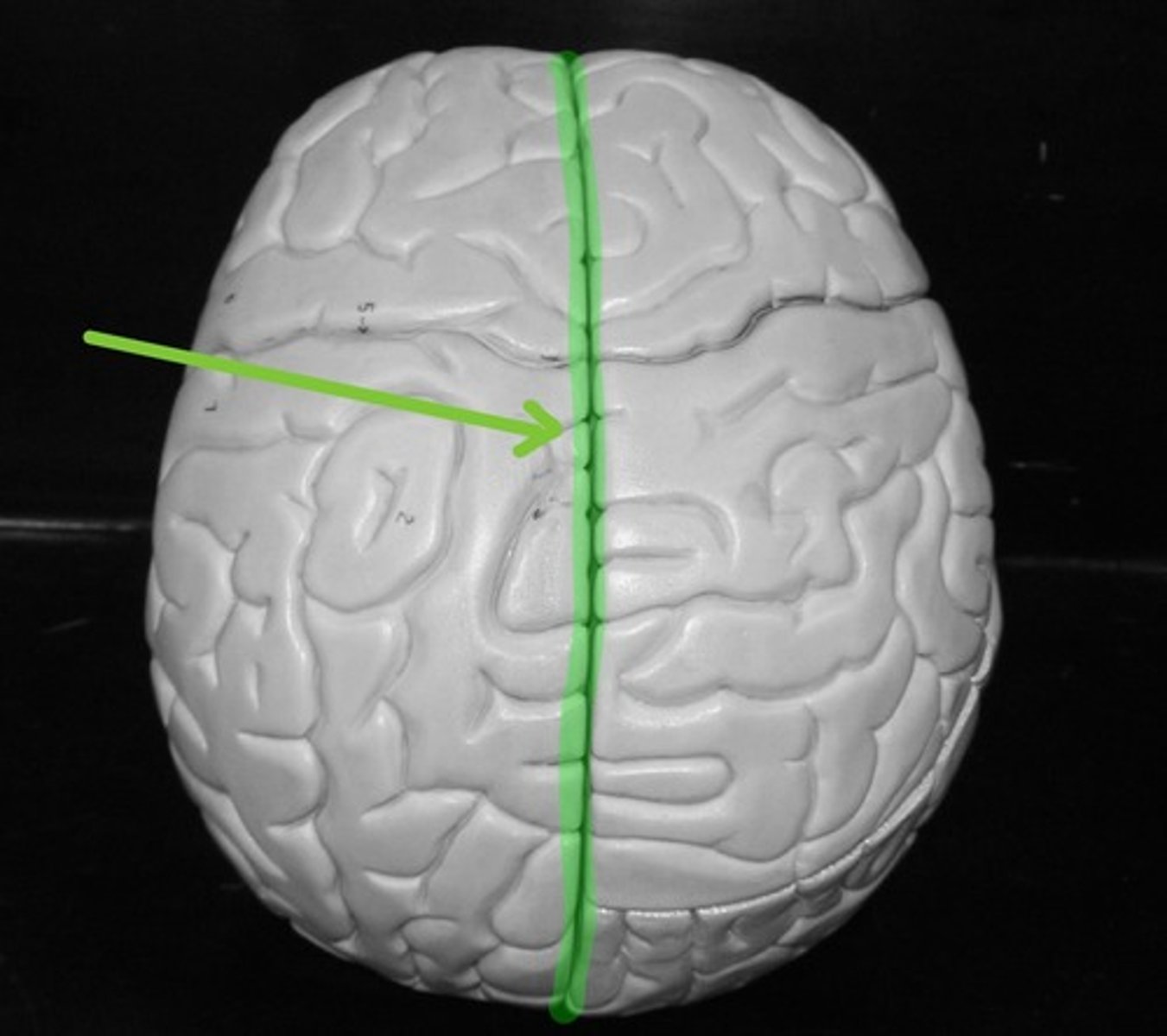
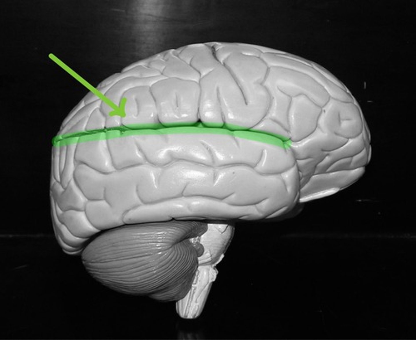
Longitudinal fissure
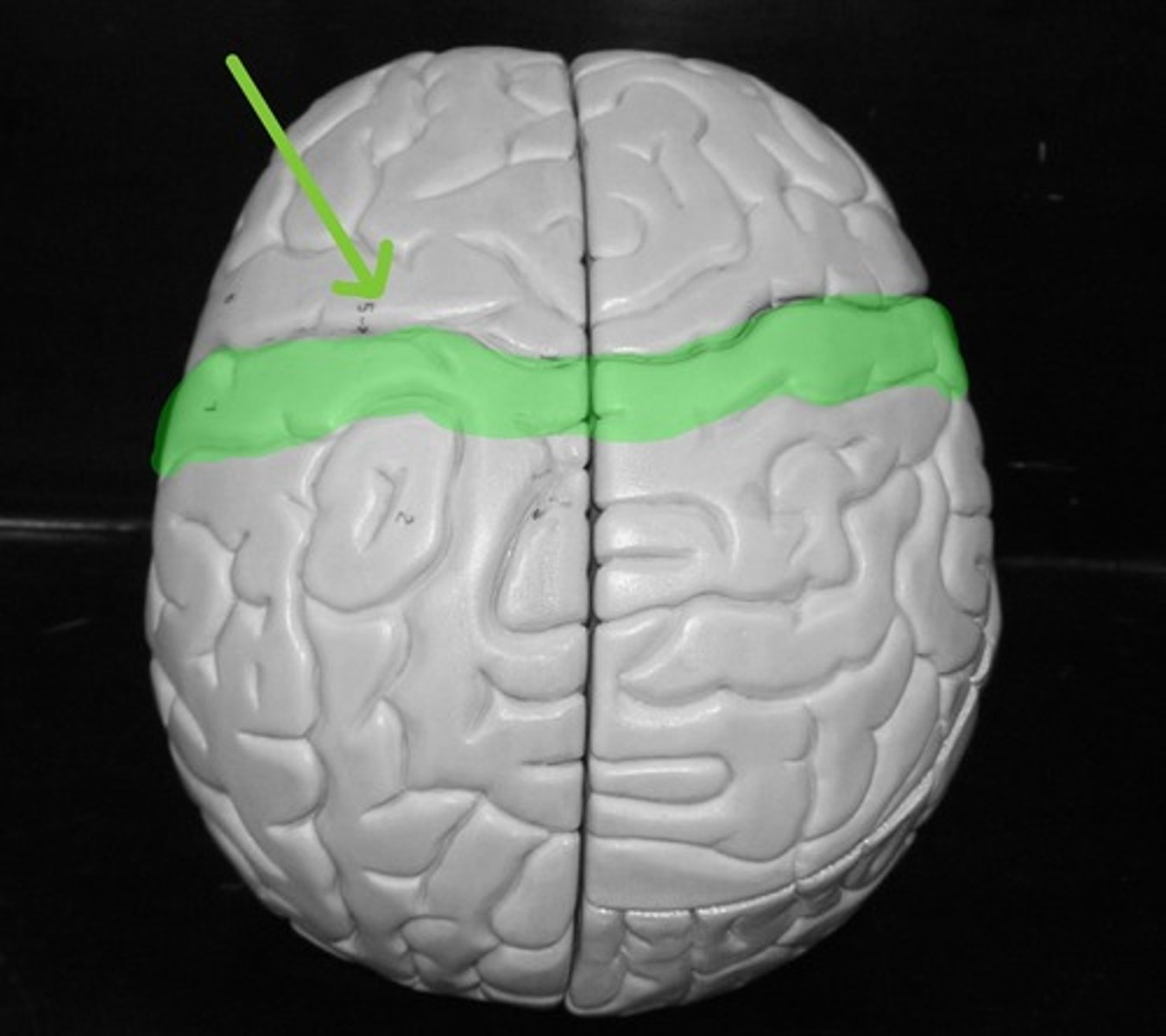
Central sulcus
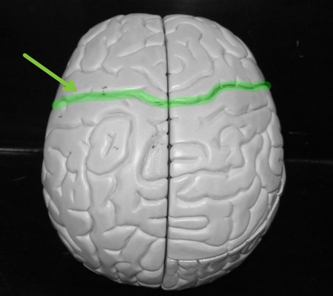
Precentral gyrus
Contains the primary MOTOR cortex
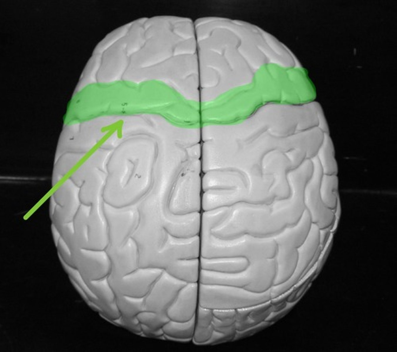
Postcentral gyrus
Contains the primary SENSORY cortex
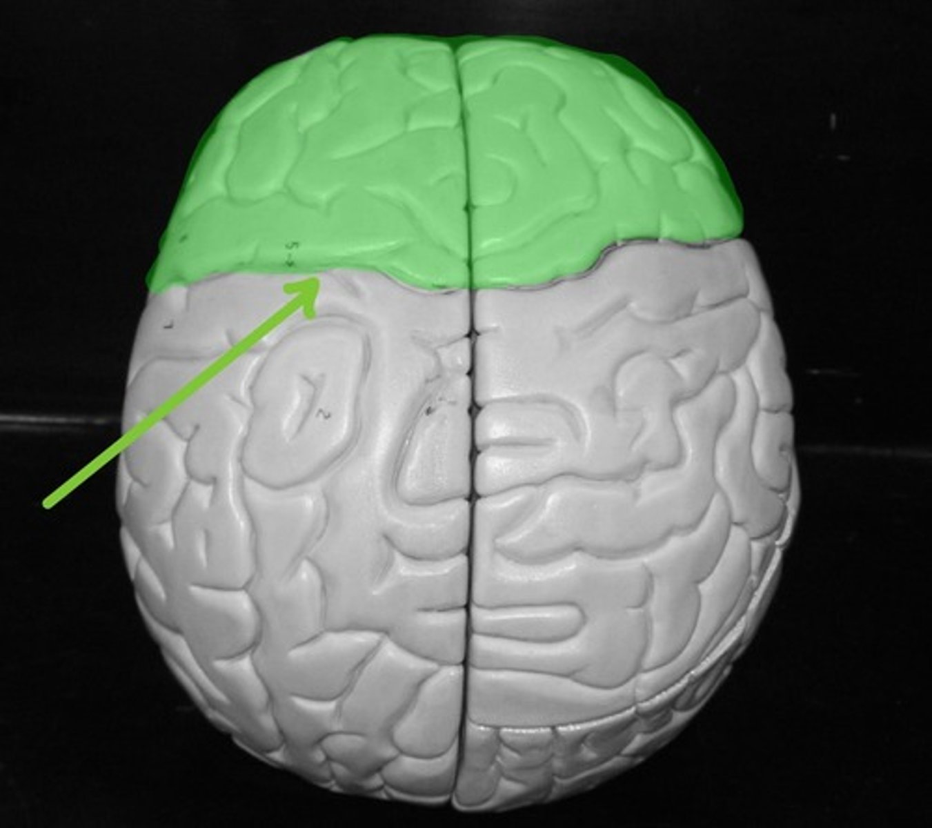
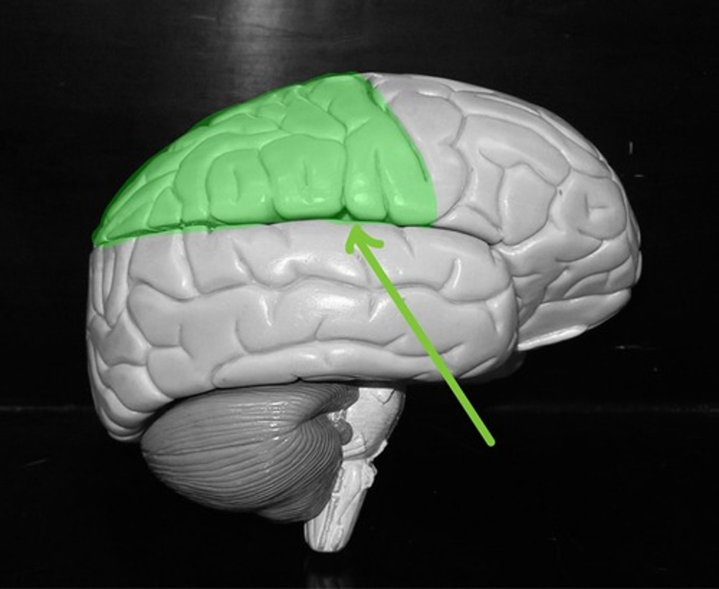
Frontal lobe
Controls voluntary skeletal muscle movement, and is where our intelligent thought comes from.
Lateral sulcus
Parietal lobe
Process general senses
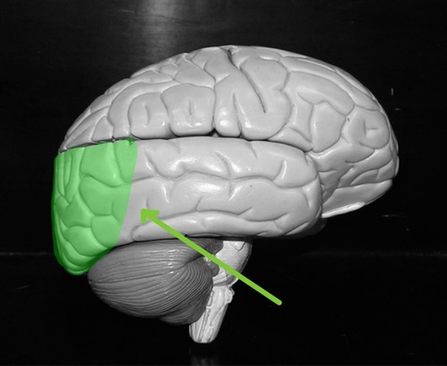
Occipital Lobe
Processes vision
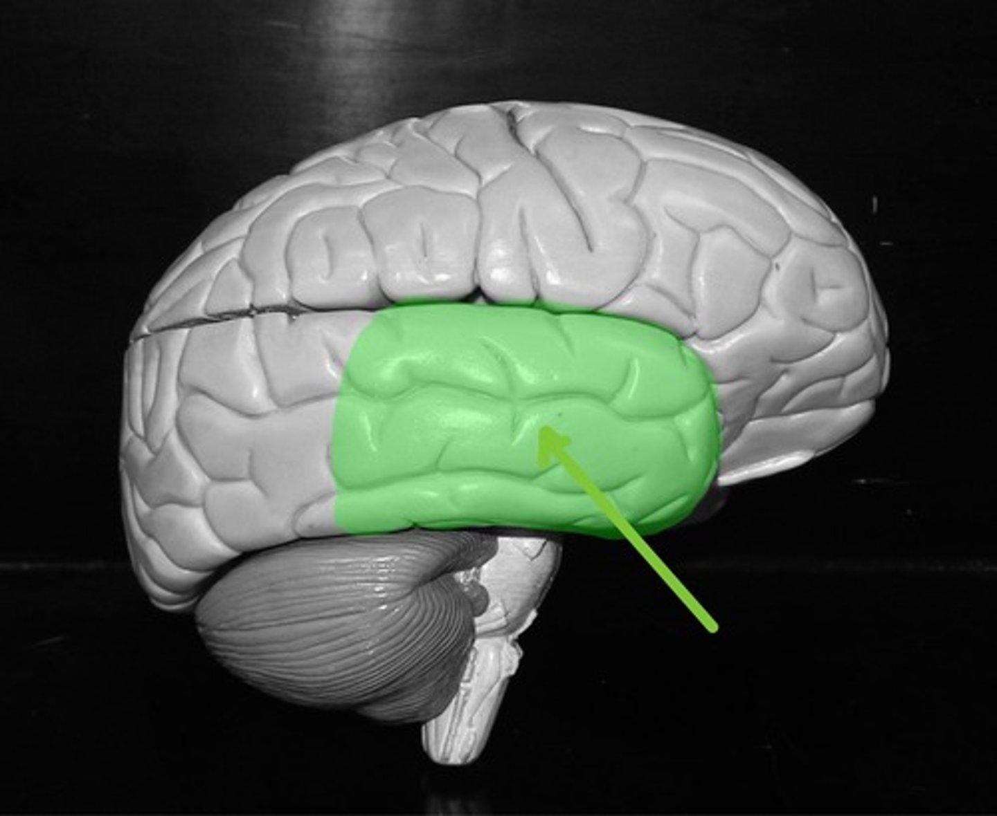
Temporal lobe
Processes hearing and equilibrium
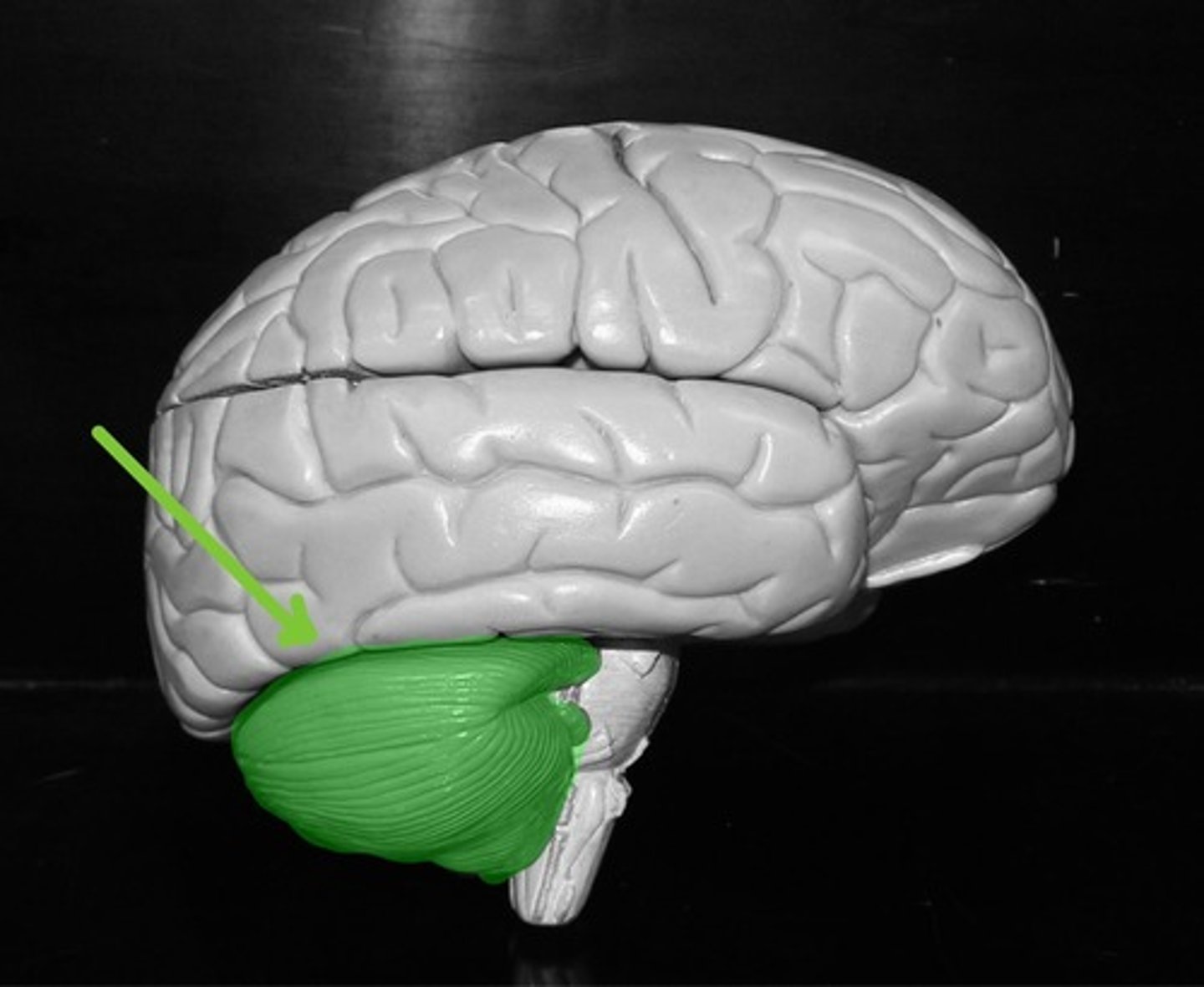
Cerebellum
Monitors and controls proprioception. Which is the awareness of where your body parts are in space and how they are moving.
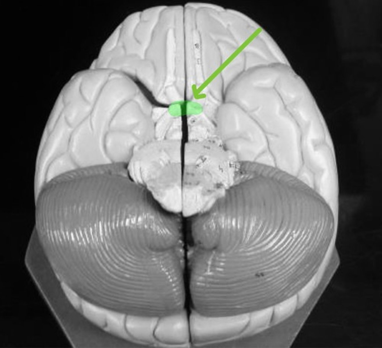
Olfactory bulb
Transmits OLFACTORY (smell) information to the brain.
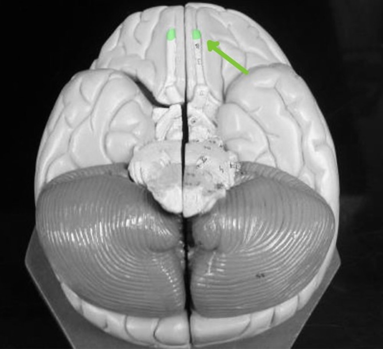
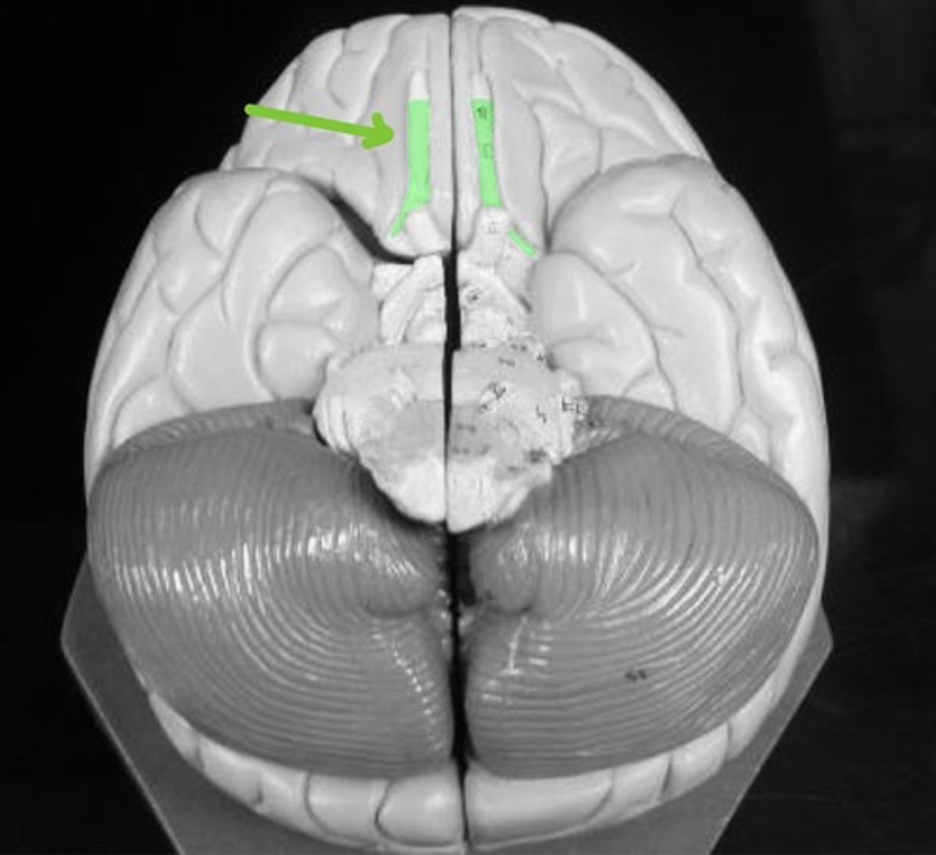
Olfactory tract
Transmits OLFACTORTY (smell) information to the brain.
Optic nerve
Transmits VISUAL infromation to the brain.
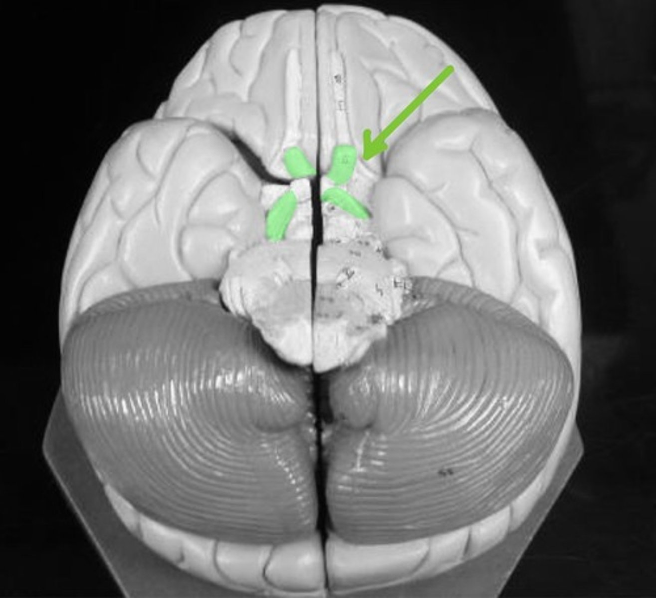
Optic chiasm
Transmit VISUAL information to the brain.
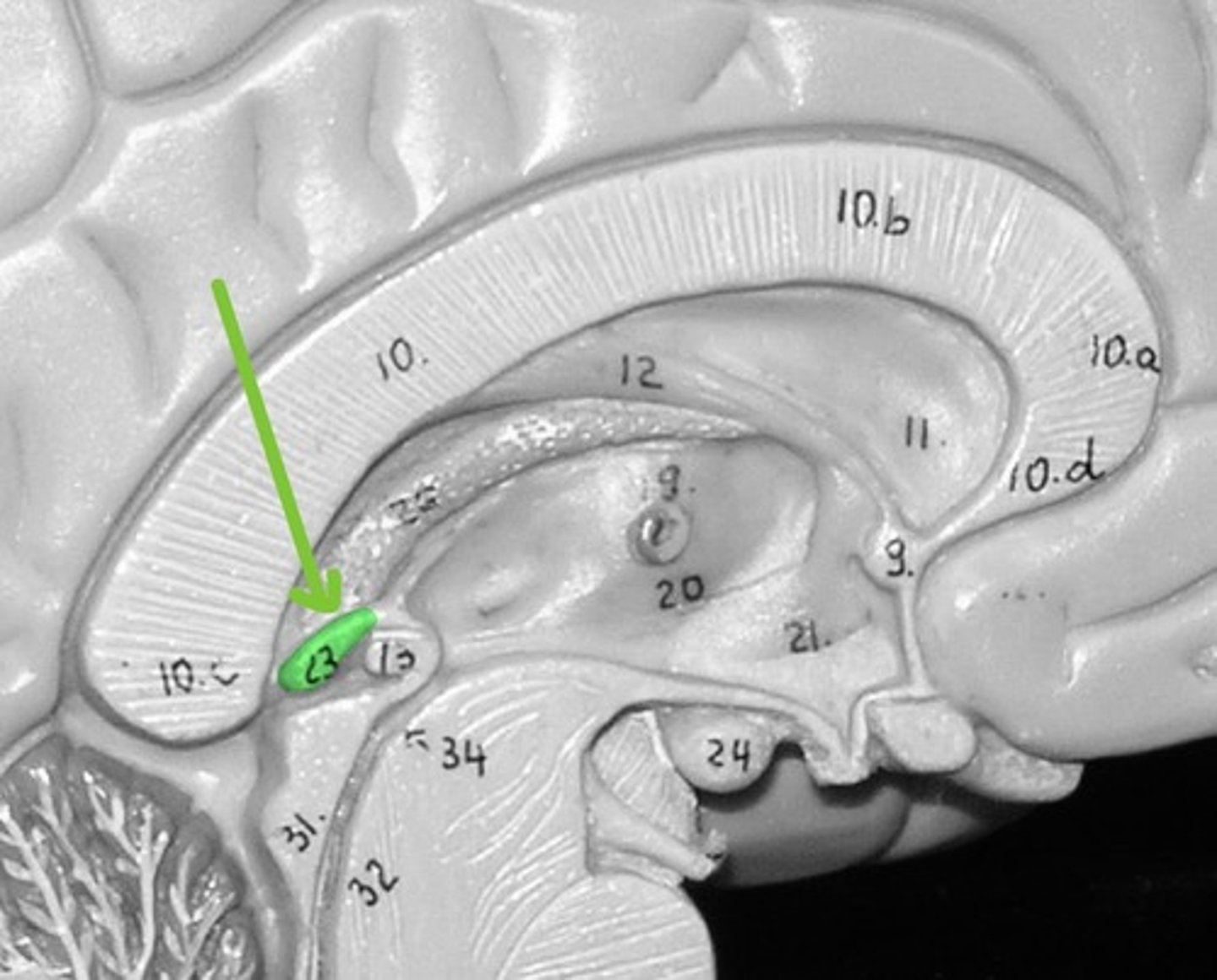
Corpus callosum
Allows for communication between the left and right brain
Midbrain
Keeps cerebrum in a state of wakefulness (RAS), and controls visual/auditory reflex
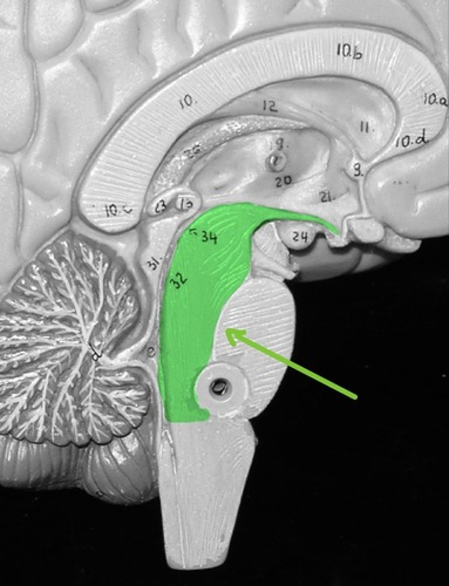
Pons
Has areas that control respiration
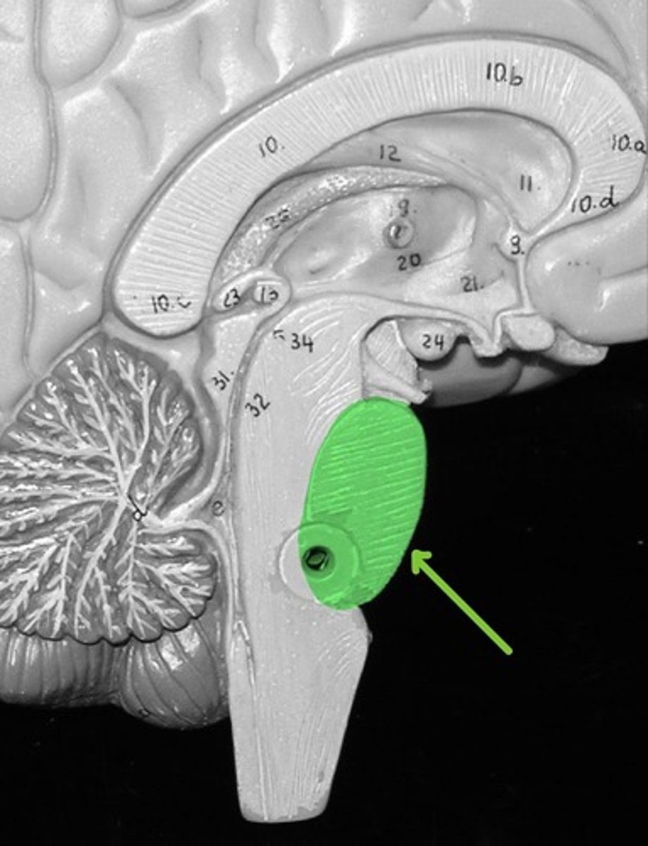
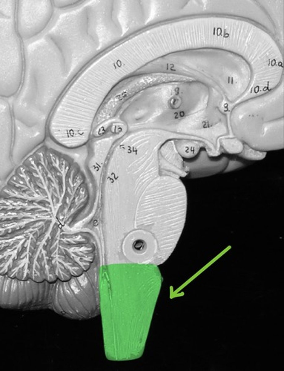
Medulla Oblongata
Contains the vital control centers for heart rate, respiratory rate and blood pressure.
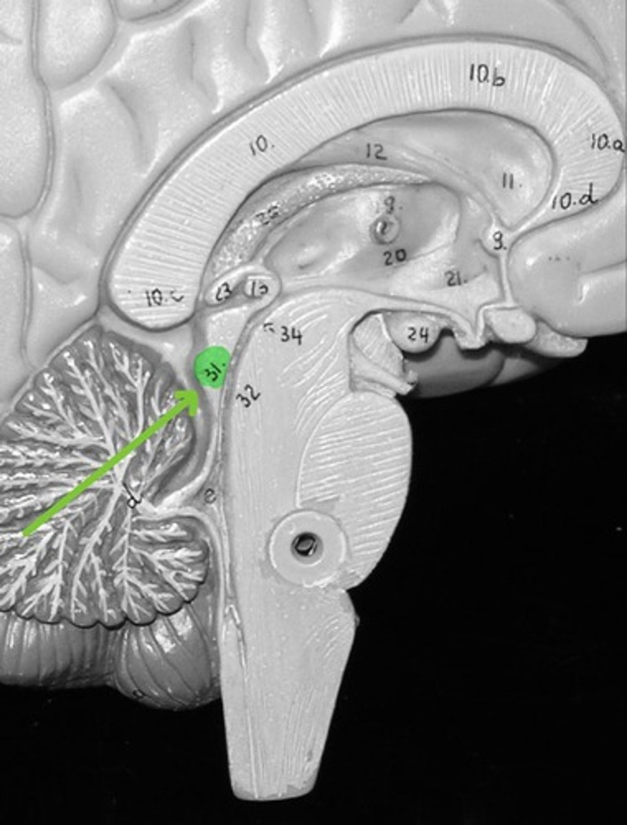
Corpora Quadrigemina - superior colliculi
Located in the midbrain and contains reflex centers for vision
Corpora Quadrigemina - Inferior colliculi
Located in the midbrain and contains reflex centers for audition/hearing
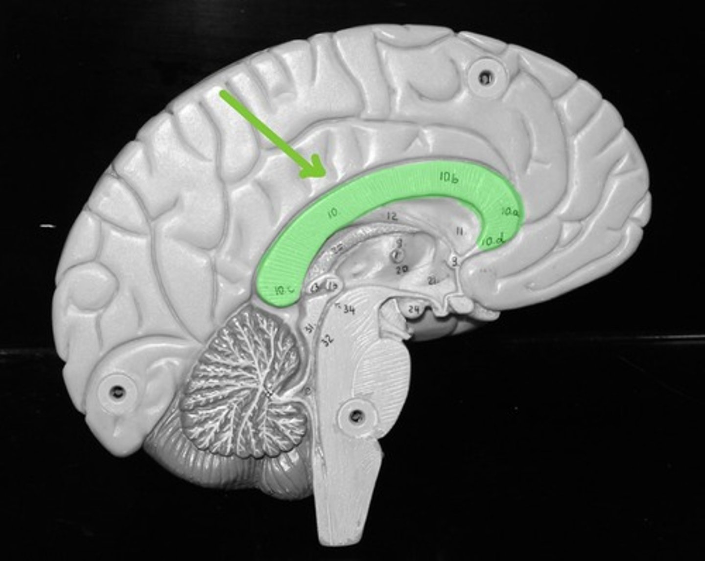
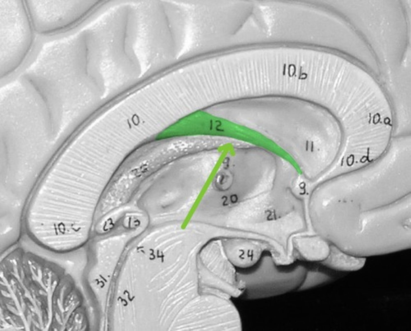
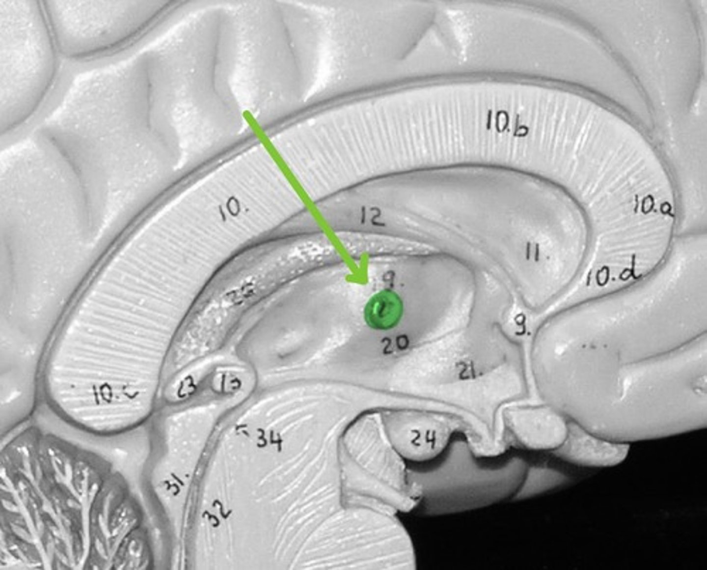
Fornix
Contains nerve tracts dealing with olfaction/sense of smell
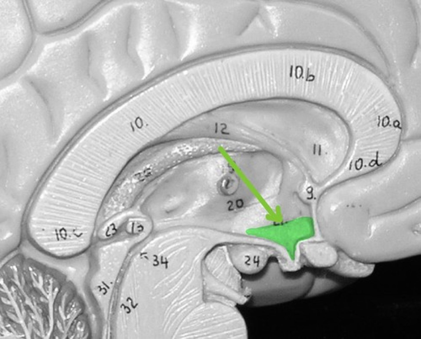
Mammillary body
Contains centers for feeding and memory

Thalamus
Processes and sends sensory information to the cerebrum
Hypothalamus
regulate hormones
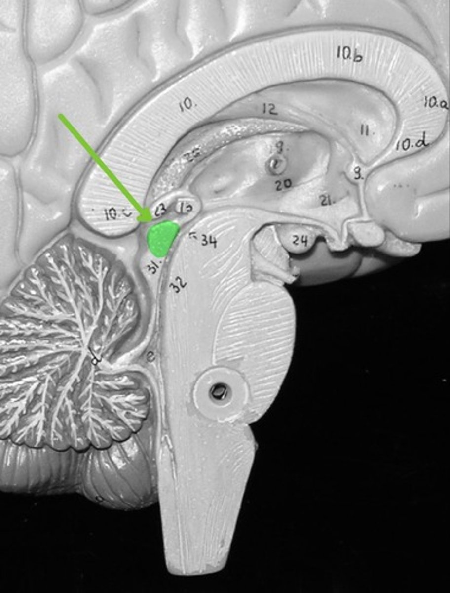
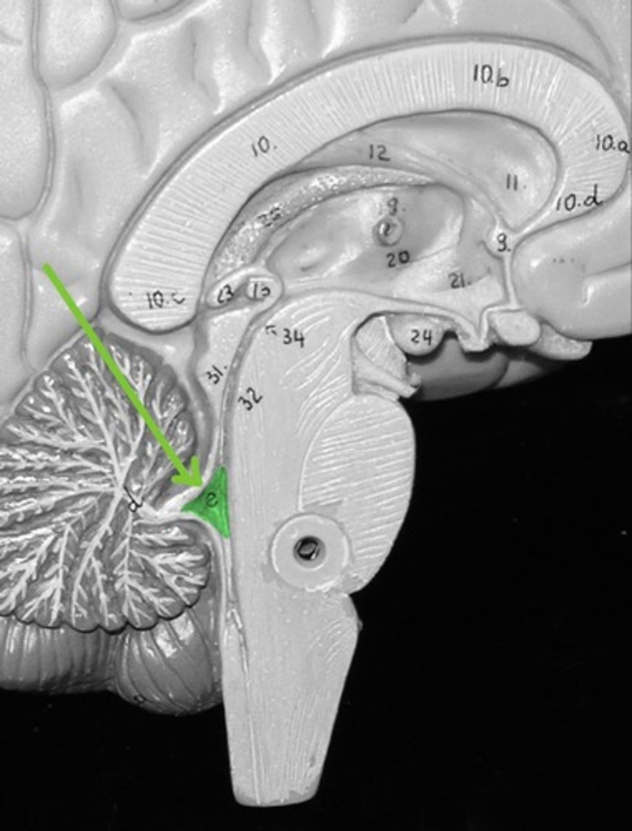
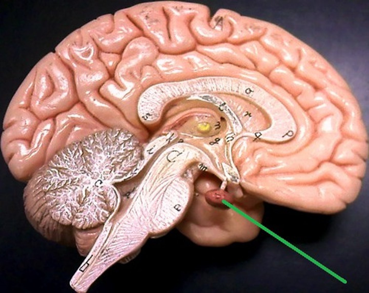
Pineal gland (or Pineal body)
Endocrine gland that secrets the hormone melatonin
Choroid plexus
Produces cerebrospinal fluid
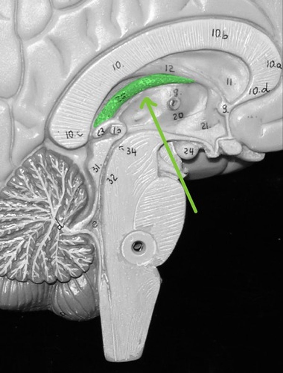
Septum Pellucidum
Cerebral Aqueduct
helps in the circulation of cerebrospinal fluid

Fourth Ventricle
A chamber containing cerebrospinal fluid
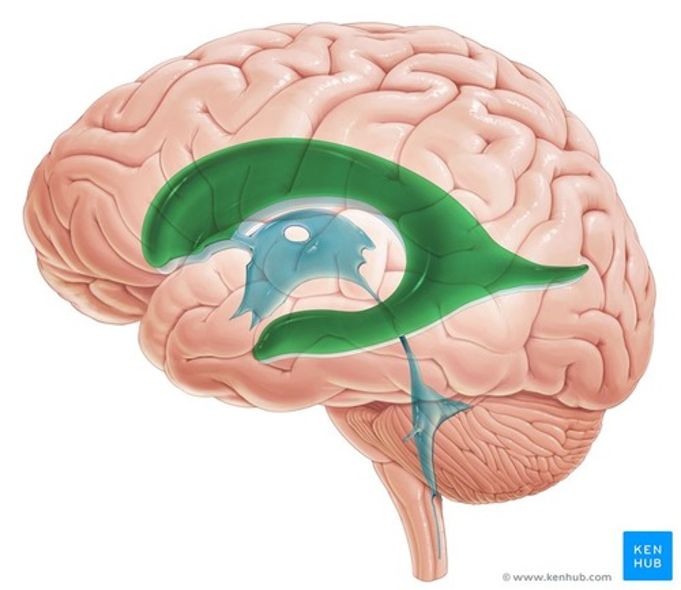
Lateral Ventricle
A chamber containing cerebrospinal fluid
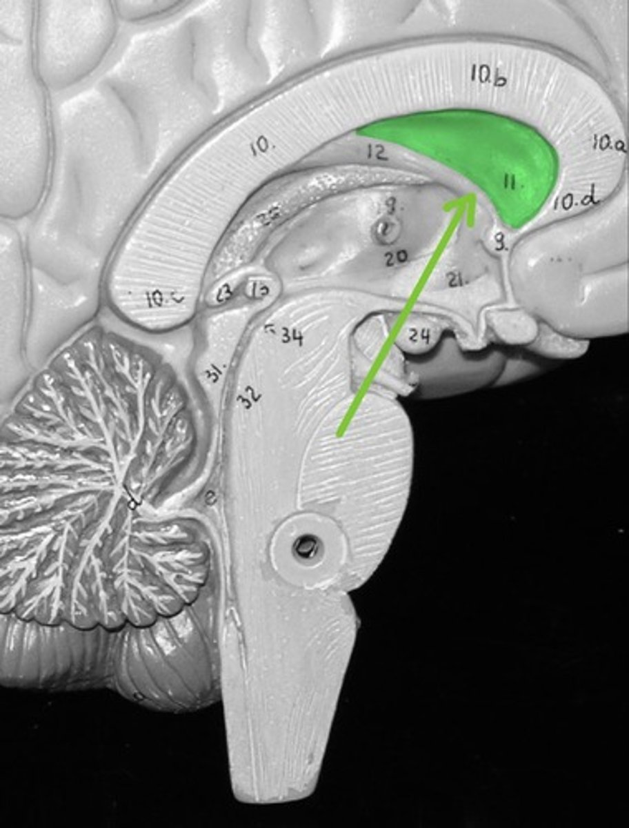
Pituitary Gland
Major endocrine gland that secretes hormones that control numerous organ system.
1. Gray commissure
2. Central canal
3. Posterior/ dorsal funiculus/ white column
4. Anterior/ ventral funiculus/ white column
1. What is the structure at the pointer?
2. What is the hole in the center near the pointer?
3. What is the lighter area of white matter above the pointer?
4. What is the lighter area of white matter below the pointer?
1. Dorsal root
2. Dorsal root ganglion
1. What is the structure at the pointer?
2. What is the large round structure to the lower left of the pointer?
1. Osteon
2. Central canal
3. Lacunae
4. Canaliculi
1. What is one entire circular unit of compact bone seen here?
2. What structure is the pointer on?
3. What are the numerous smaller black "holes" seen in the slide?
4. what are the black lines or "cracks" seen in this slide?
1. Perforating canal
2. Lamellae
1. What is the structure at the pointer?
2. What are the areas of hard bone matrix called?
1. Multipolar neuron
2. Neuroglial cells or glial cells
1. What type of cell is seen at the pointer?
2. What type of cell is seen surrounding the cell at the pointer (smaller blue dots)?

1. Cross section of a nerve
2. Perineurium
3. Nerve fascicles
1. What are you looking at in this slide?
2. What connective tissue layer is the pointer on?
3. What are the large round structures called?

1. Axon
2. Myelin sheath
3. Endoneurium
1. What is the structure at the pointer (pink dot)?
2. What is the white area surrounding the pink dot?
3. What connective tissue layer would be found surrounding #1 and #2?
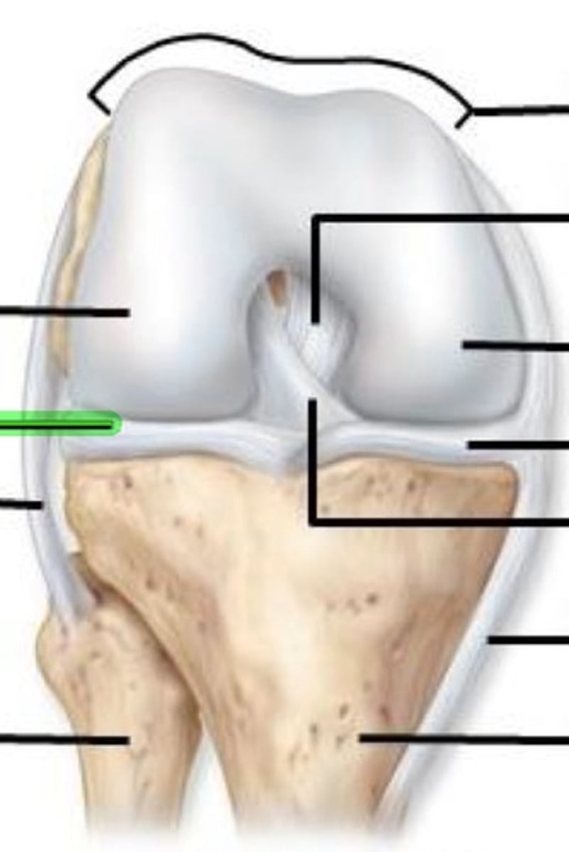
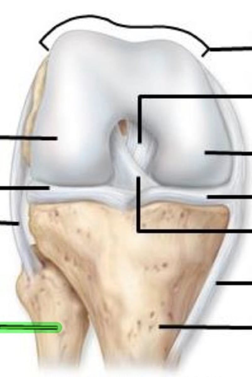
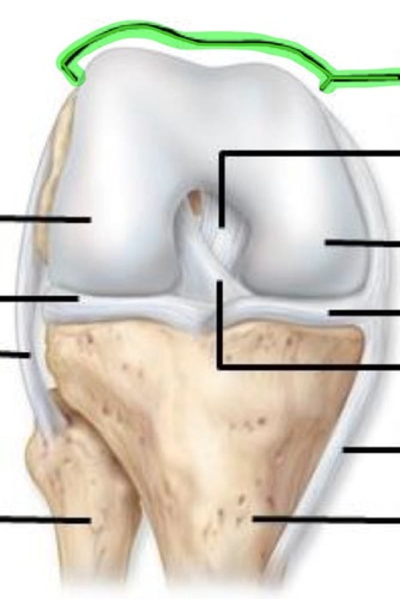
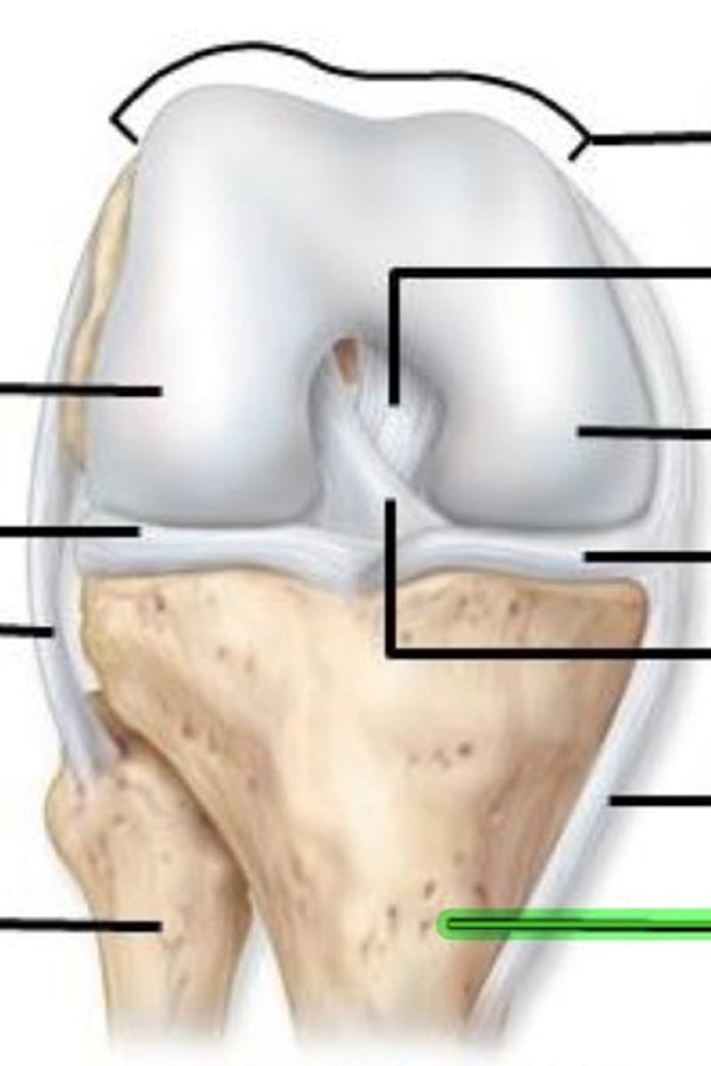
Fibular/lateral collateral ligament
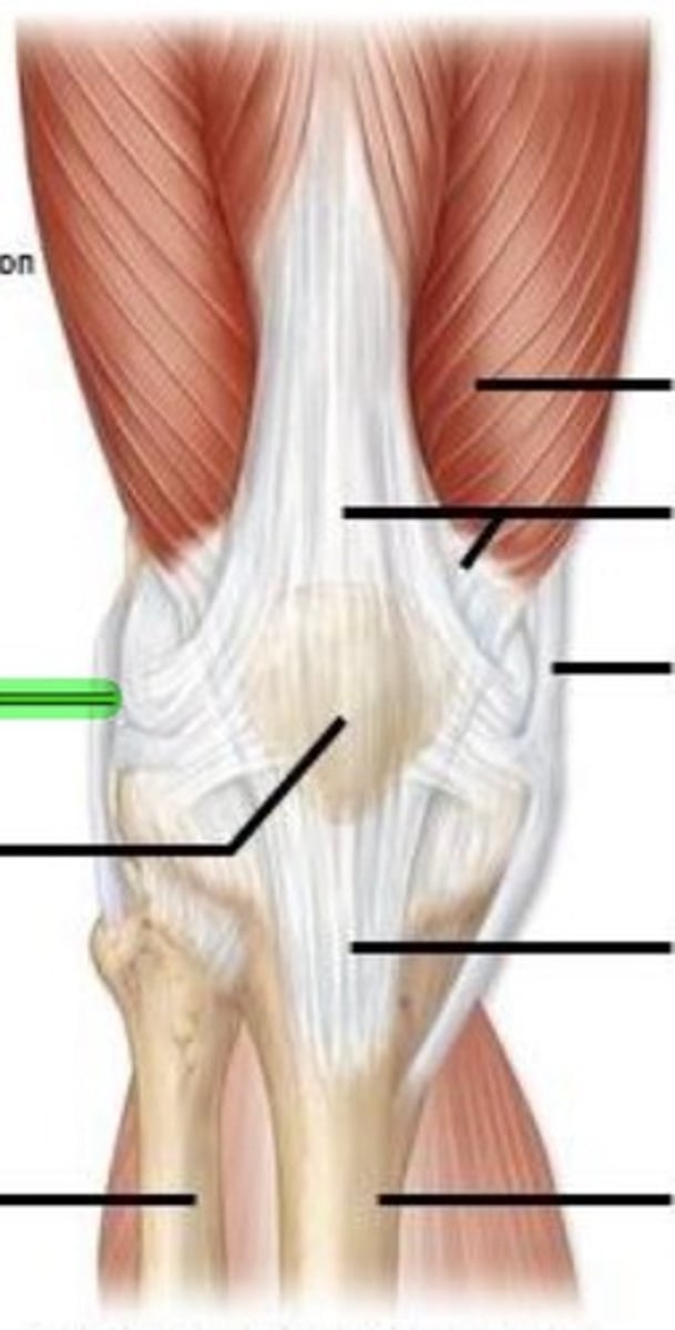
Patella
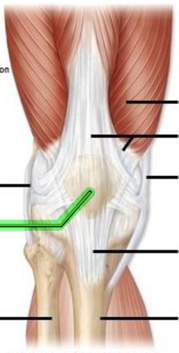
Fibula
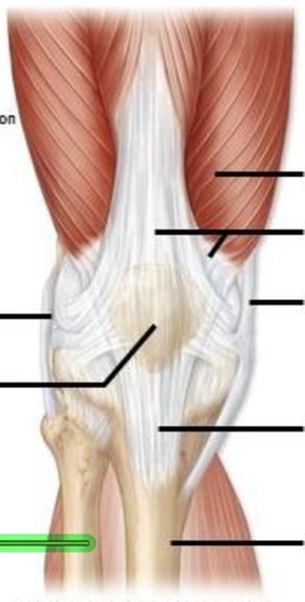
Quadriceps femoris muscle

Quadriceps femoris tendon
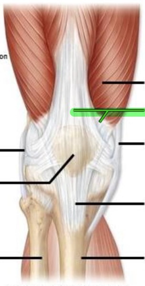
Tibial/medial collateral ligament
Patellar ligament
Tibia
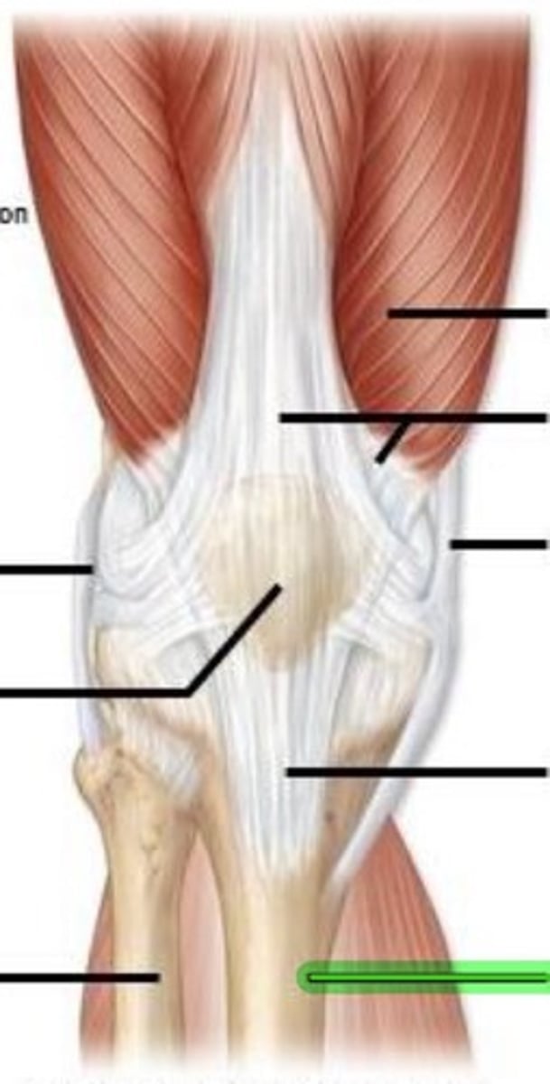
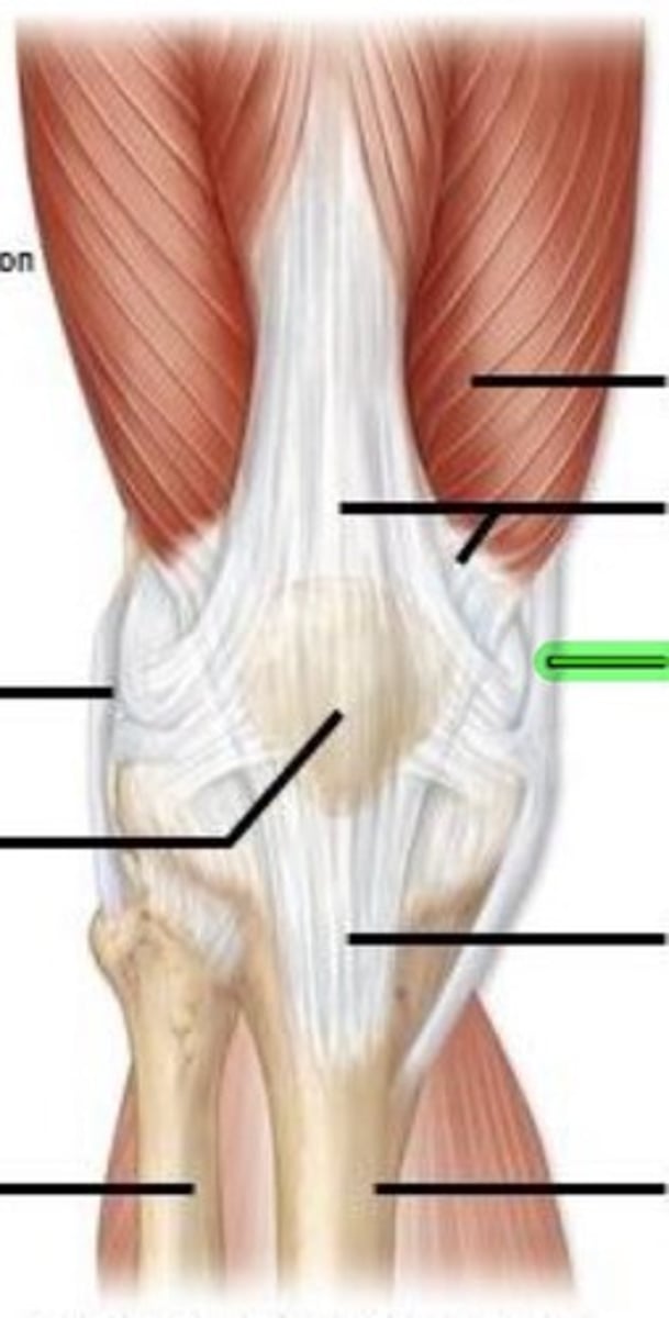
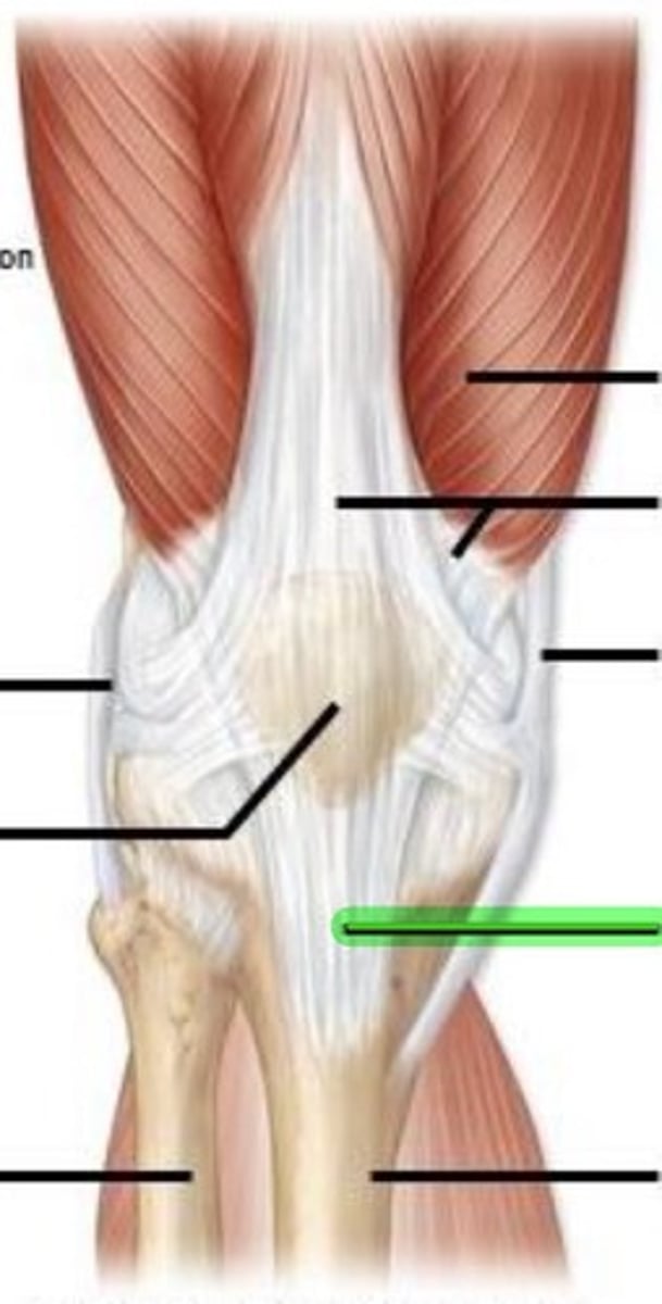
Lateral condyle
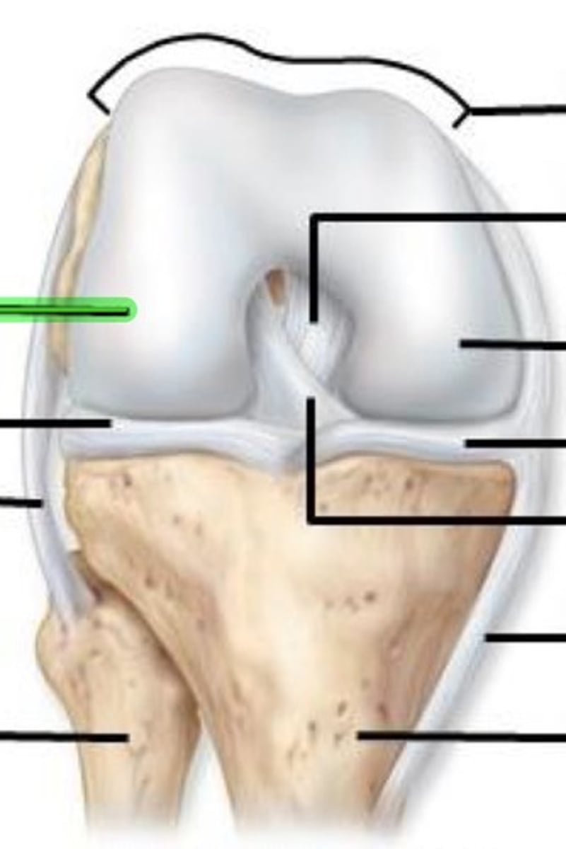
Lateral meniscus
Fibular/lateral collateral ligament

Fibula
Articular cartilage
Posterior cruciate ligament
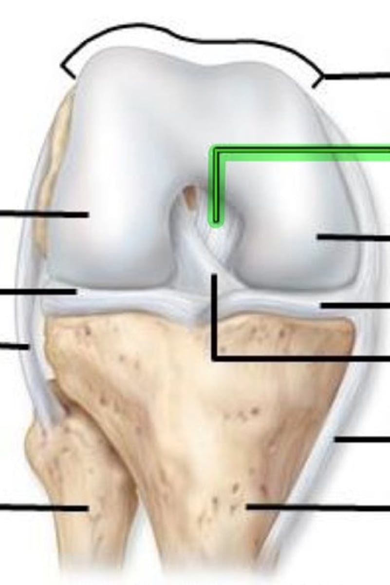
Medial condyle
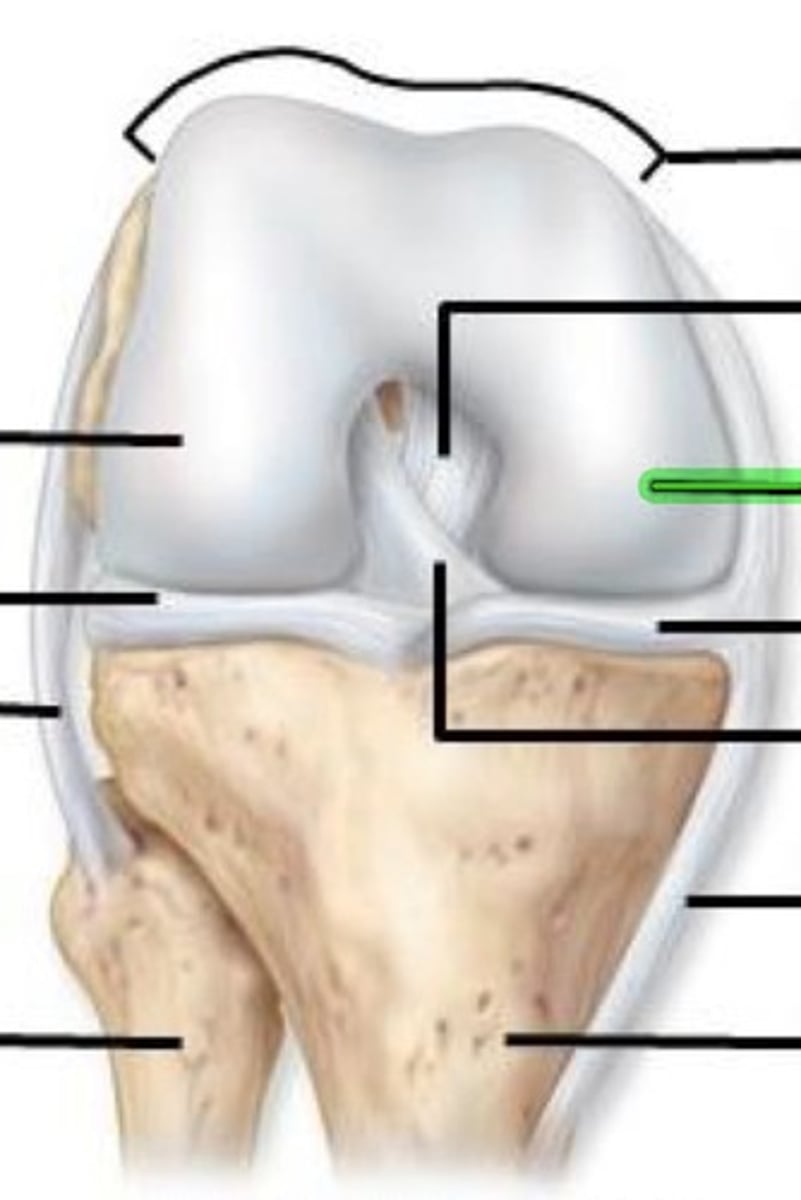
Medial meniscus
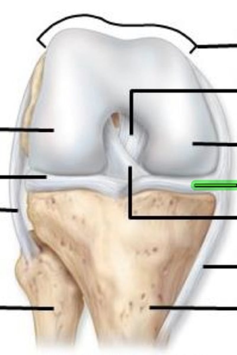
Anterior cruciate ligament
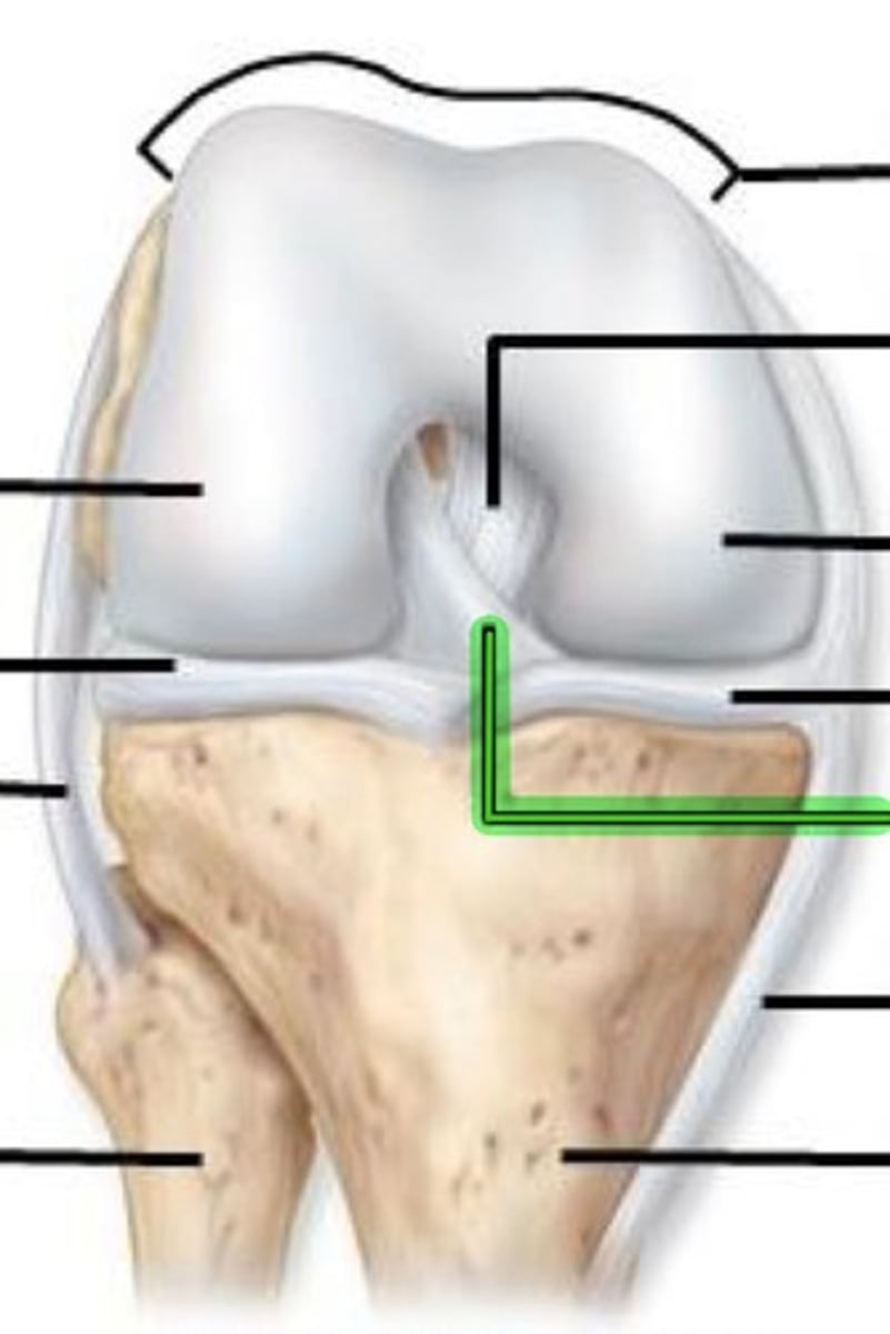
Tibial/medial collateral ligament
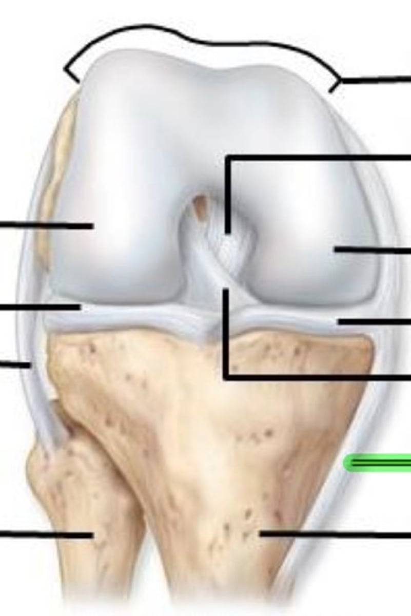
Tibia
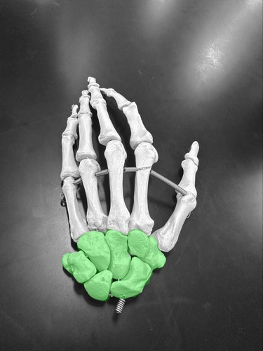
Phalanges (finger bones)
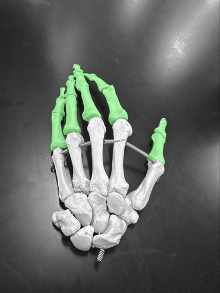
proximal phalange (phalanx
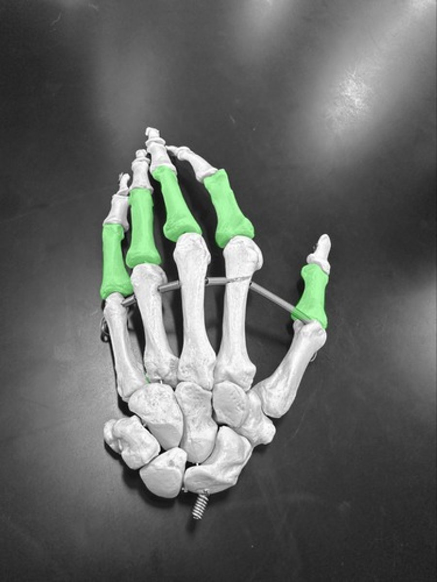
middle phalange (phalanx

distal phalange (phalanx)
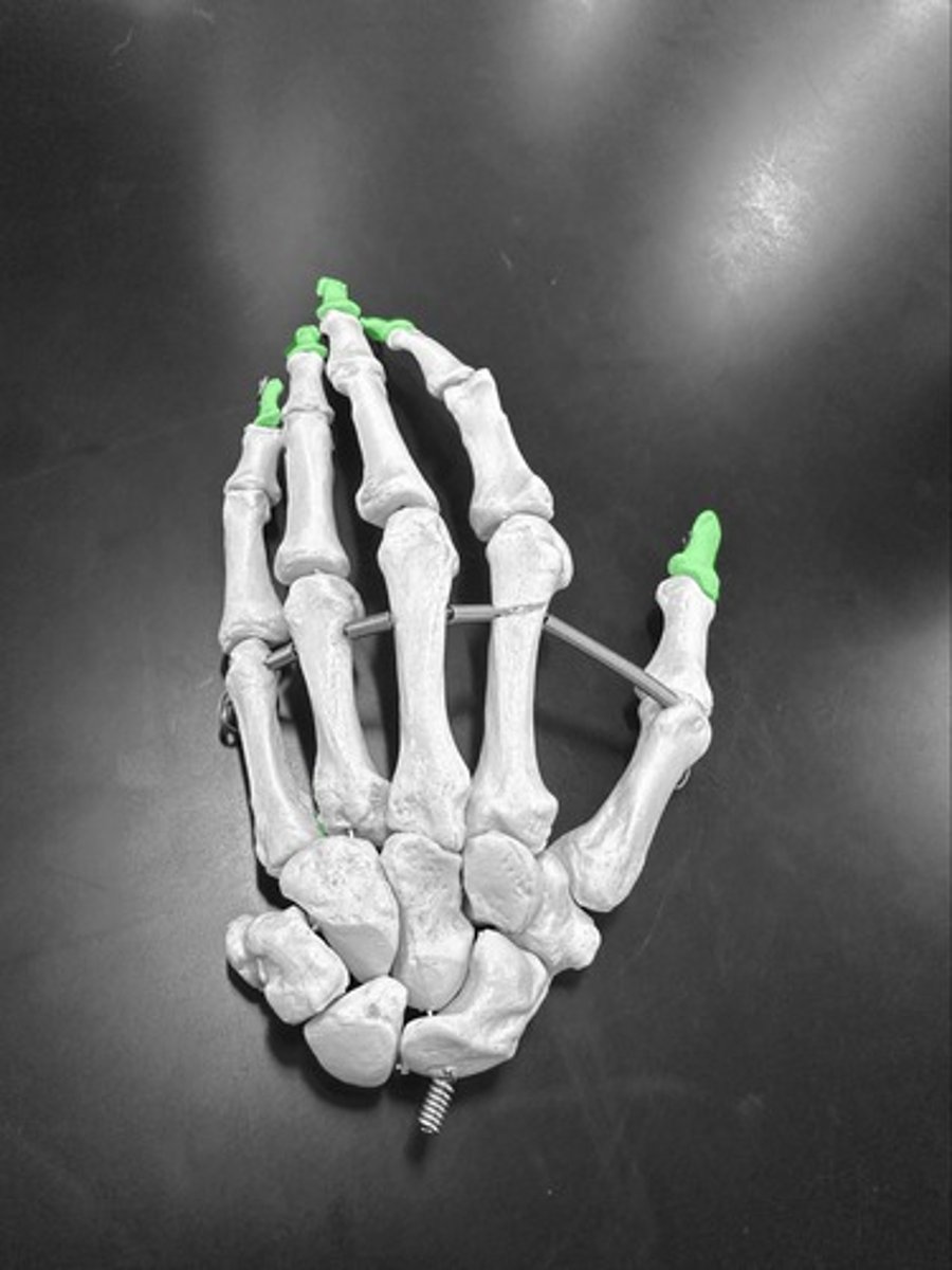
Metacarpals (hand bones)

Metacarpals #1
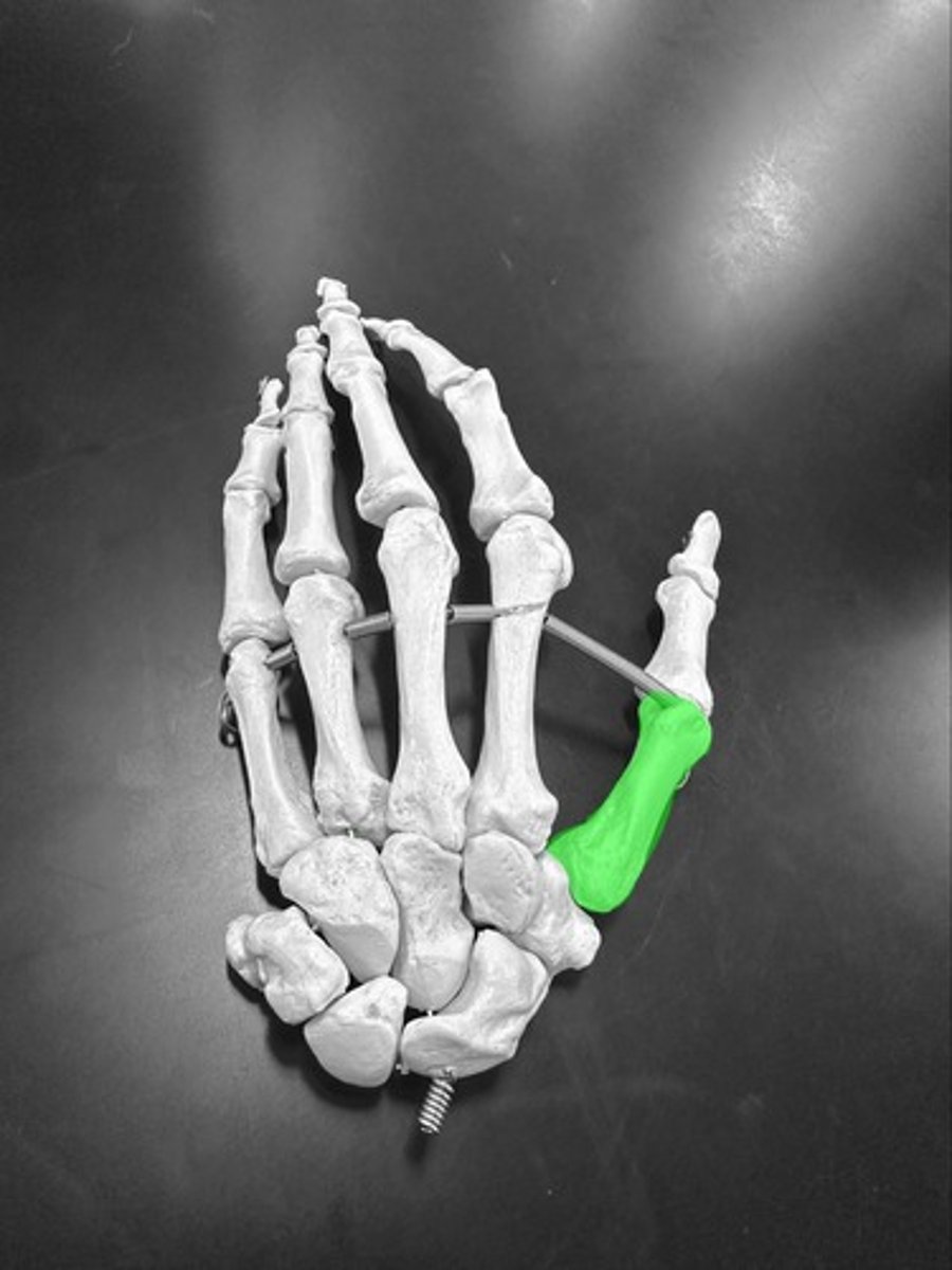
Metacarpals #2
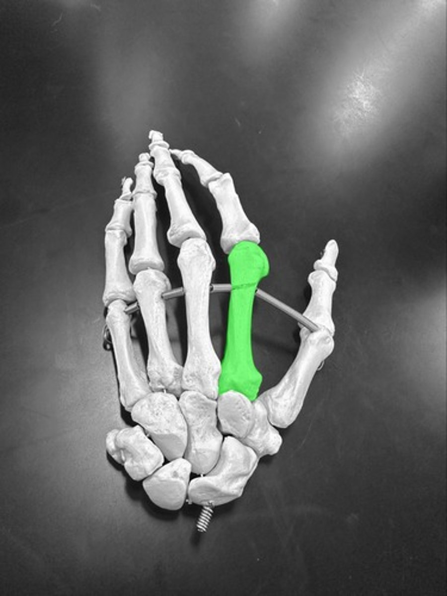
Metacarpals #3
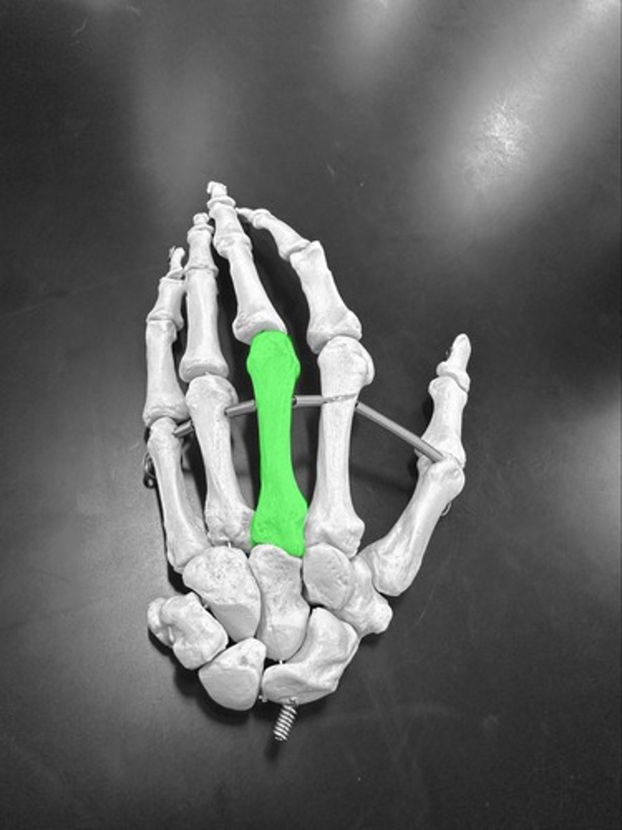
Metacarpals #4
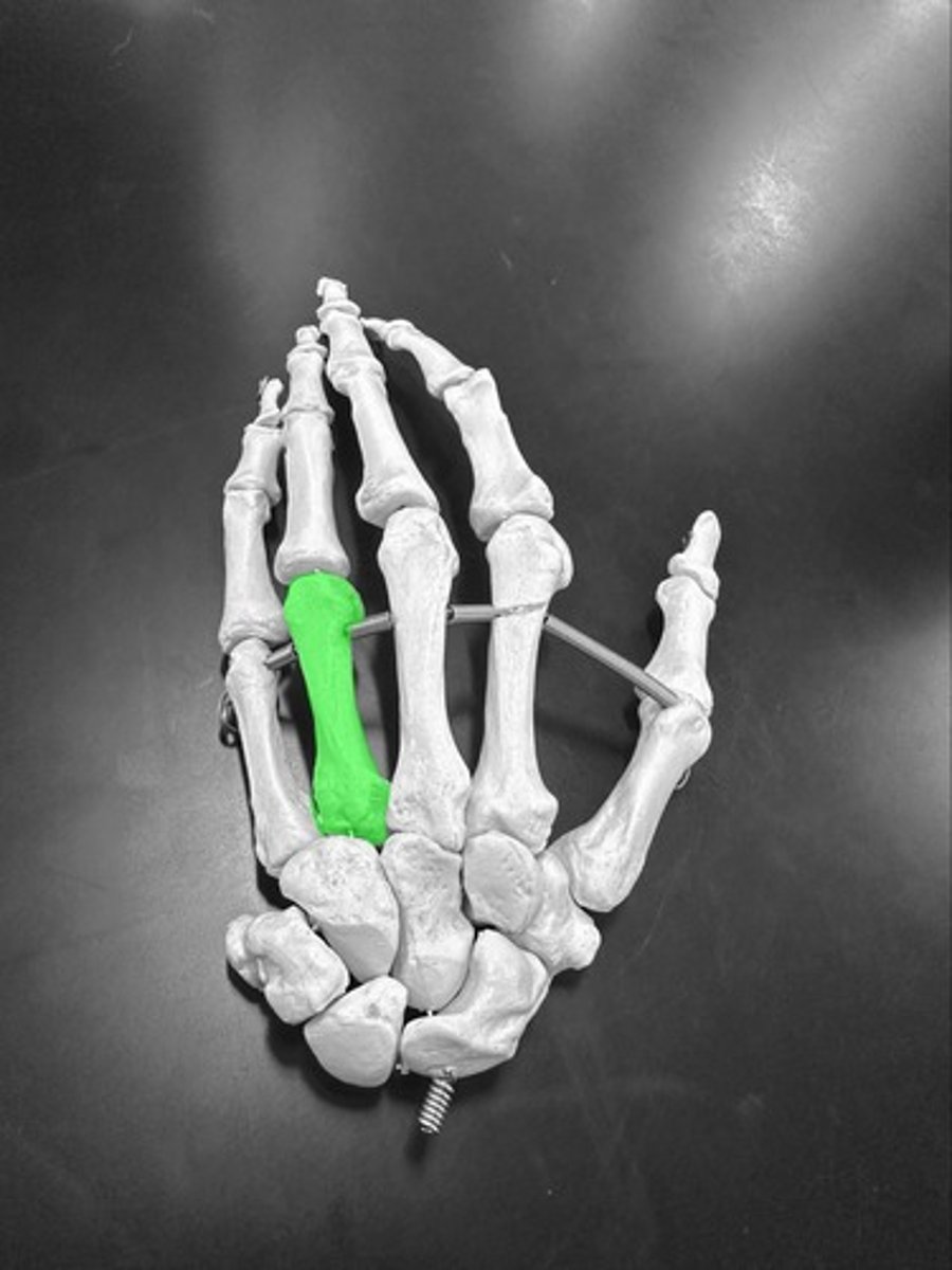
Metacarpals #5
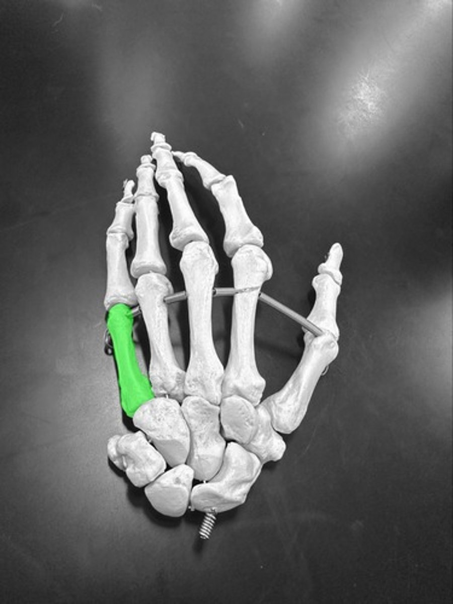
Carpals (wrist bones)