ACTIVITY 9 : HISTOLOGY cardiac muscle/ LM: high magnification
1/3
There's no tags or description
Looks like no tags are added yet.
Name | Mastery | Learn | Test | Matching | Spaced | Call with Kai |
|---|
No analytics yet
Send a link to your students to track their progress
4 Terms
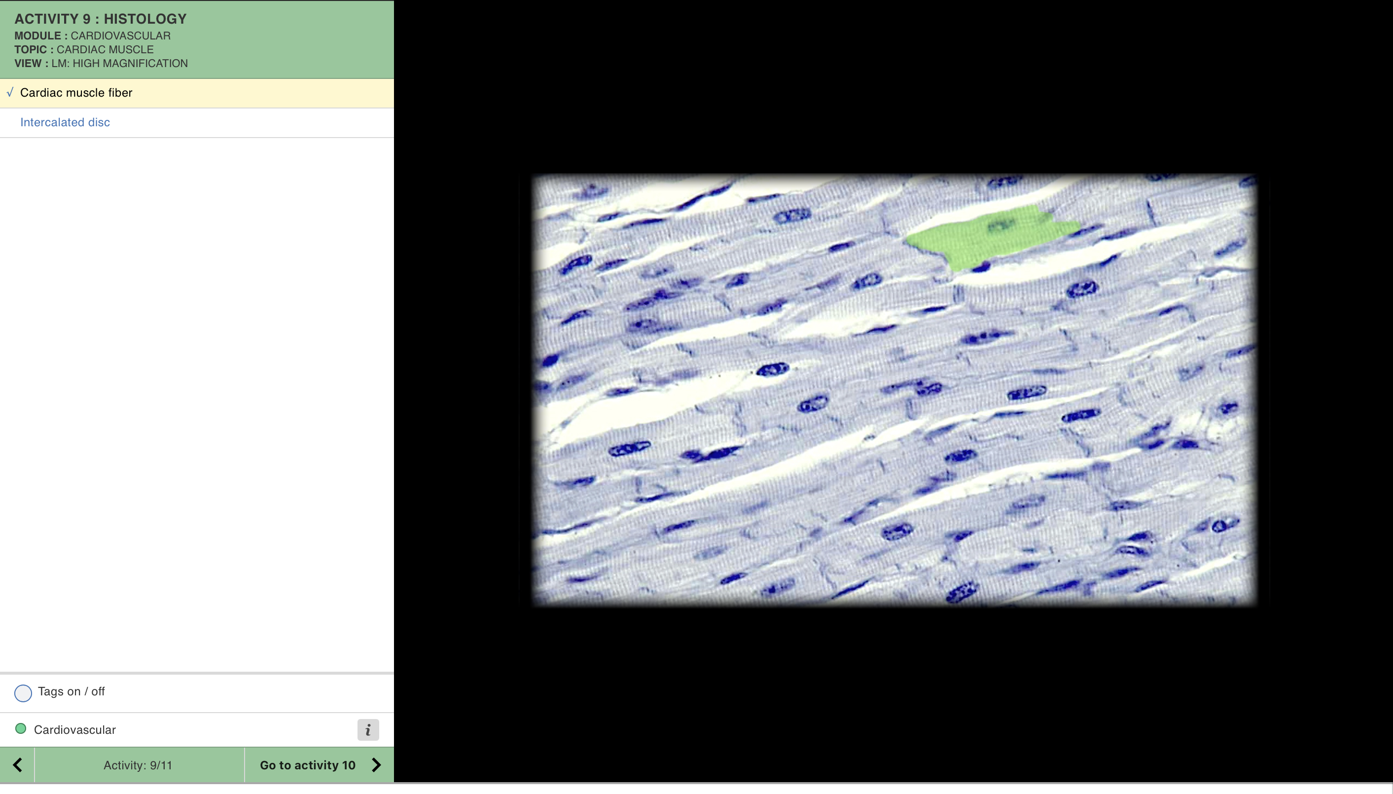
Cardiac muscle fiber image (green highlight)
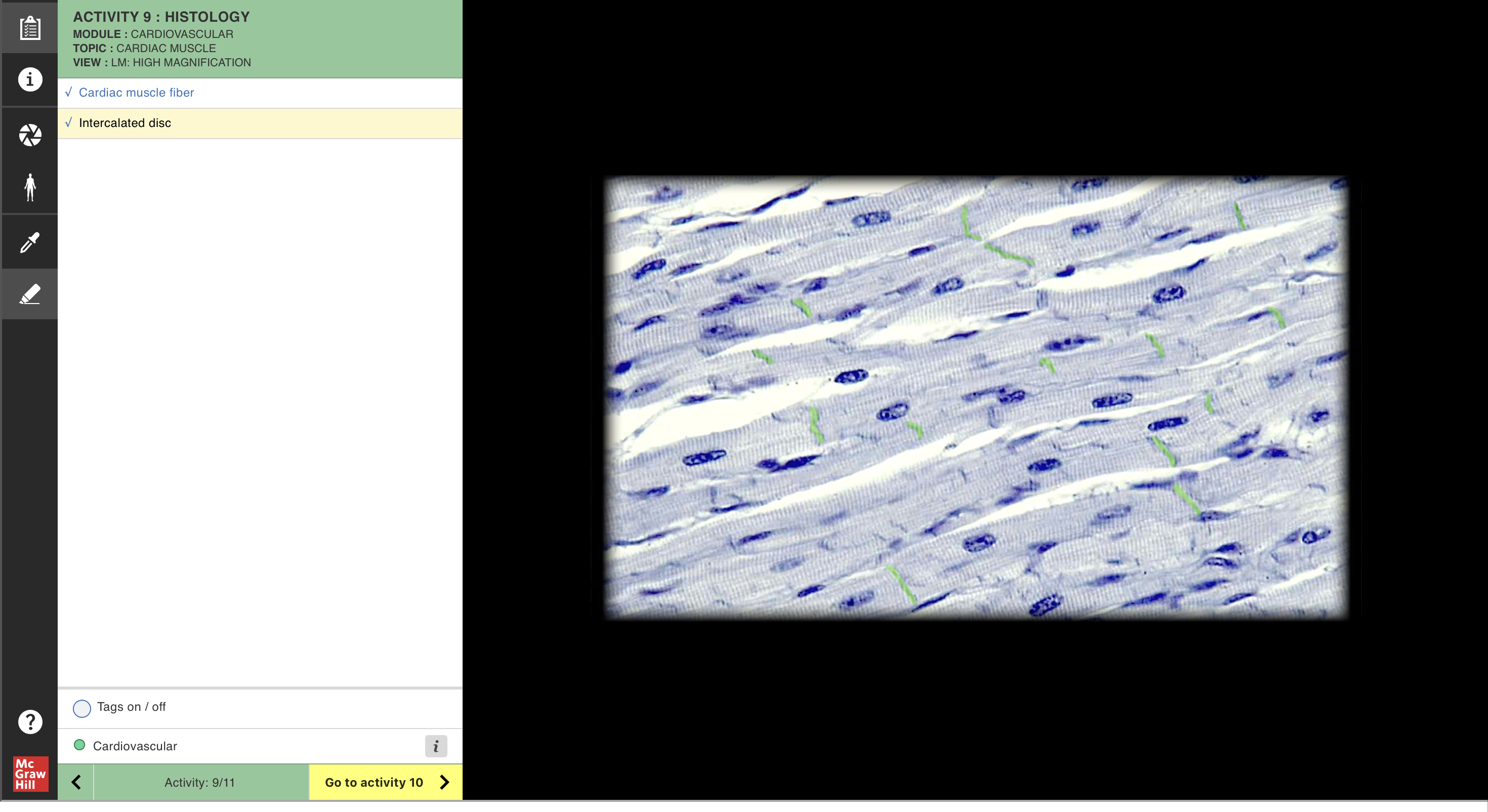
Intercalated disc image (green highlight)
Cardiac muscle fiber
Location:
Myocardium (muscular walls of heart)
Walls of pulmonary veins and superior vena cava
Description:
Short, branched muscle fibers joined end-to-end by intercalated discs
Striated, involuntary muscle
Contains one or two centrally placed nuclei
Perinuclear cytoplasm apparent
Function:
Involuntary contraction of myocardium
Contraction may be modulated by autonomic nervous system
Comment:
Intercalated discs contain numerous desmosomes and gap junctions
Cardiac muscle fiber also known as cardiac muscle cell or cardiomyocyte
Intercalated disc
Location:
Between cardiac muscle fibers
Description:
Interdigitating folds of cell membrane that are sites of adhering (mechanical) junctions and gap (electrical) junctions
Function:
Mechanical connection between adjacent cardiac muscle cells
Electrical connection between adjacent cardiac muscle cells, providing for spread of polarization impulses throughout myocardium
Comment:
Adhering junctions also known as desmosomes
Cardiac muscle fiber also known as cardiac muscle cell or cardiomyocyte