Medical physics
1/23
There's no tags or description
Looks like no tags are added yet.
Name | Mastery | Learn | Test | Matching | Spaced | Call with Kai |
|---|
No analytics yet
Send a link to your students to track their progress
24 Terms
What function does a piezoelectric crystal provide, how does it do so?
Used in production and detection of ultrasound waves as part of a piezo-electric transducer
when a p.d. is applied across it, it changes shape
when its shape changes, it generates an e.m.f.
Explain the main principles behind the use of ultrasound to obtain diagnostic information about internal body structures
pulses of ultrasound are produced by piezo-electric crystals when they are deformed
waves are reflected at boundaries between media
reflected pulses are detected by ultrasound generator
the time delay between transmission and receipt gives information about depth
intensity of reflected pulse gives information about nature of the boundary
gel is used to minimise reflection at the skin
the degree of reflection depends on impedances of two media at the boundary
What is the specific acoustic impedance of a medium?
Z = ρc
p density of medium
c speed of sound in the medium
Z specific acoustic impedance
What is the intensity reflection coefficient. Give the equation to find it.
the proportion of incident ultrasound signal that is reflected back
I reflected / I incident = (Z1 – Z2)² /(Z1 + Z2)²
If Z is the same, the wave is not reflected
If there is a greater difference between Z1 and Z2, more of the wave is reflected back
What is a coupling medium and why is it used in ultrasound
The intensity reflection coefficient between air and skin is very high. A coupling medium (e.g. gel) must be used between transducer and the body so ultrasound is not mostly reflected before entering the body.
removes the air between the transducer and body
usually oil or gel
minimises the difference in acoustic impedances
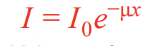
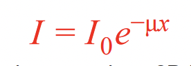
What is attenuation.?
When ultrasound waves are absorbed and scattered, which decreases their amplitude.
What is the linear attenuation coefficient
a measure of how easily an ultrasound wave can pass through a given material
describes the rate of energy loss per unit thickness
The larger the coefficient, the quicker the intensity of the ultrasound will decrease as the wave passes through a medium
What equation gives the intensity of an ultrasound after passing a distance through a medium?
What is thermionic emission?
When is it used?
a metal is heated until free electrons on the surface gain enough energy to be emitted
used in the production of X-Rays
Describe the process of production of X-Rays
electrons emitted from a filament by thermionic emission and are accelerated through a potential difference towards anode (metal target)
once they collide, they decelerate and emit part of their energy as EM radiation
this is in the form of X-ray photons - braking radiation. Forms a continuous spectrum of X-ray radiation
some electrons collide with orbital electrons of the target atoms and ionise the atoms, causing electrons to de-excite, releasing X-ray photons (energy)
this energy depends on the difference between energy levels of the metal’s atoms so will depend on the material
“Outline the principles of computed tomography (CT) scanning”. [5]
X-rays are used
object is scanned in sections
scans taken at many angles, images of each section are 2-dimensional
scans of many sections are combined
to give 3-dimensional image of whole structure
“By reference to an ultrasound wave, explain what is meant by specific acoustic impedance'“ [2]
Product of density and speed
Speed of ultrasound in medium
“State what is meant by the attenuation of an ultrasound wave” [1]
Loss of intensity/amplitude/power of the wave
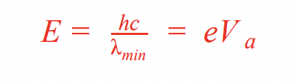
How to calculate the minimum wavelength of X-rays produced from the accelerating p.d.?
Product of charge of an electron and accelerating voltage = max energy because this is the value of the KE as the electrons hit the target.
Describe two methods of controlling beam intensity of X-ray
increasing accelerating voltage. Electrons gain more KE, so photons will have higher energies
increasing current passing through filament emitting electrons. More electrons released per second, therefore more X-ray photons produced per second
What is the meaning of contrast in X-ray scanning
difference in degree of blackening between adjacent structures
allows tissues to be differentiated
impacted by how much of the X-ray is absorbed - linear attenuation coefficient is an indicator of how much will be absorbed
What is the equation to find the intensity of X-rays after they have passed through a medium?
In X-ray imaging, what is sharpness vs contrast?
Sharpness - ease with which edges can be distinguished
Contrast - difference in degrees of blackening
Define the intensity of an X-ray beam
The total energy emitted per second per unit area passing through a surface perpendicular
What are some advantages of a CT scanner
non-invasive
high quality image
full cross-sectional area
What are some disadvantages of a CT scanner
patient exposed to large dose of ionising radiation
expensive
contrast between materials of similar densities is small
What is a tracer
A substance containing radioactive nuclei that can be introduced into the body and is then absorbed by the tissue being studied
Decays by Beta-plus decay
Used in positron emission tomography
What is annihilation. When does it occur
when a particle interacts with its anti-particle
minimum energy of each photon emitted is equal to rest energy of electron/positron
mass-energy and momentum are conserved
energy of electron and positron is shared equally between gamma ray photons
Describe PET (Positron emission tomography) scanning
used to form 3D and cross-sectional images of body
patient injected with tracer
patient left to allow the radionuclide to move to region of interest
radionuclide will be absorbed and broken down, releasing positrons which collide with electrons present in the tissue and are annihilated
this releases two gamma rays moving in opposite directions, recorded by detectors
signals sent to computer
an image of the radioactivity in that region is formed by processing the arrival times of the gamma-ray photons