Biology Study Material - BFUN Lab 2: Vocabulary and Definitions Related to Heart Anatomy
1/72
There's no tags or description
Looks like no tags are added yet.
Name | Mastery | Learn | Test | Matching | Spaced |
|---|
No study sessions yet.
73 Terms
Fibrous Pericardium
Name the Layer
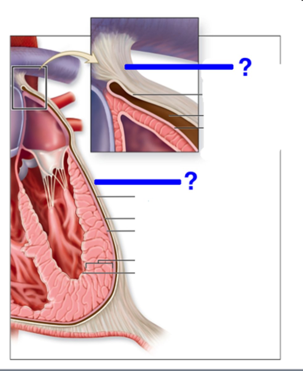
Parietal Pericardium
Name the Layer
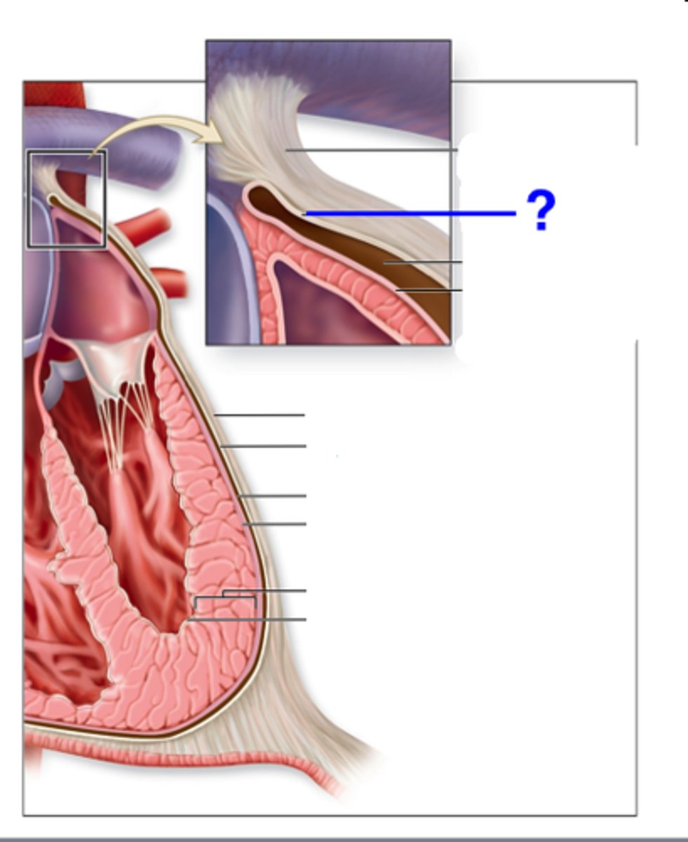
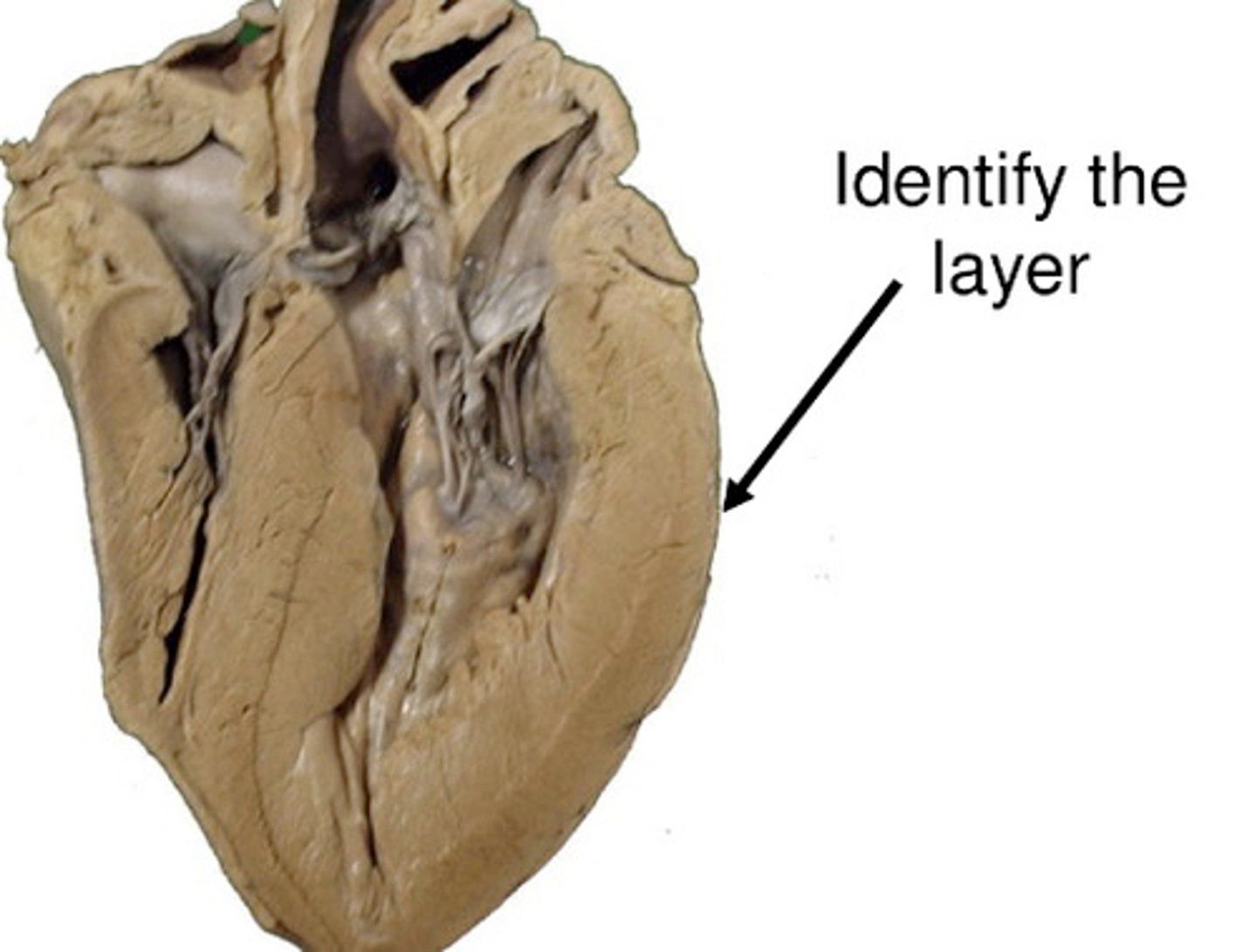
Epicardium (AKA Visceral Pericardium)
Name the Layer
Pericardial Cavity
Note: in between epicardium/visceral pericardium & parietal pericardium
Name the Blue Space
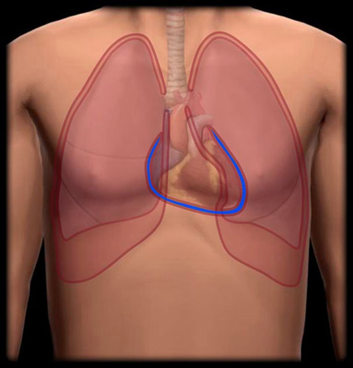
Oblique Pericardial Sinus
Name the Bottom Arrow
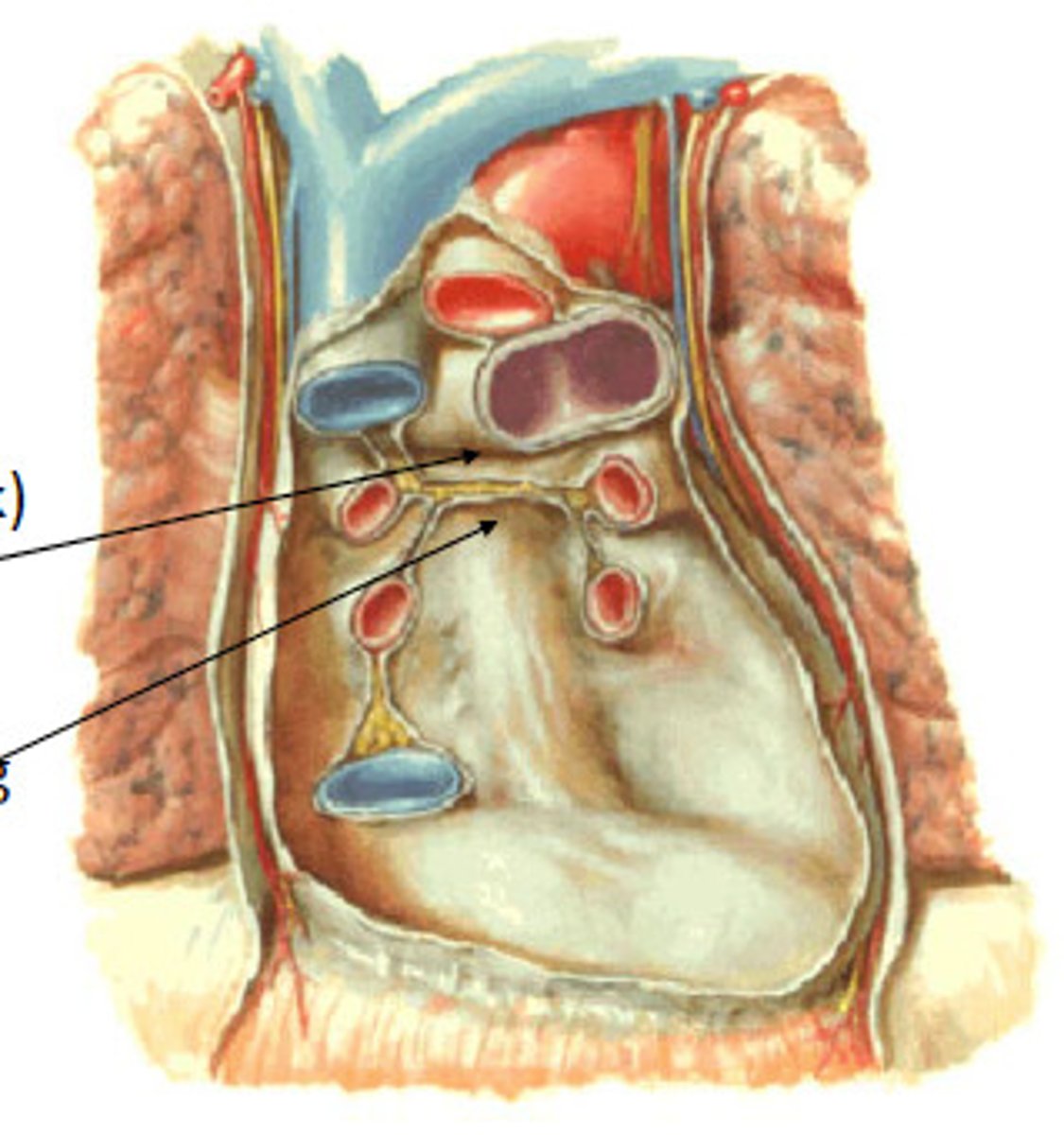
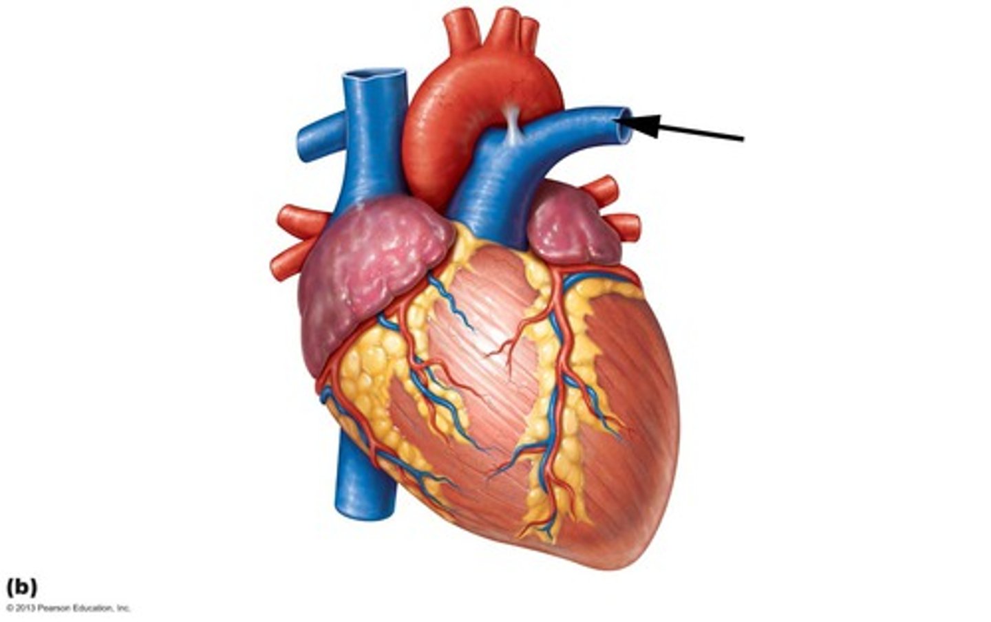
Transverse Pericardial Sinus
Name the Top Arrow

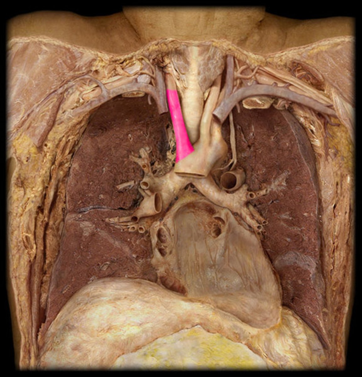
Superior Vena Cava
Name the Vein
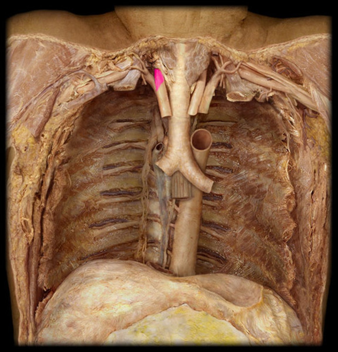
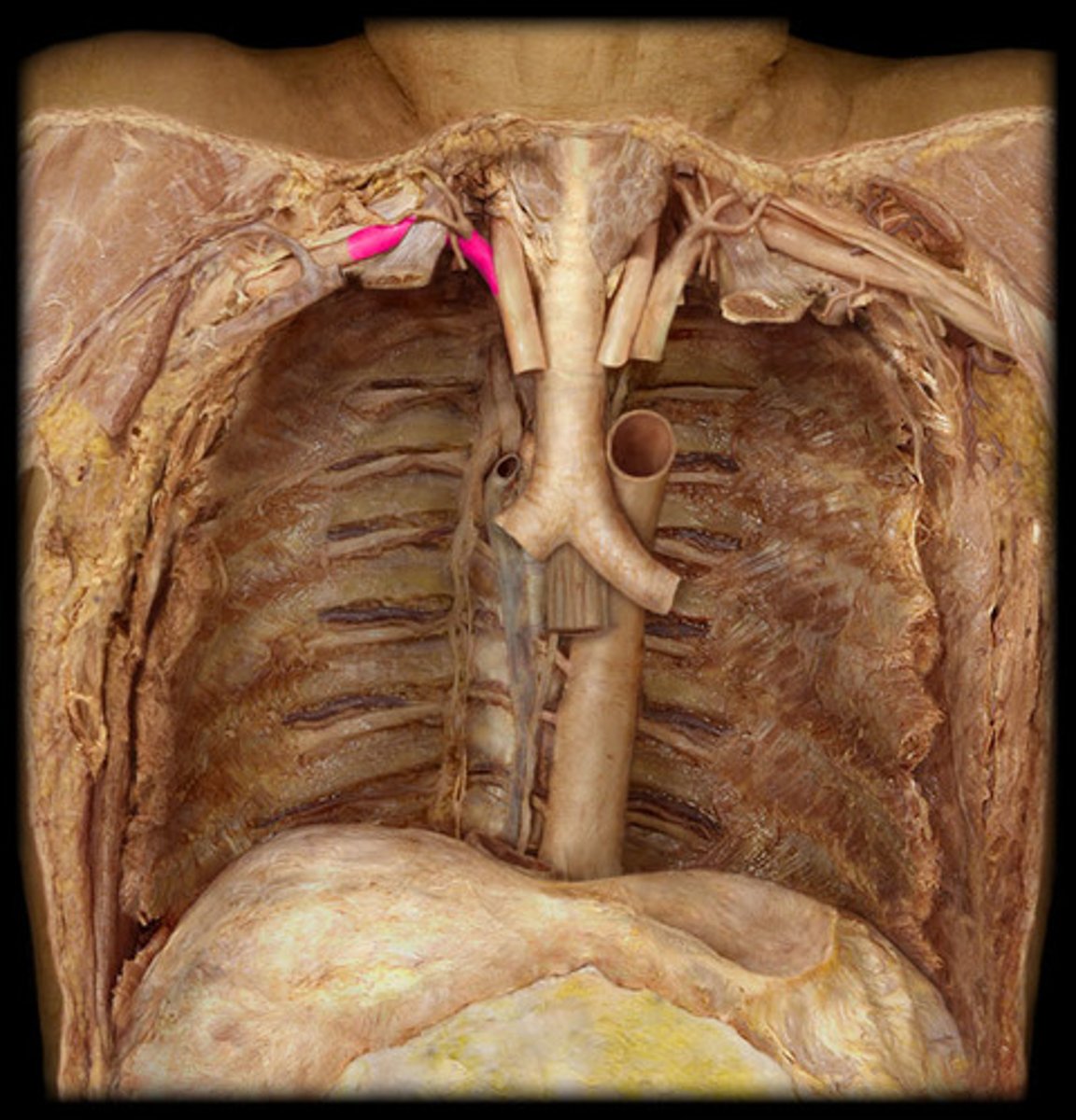
Right Brachiocephalic Vein
Name the Vein
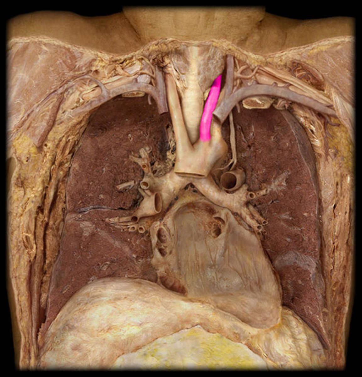
Left Brachiocephalic Vein
Name the Vein
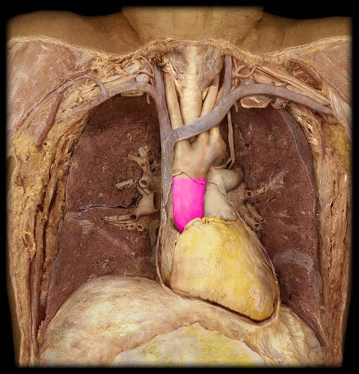
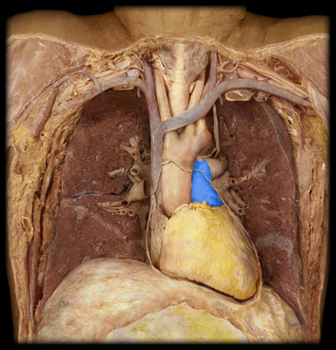
Ascending Aorta
Name the Artery
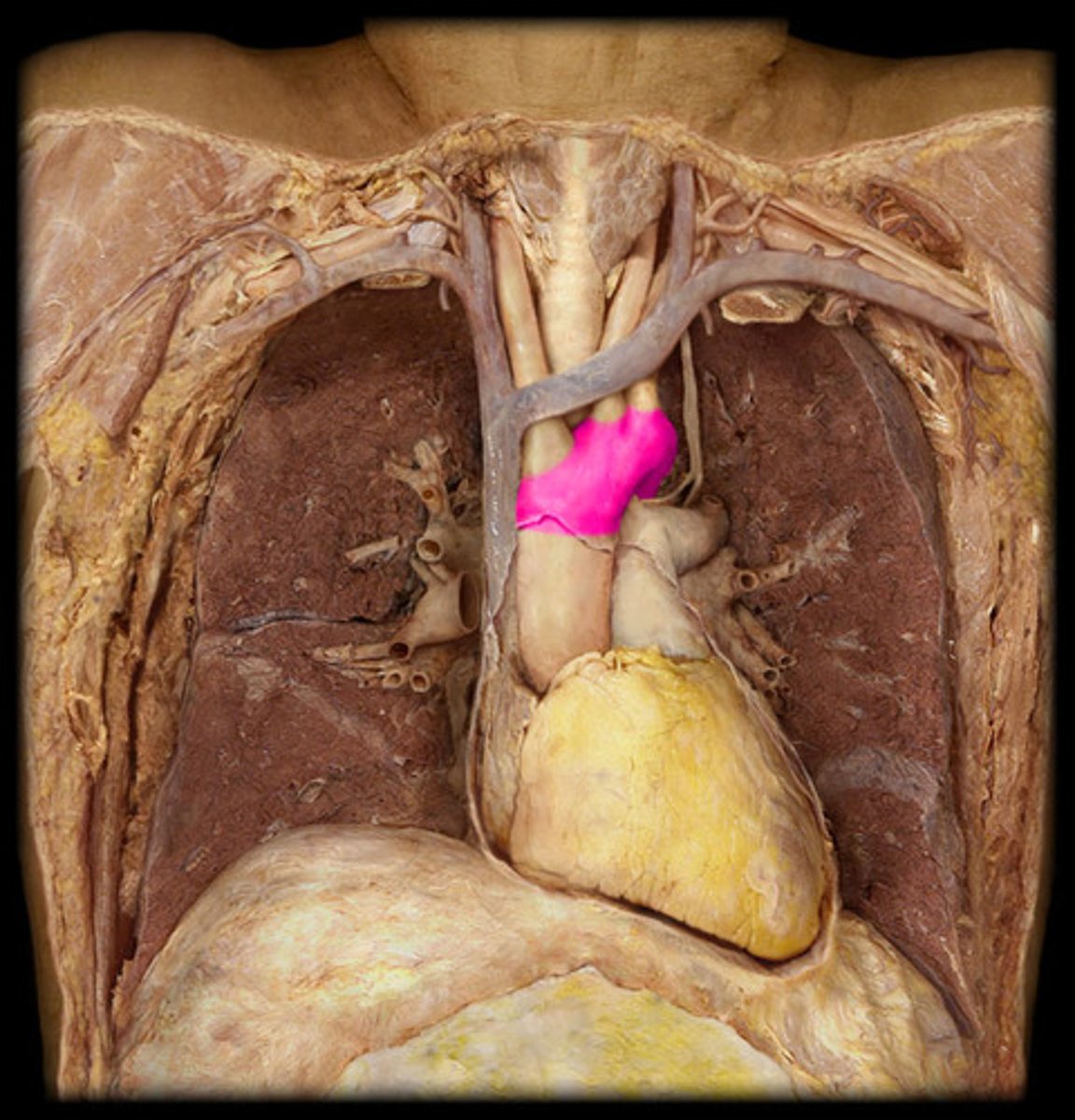
Arch of Aorta
Name the Artery
Brachiocephalic Trunk
Name the Artery
Right Common Carotid
Name the Artery
Right Subclavian
Name the Artery
Left Common Carotid
Name the Artery
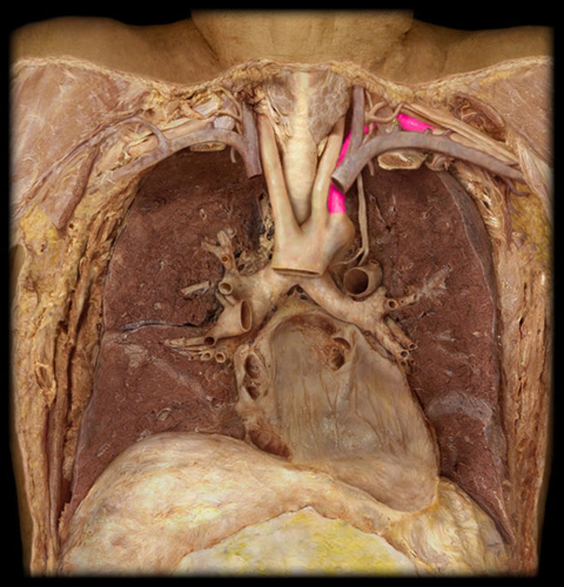
Left Subclavian
Name the Artery
Pulmonary Trunk
Name the Artery
Pulmonary Artery
Note: there is a left and right pulmonary artery, which are both branches of pulmonary trunk...the arrow here only points to left pulmonary artery
Name the Artery
Atrioventricular Sulcus (Anterior View)
Note: location of RCA & LCX
Name the Groove
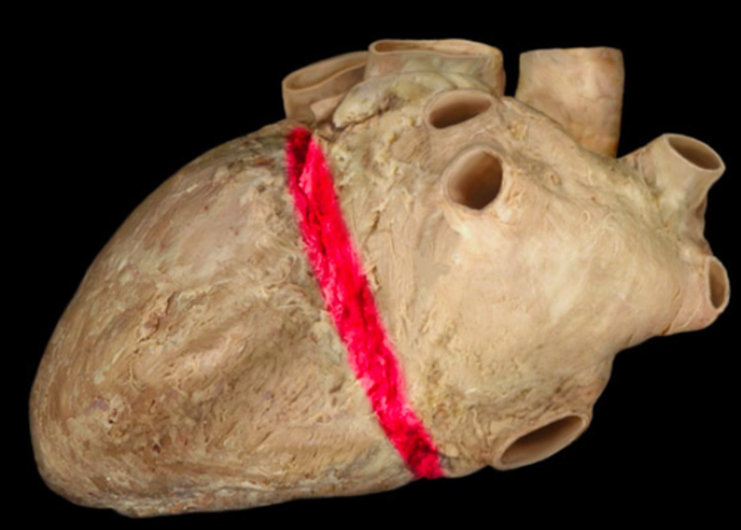
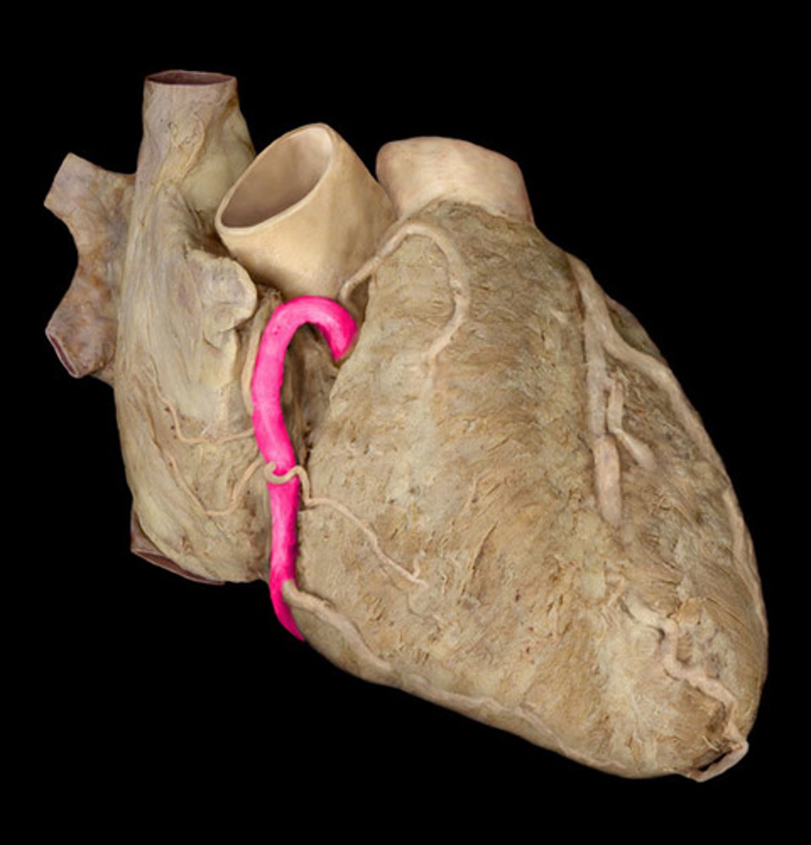
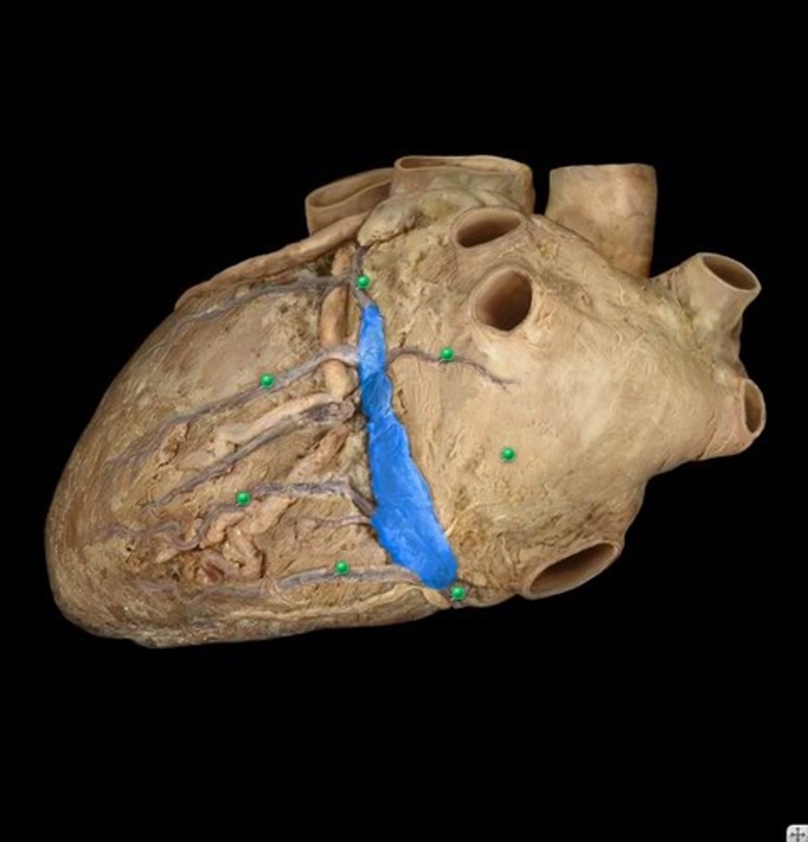
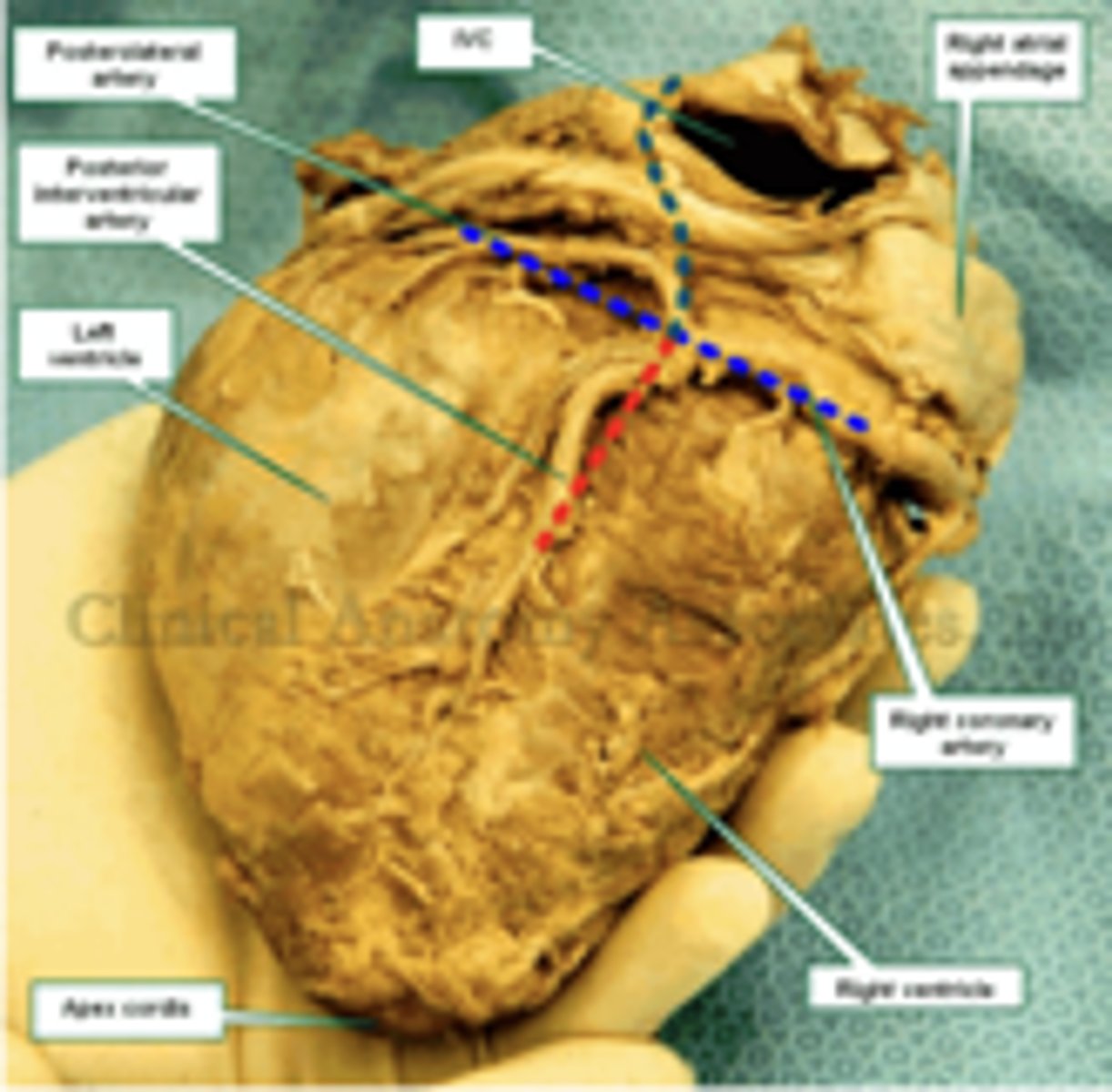
Atrioventricular Sulcus (Posterior View)
Name the Groove
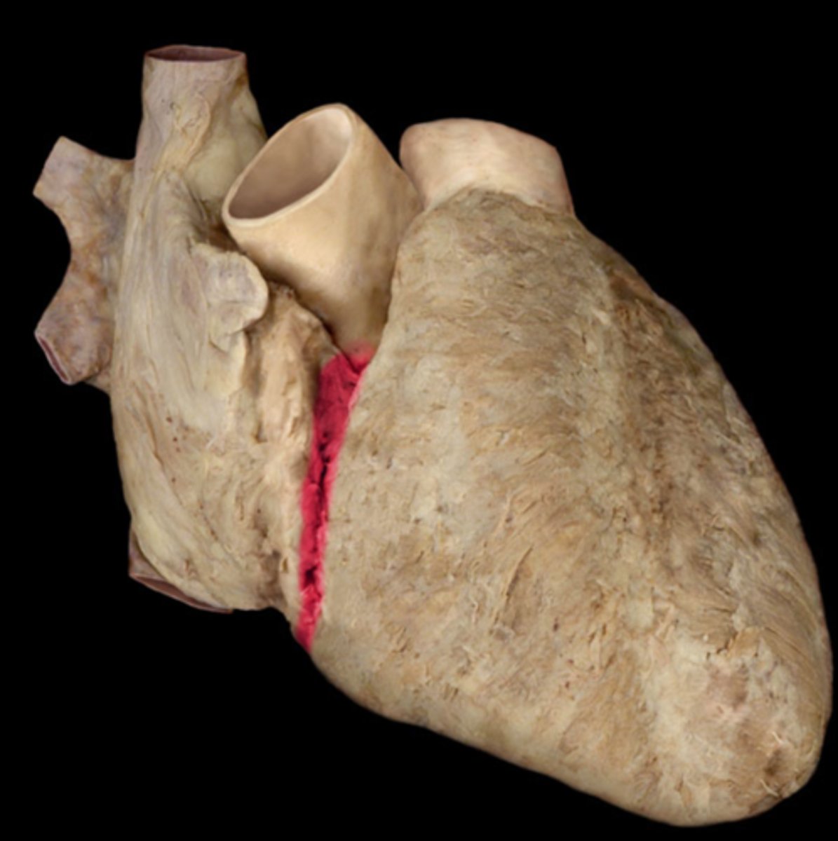
Interventricular Sulcus (Anterior View)
Note: location of LAD & posterior descending, as well as great & middle cardiac veins
Name the Groove
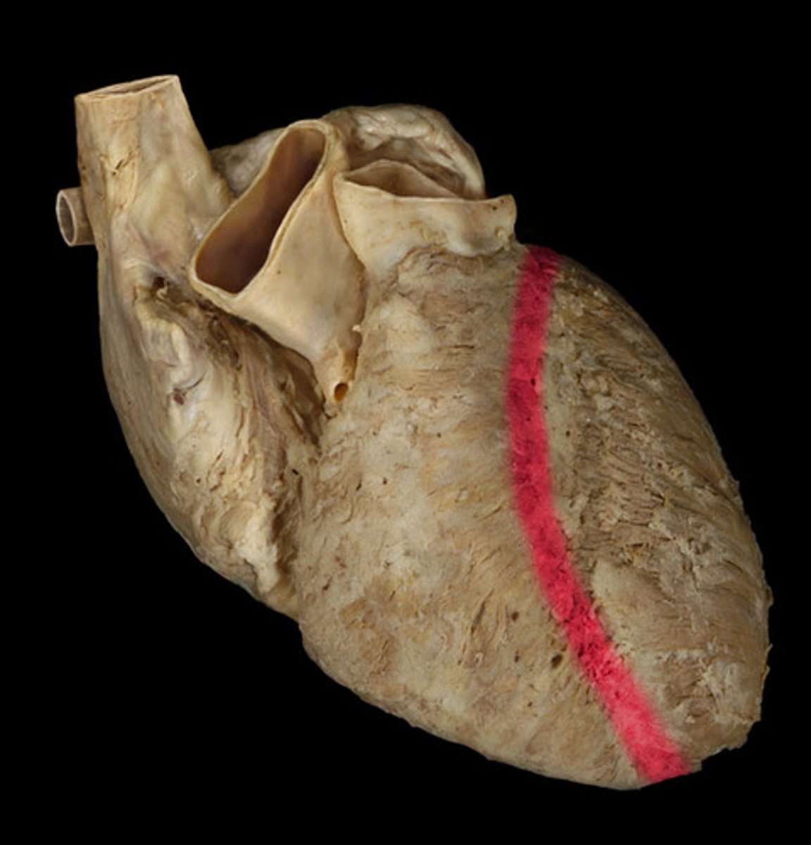
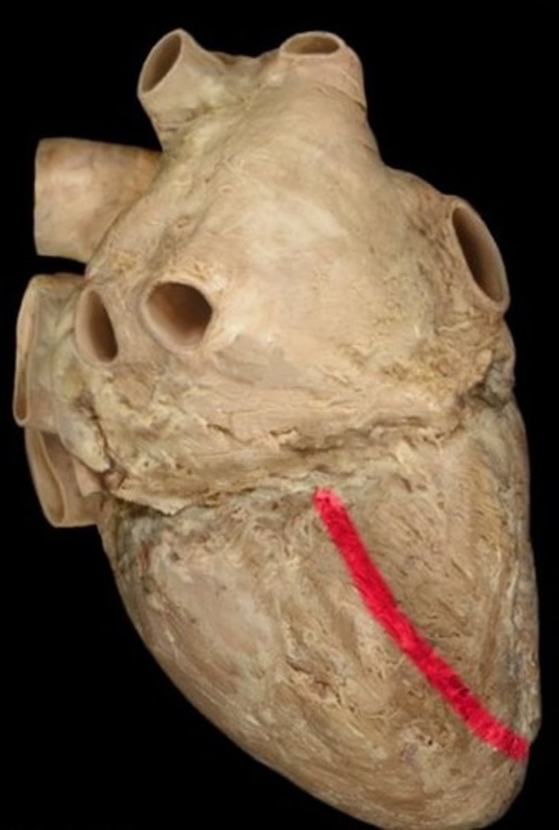
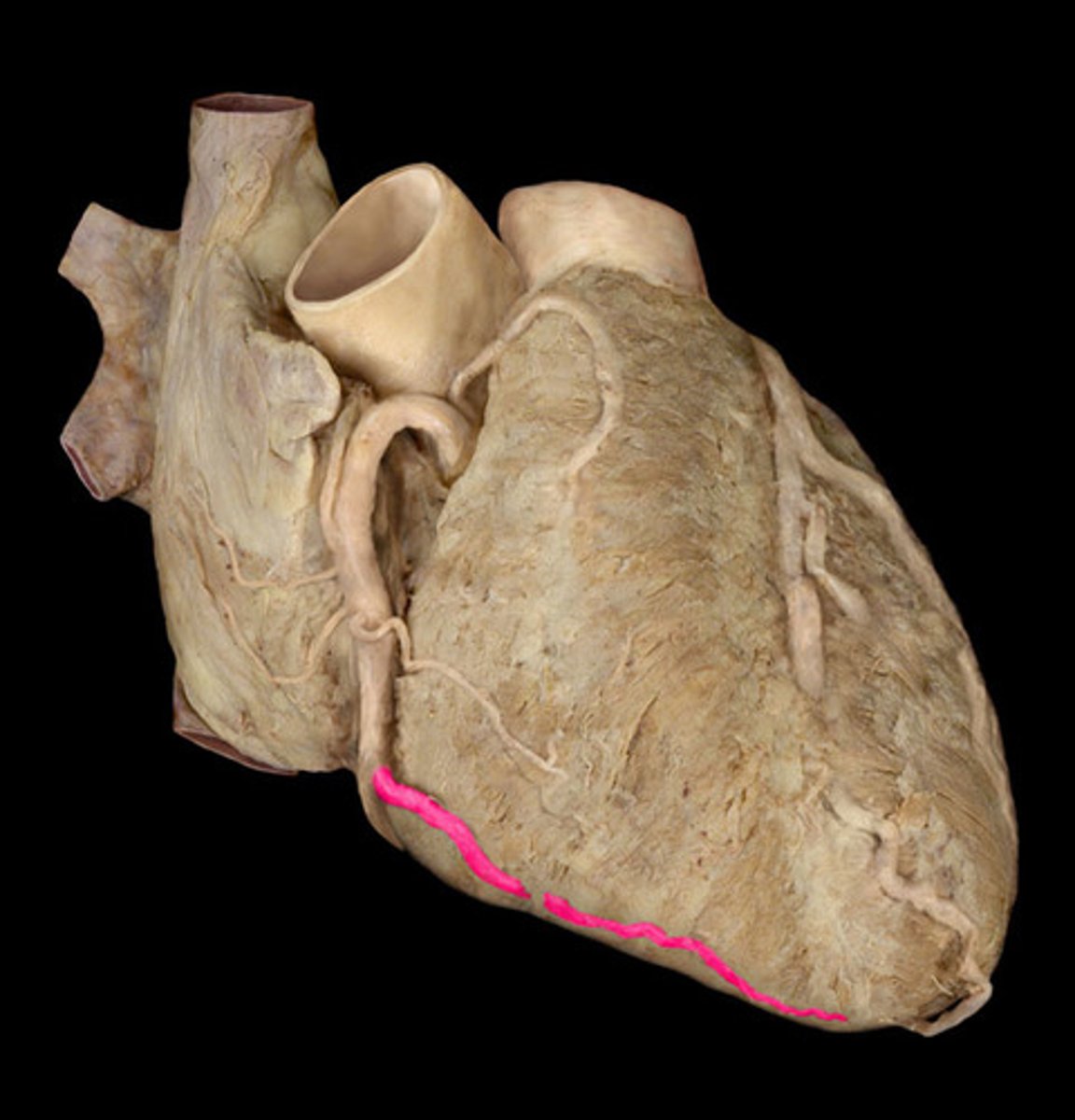
Interventricular Sulcus (Posterior View)
Name the Groove
Right Coronary Artery (RCA)
Name the Artery
Right Marginal Artery
Note: branch of RCA
Name the Artery
Left Coronary Artery (LCA)
Note: very short (splits into LCX and LAD), hard to find good pictures
Name the Artery
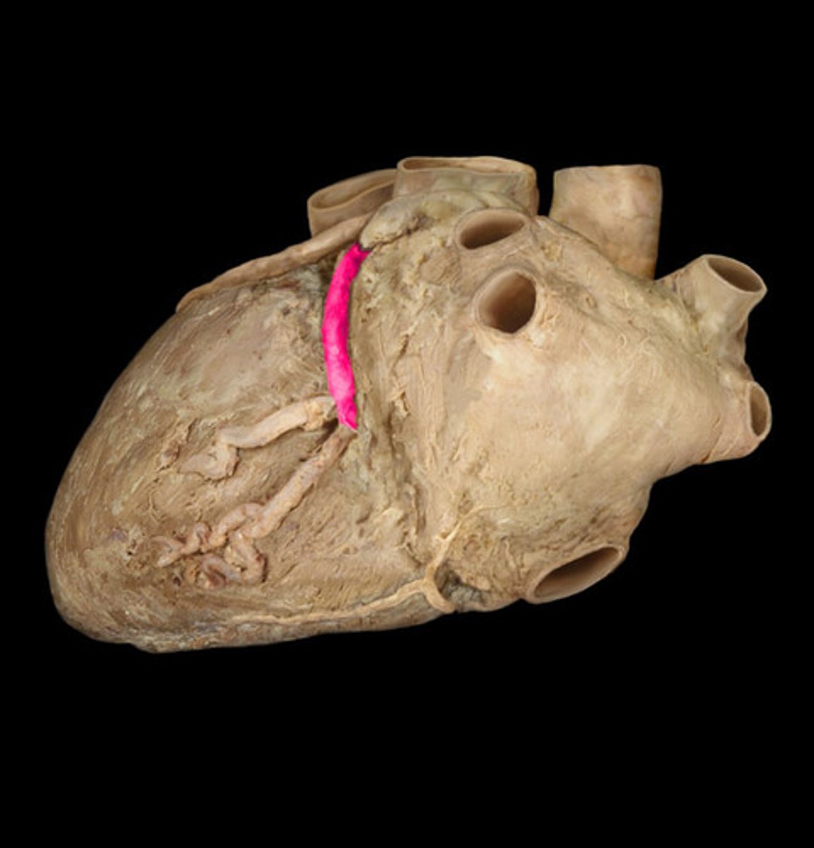
Left Circumflex Artery (Posterior)
Note: branch of LCA, travels alongside coronary sinus in atrioventricular groove
Name the Artery
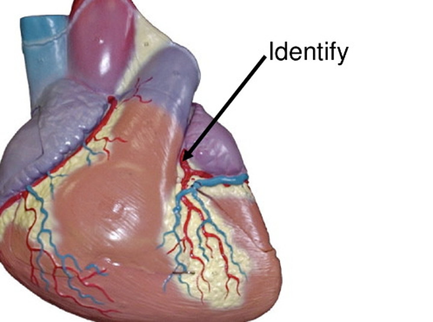
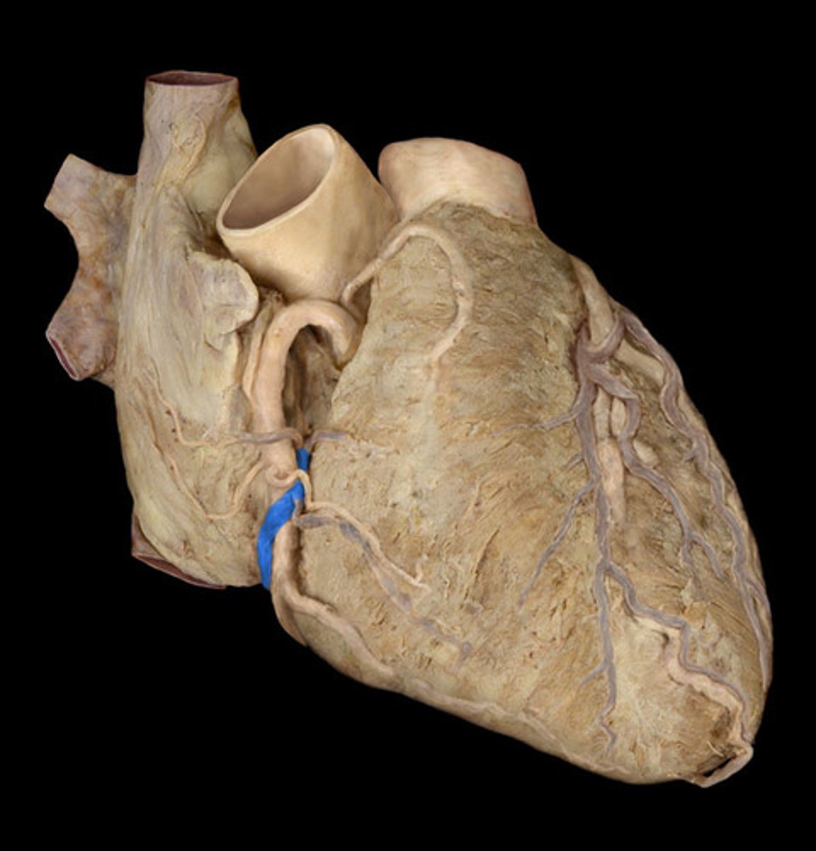
Left Circumflex Artery (Anterior)
Name the Artery (C)
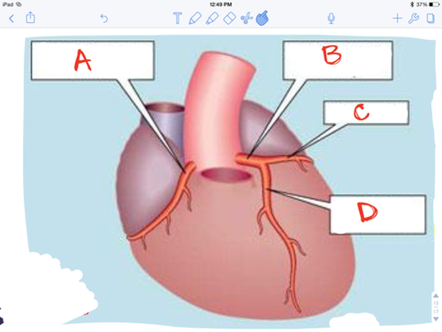
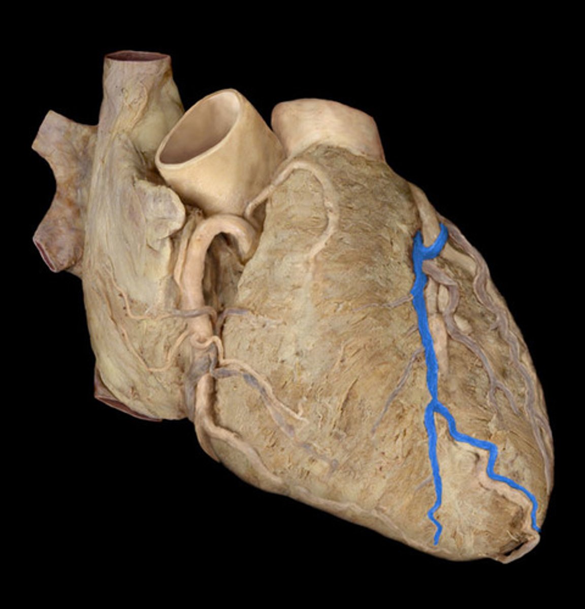
Left Anterior Descending Artery
Note: branch of LCA, travels alongside great cardiac vein in the interventricular groove
Name the Artery
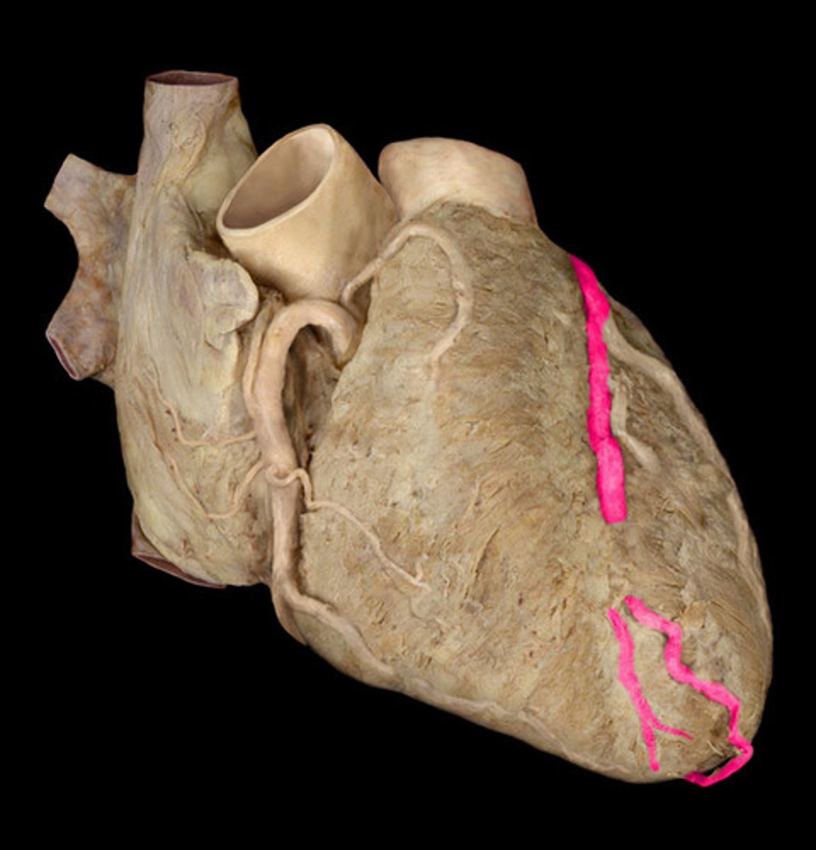
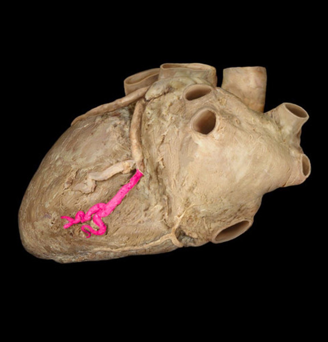
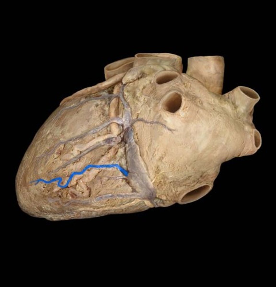
Posterior Descending Artery
Note: travels in interventricular groove, can come off LCX or RCA (in this picture I think it is LCX, making this a left dominant heart)
Name the Artery (HINT: it's a posterior view)
Great Cardiac Vein
Note: runs along LAD in interventricular groove, drains into coronary sinus
Name the Vein
Middle Cardiac Vein
Note: runs along PDA in interventricular groove, drains into coronary sinus
Name the Vein (HINT: it's a posterior view)
Small Cardiac Vein
Note: travels alongside right marginal artery
Name the Vein
Small Cardiac Vein (Posterior View)
Name the Vein

Coronary Sinus
Note: drains into RA, collects from all coronary veins on heart surface except anterior cardiac veins
Name the Space
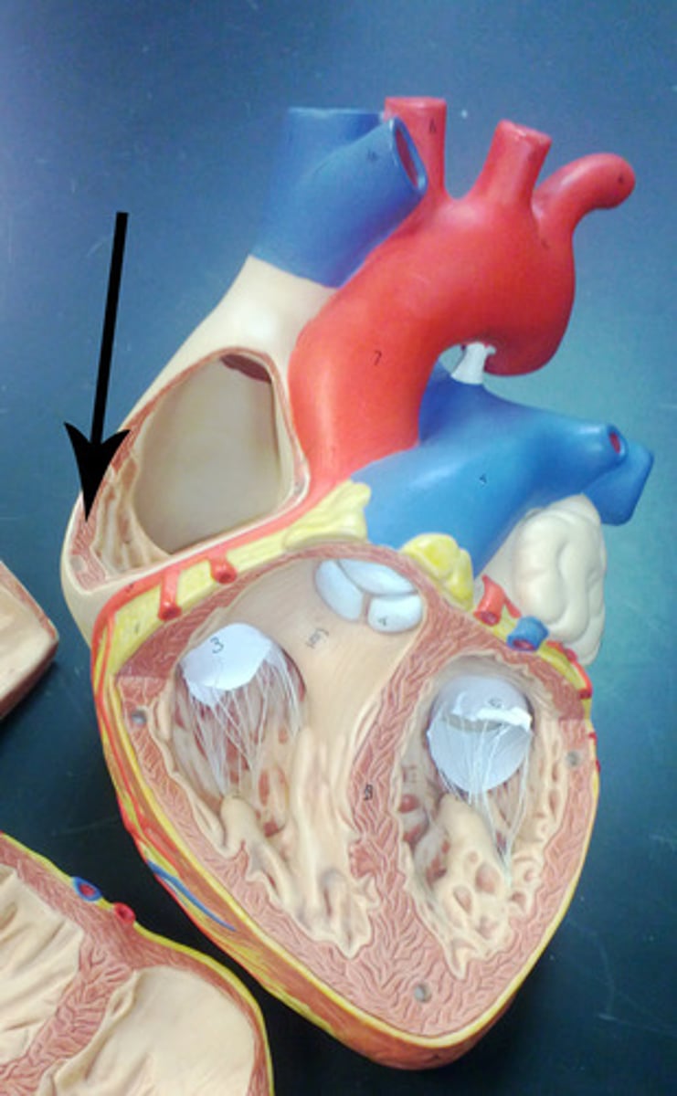
Right Auricle
Name the Structure of the Right Atrium
Opening of Superior Vena Cava
Name the Structure of the Right Atrium
Opening of Inferior Vena Cava
Name the Structure of the Right Atrium
Crista Terminalis
Note: ridge between smooth-walled portion of right atrium and pectinate muscles
Name the Structure of the Right Atrium
Pectinate Muscles
Note: helps contractions in diastole, they look translucent in lab (you can see light shine through them)
Name the Structure of the Right Atrium
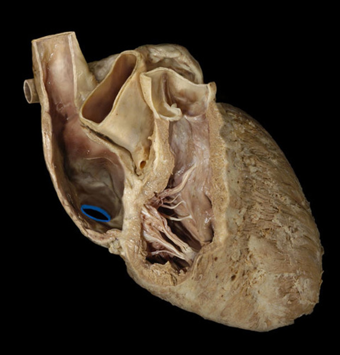
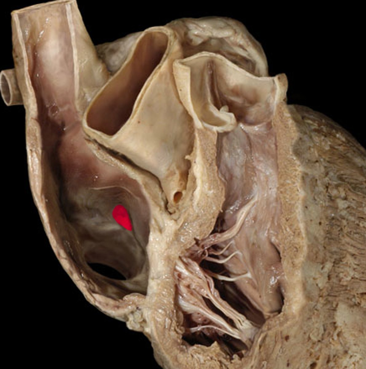
Fossa Ovalis
Note: remnant of foramen ovale
Name the Structure of the Right Atrium
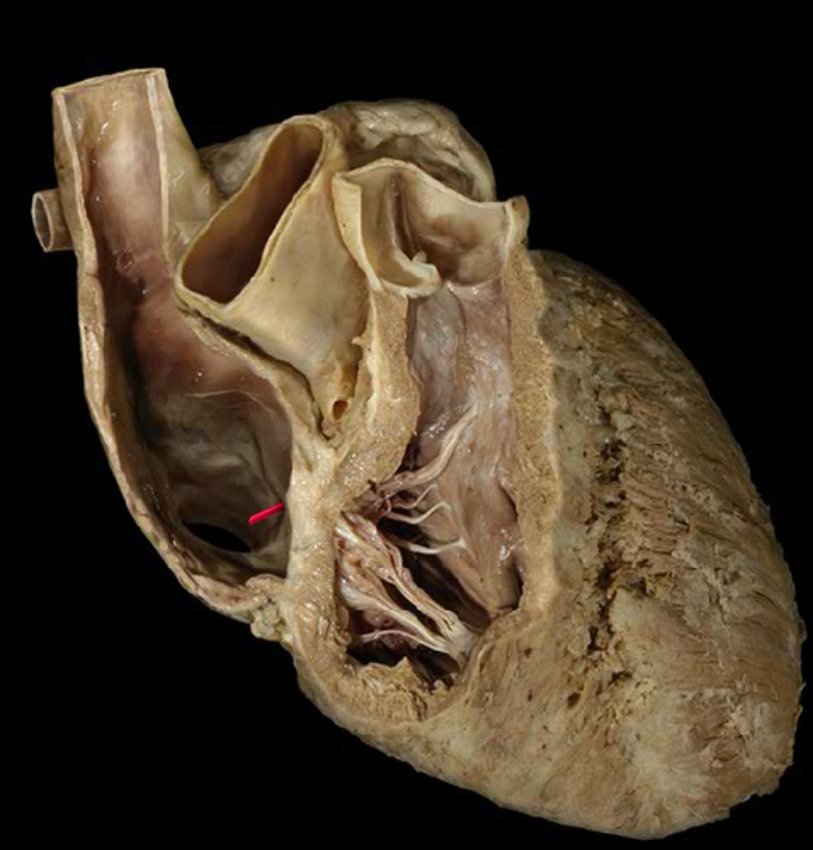
Opening of Coronary Sinus (AKA Thebesian Valve)
Name the Structure of the Right Atrium
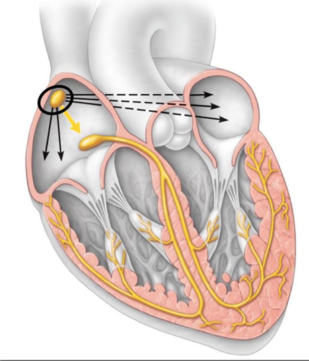
SA Node (AKA pacemaker of heart)
Note: can't actually see it, but is between SVC and crista terminalis
Name the Structure of the Right Atrium
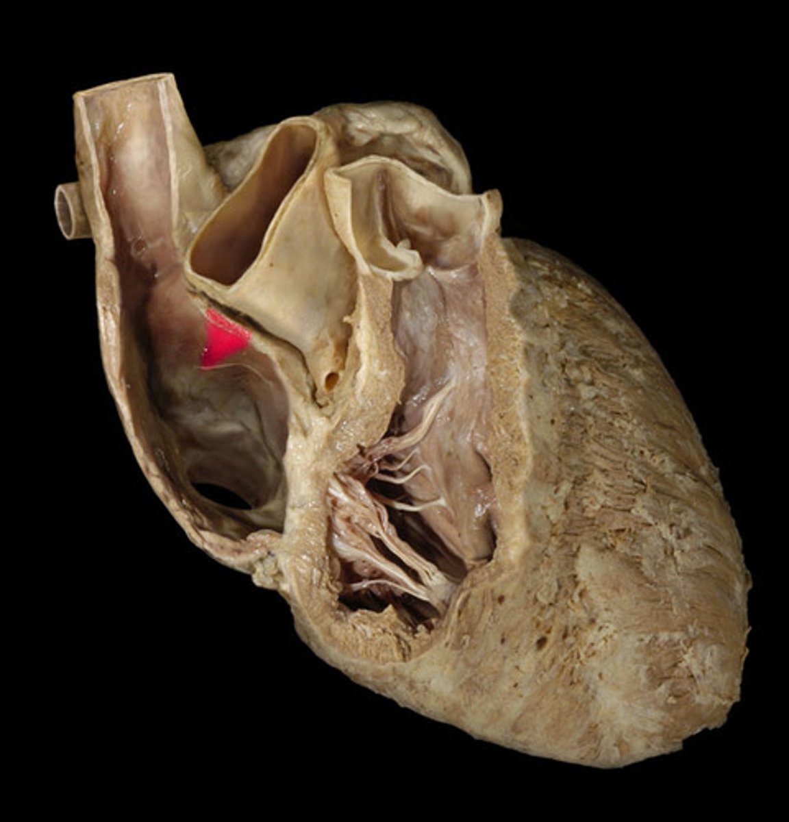
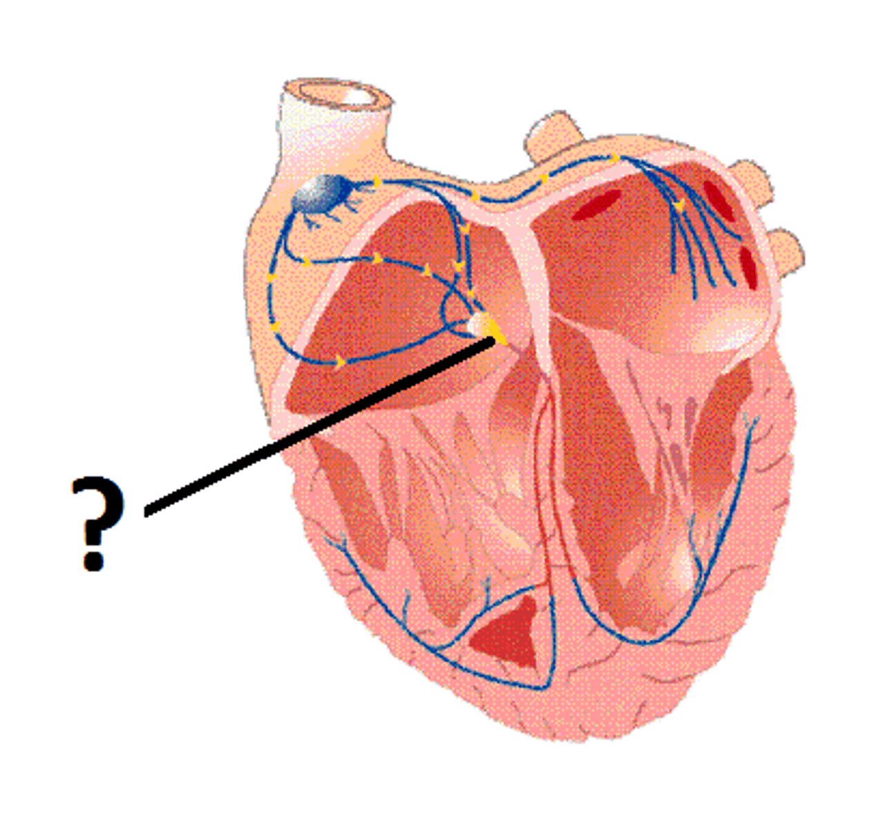
AV Node
Note: can't actually see it, but is within interatrial septum between opening of coronary sinus and septal leaflet of tricuspid
Name the Structure of the Right Atrium
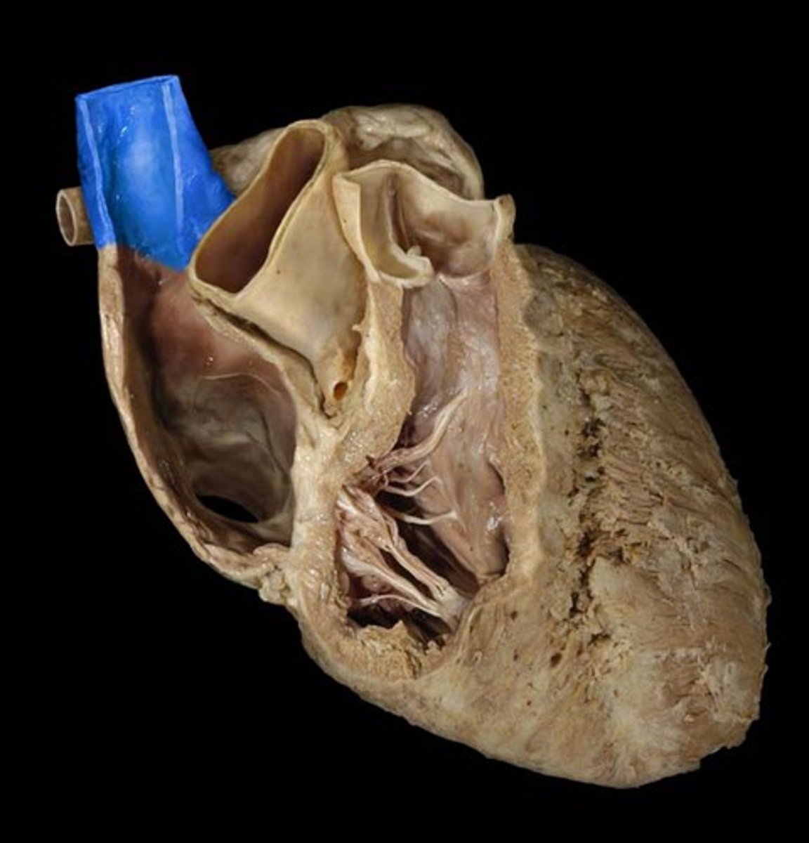
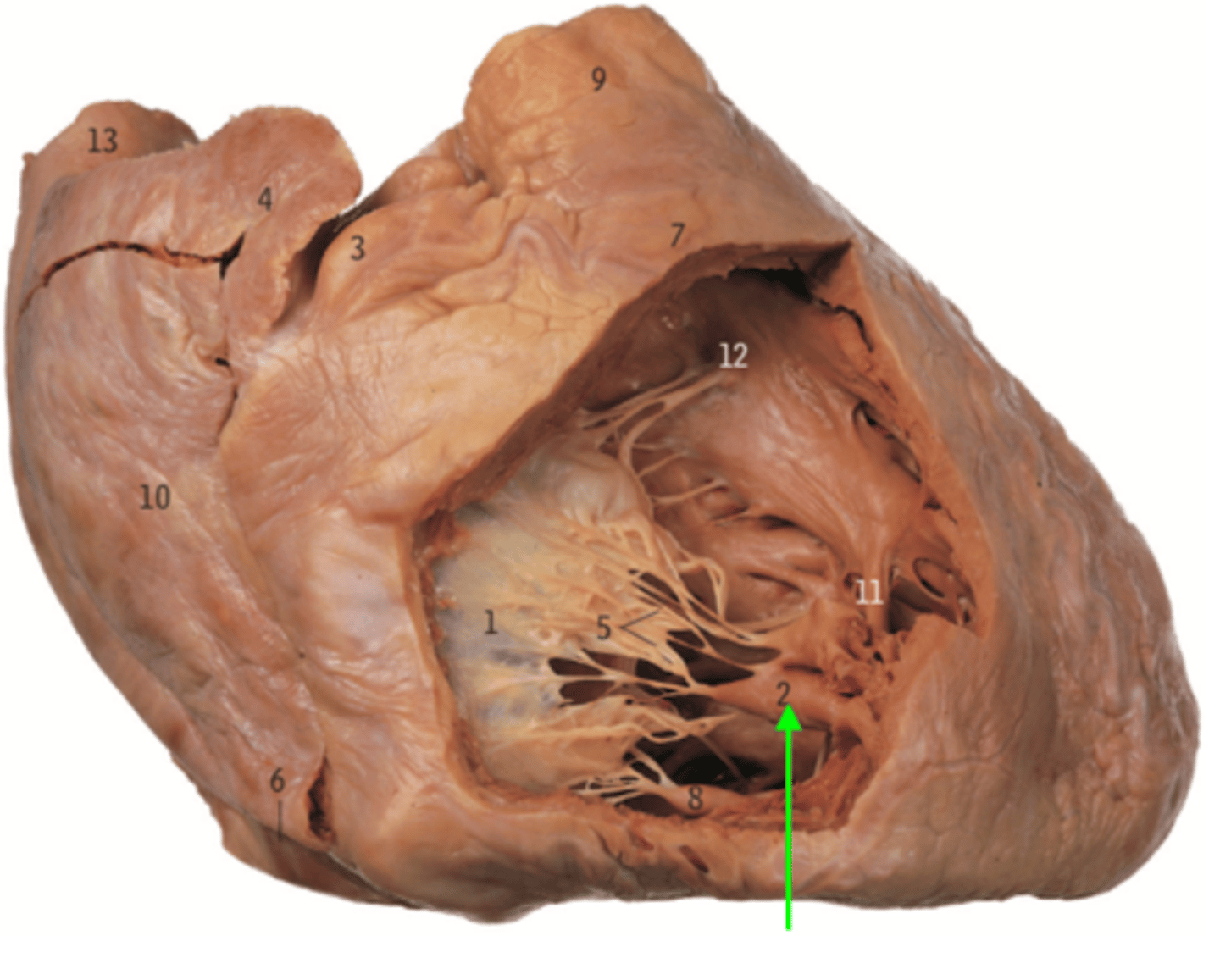
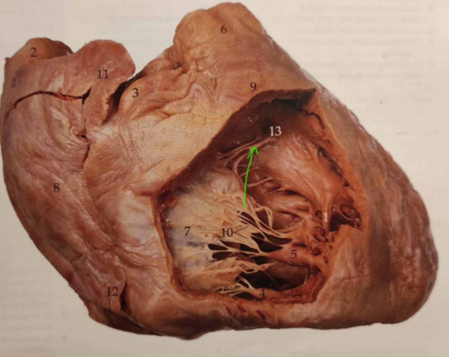
Tricuspid Valve (AKA right atrioventricular valve)
Note: contains 3 leaflets - anterior, posterior, and septal, which connect to papillary muscles via chordae tendineae
Name the Structure of the Right Ventricle
Anterior Cusp of Tricuspid
Name Structure 3 (Part of Right Ventricle)
Septal Leaflet of Tricuspid
Name Structure 4 (Part of Right Ventricle)

Posterior Leaflet of Tricuspid
Name Structure 5 (Part of Right Ventricle)Name Structure 5 (Part of Right Ventricle)

Anterior Papillary Muscle
Name the Muscle of the Right Ventricle
Posterior Papillary Muscle
Name the Muscle of the Right Ventricle
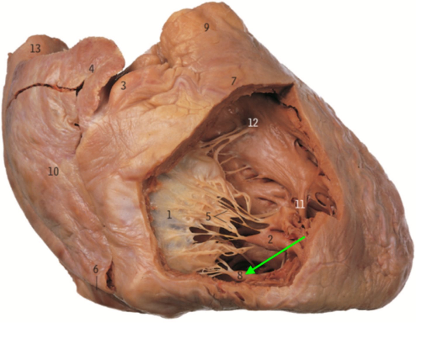
Septal Papillary Muscle
Note: attached to interventricular septum
Name the Muscle of the Right Ventricle
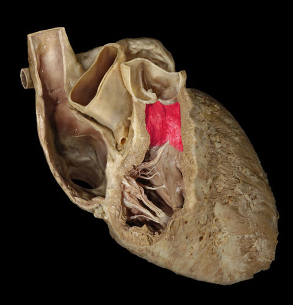
Interventricular Septum
Name the Structure of the Right Ventricle
Moderator Band (septomarginal trabecula)
Note: connects anterior papillary muscle with interventricular septum
Name the Structure of the Right Ventricle
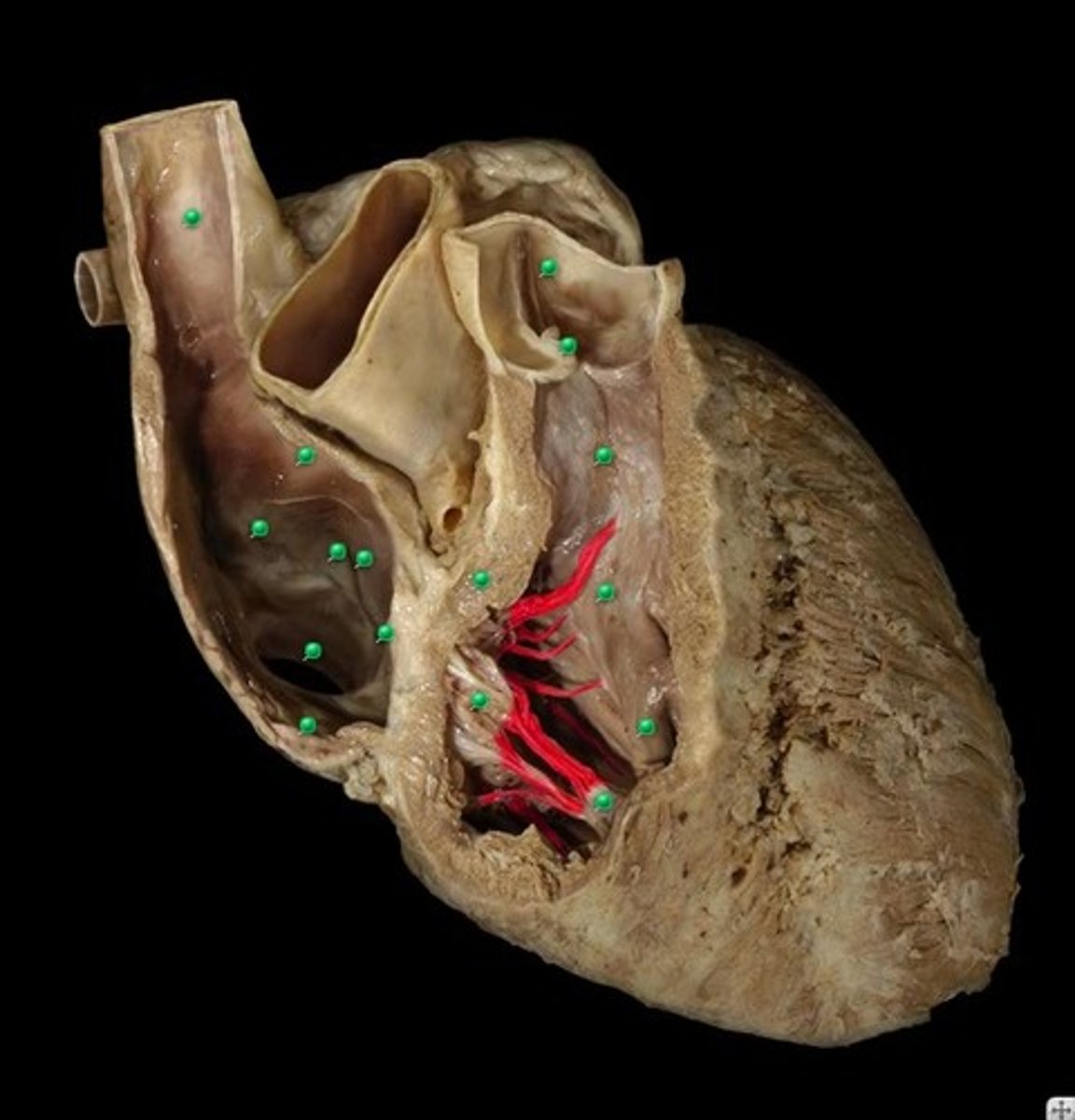
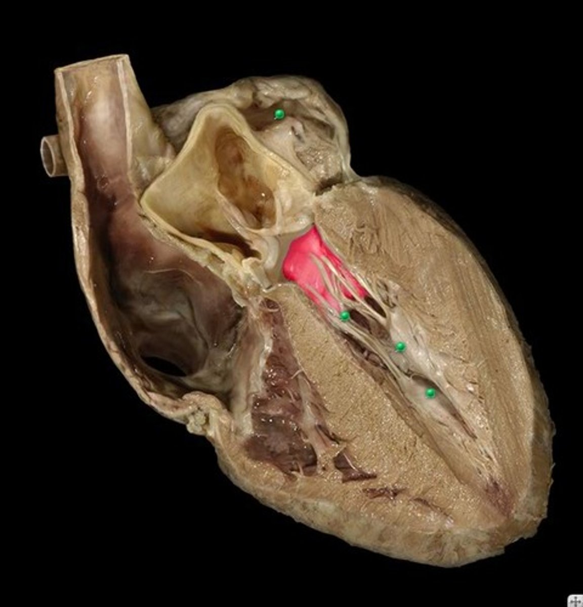
Chordae Tendineae
Note: exist for left and right ventricles (don't have a separate image for left ventricle)
Name the Structure
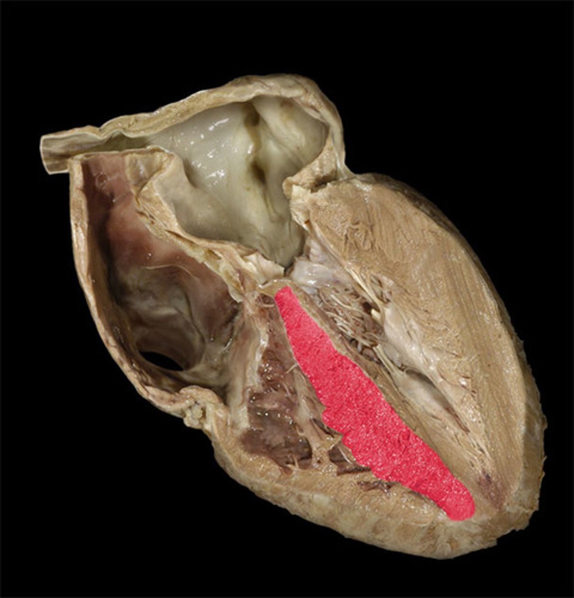
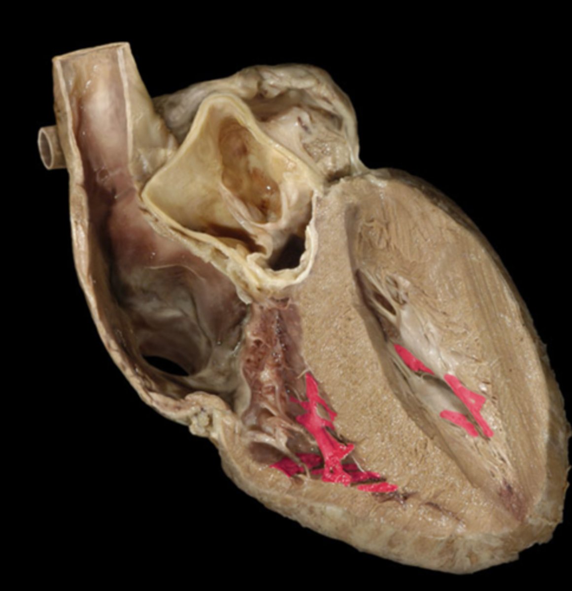
Trabaculae Carneae
Note: these are ridges of muscles in right and left ventricles
Name the Structure of the Right Ventricle
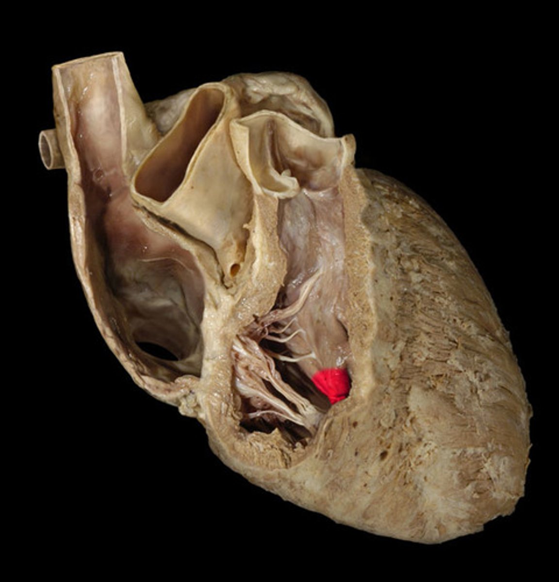
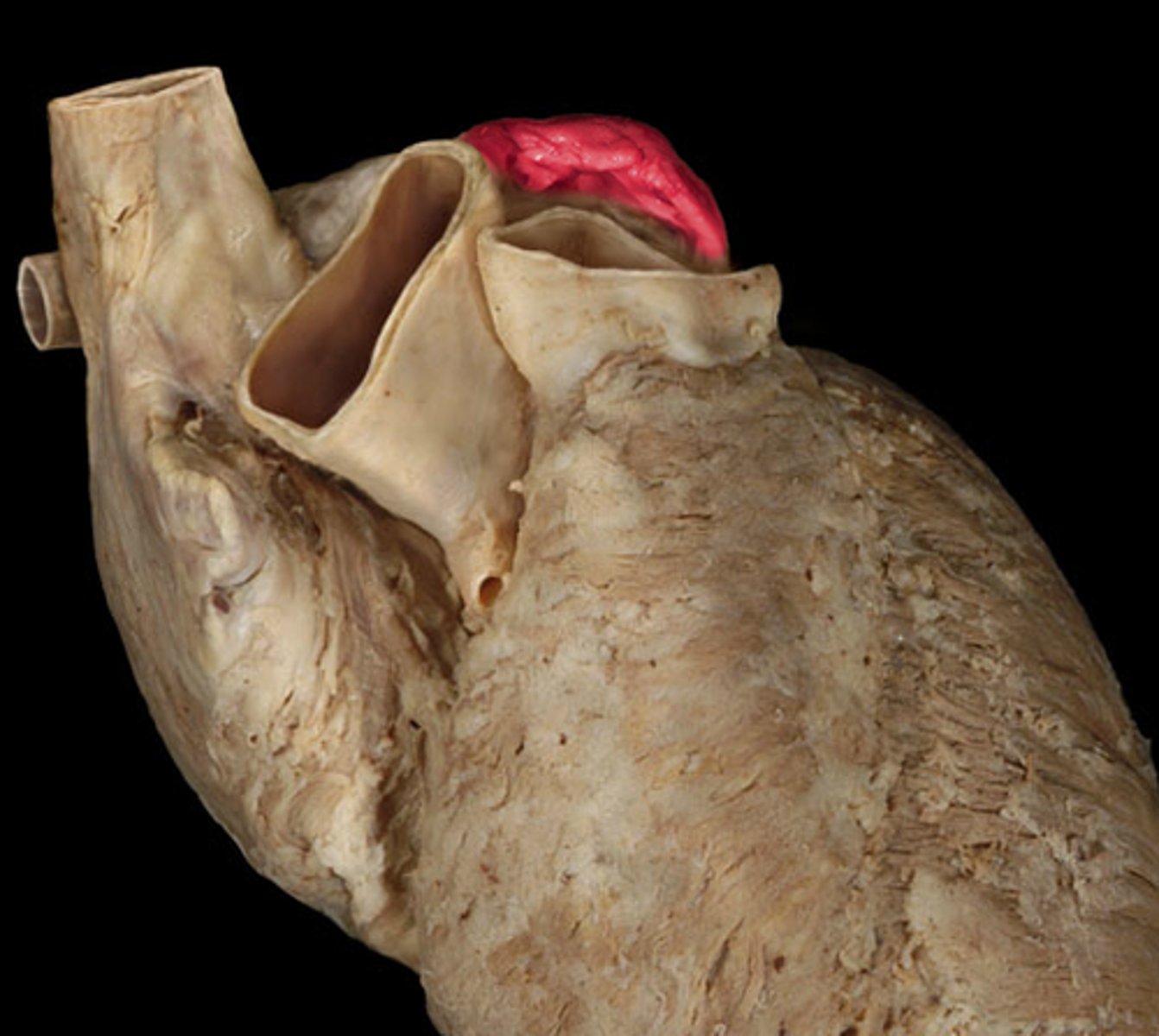
Conus Arteriosus (AKA Infundibulum)
Note: smooth space immediately inferior to the pulmonary valve...exists for left ventricle right before aortic valve too, but is only called infundibulum for left ventricle (do not have a separate image for this)
Name the Structure of the Right Ventricle
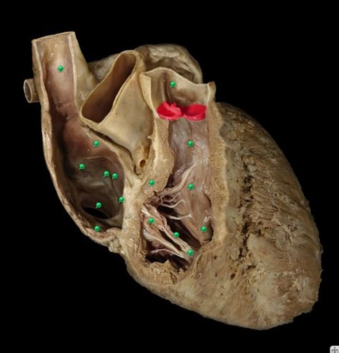
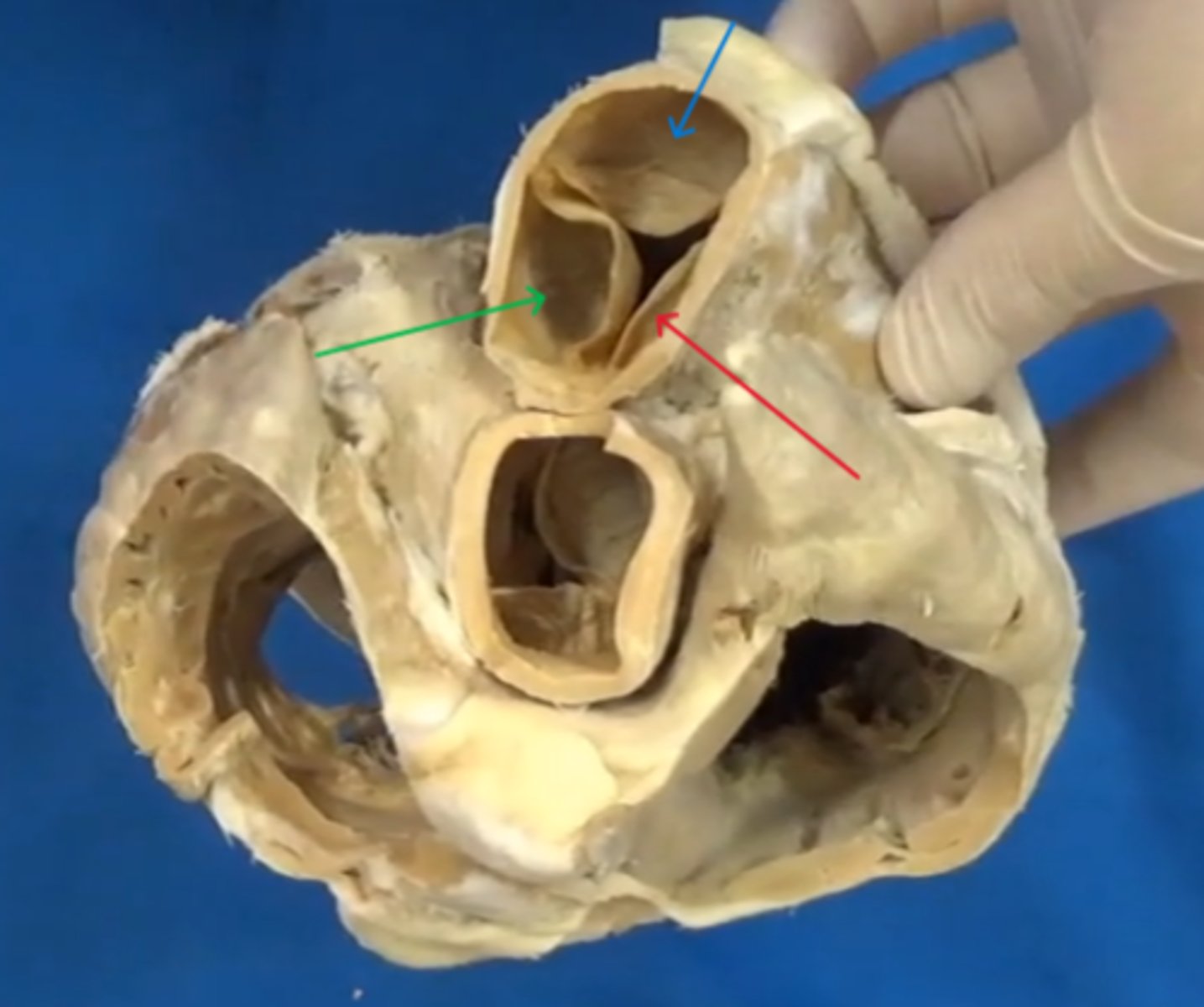
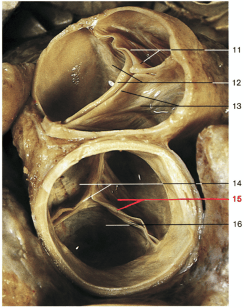
Pulmonary Valve
Note: contains 3 semilunar valves - anterior, right, left
Name the Structure of the Right Ventricle
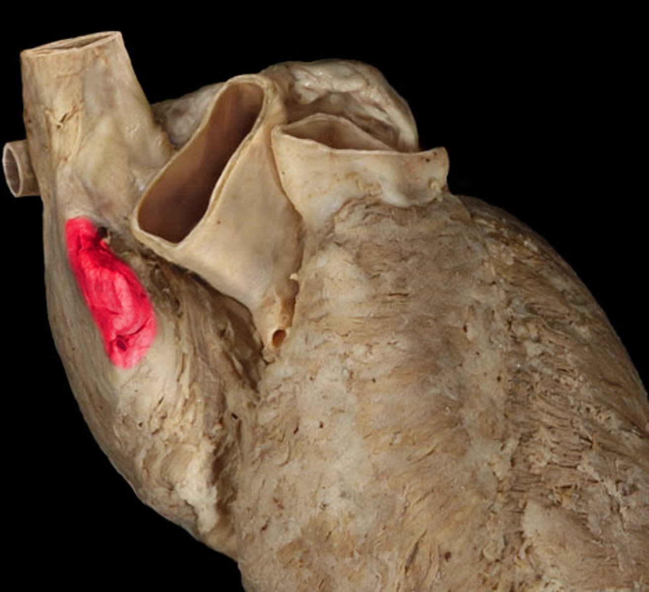
Anterior Cusp of Pulmonary Valve
Name the Blue Arrow (Part of Right Ventricle)

Right Cusp of Pulmonary Valve
Name the Red Arrow (Part of Right Ventricle)

Left Cusp of Pulmonary Valve
Name the Green Arrow (Part of Right Ventricle)
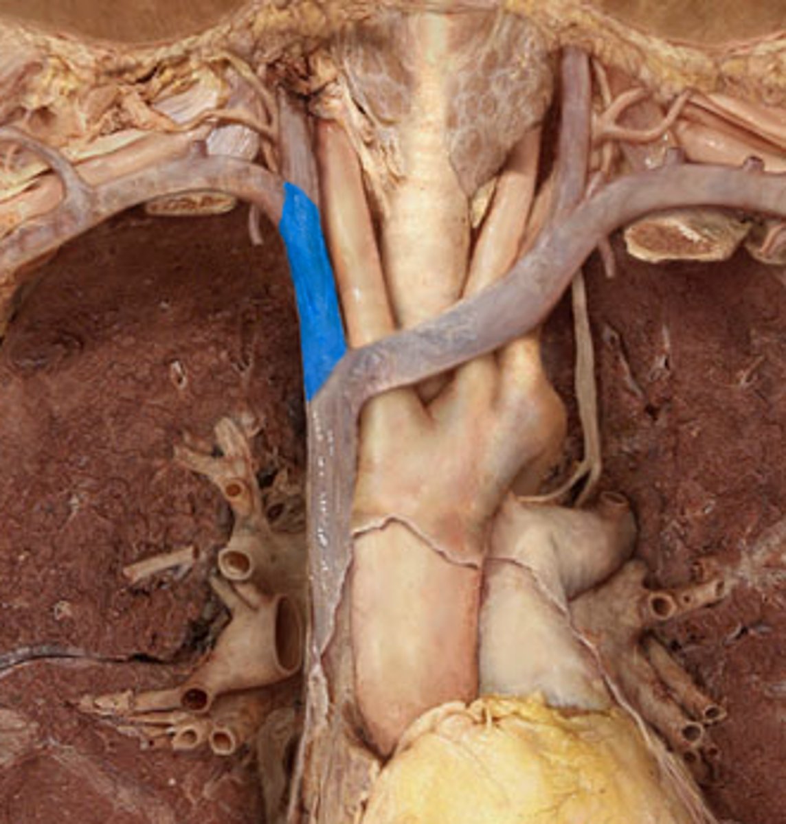
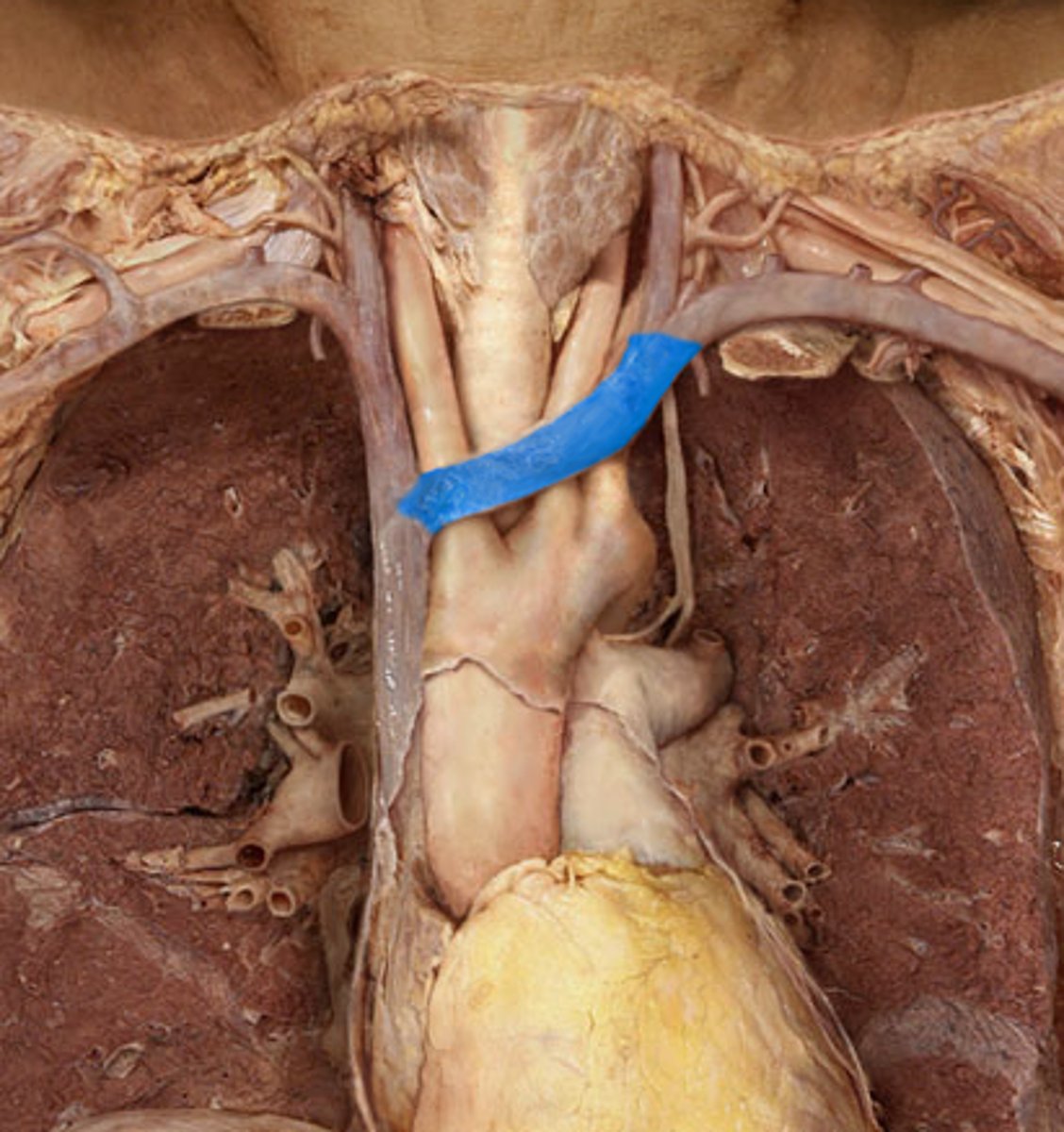
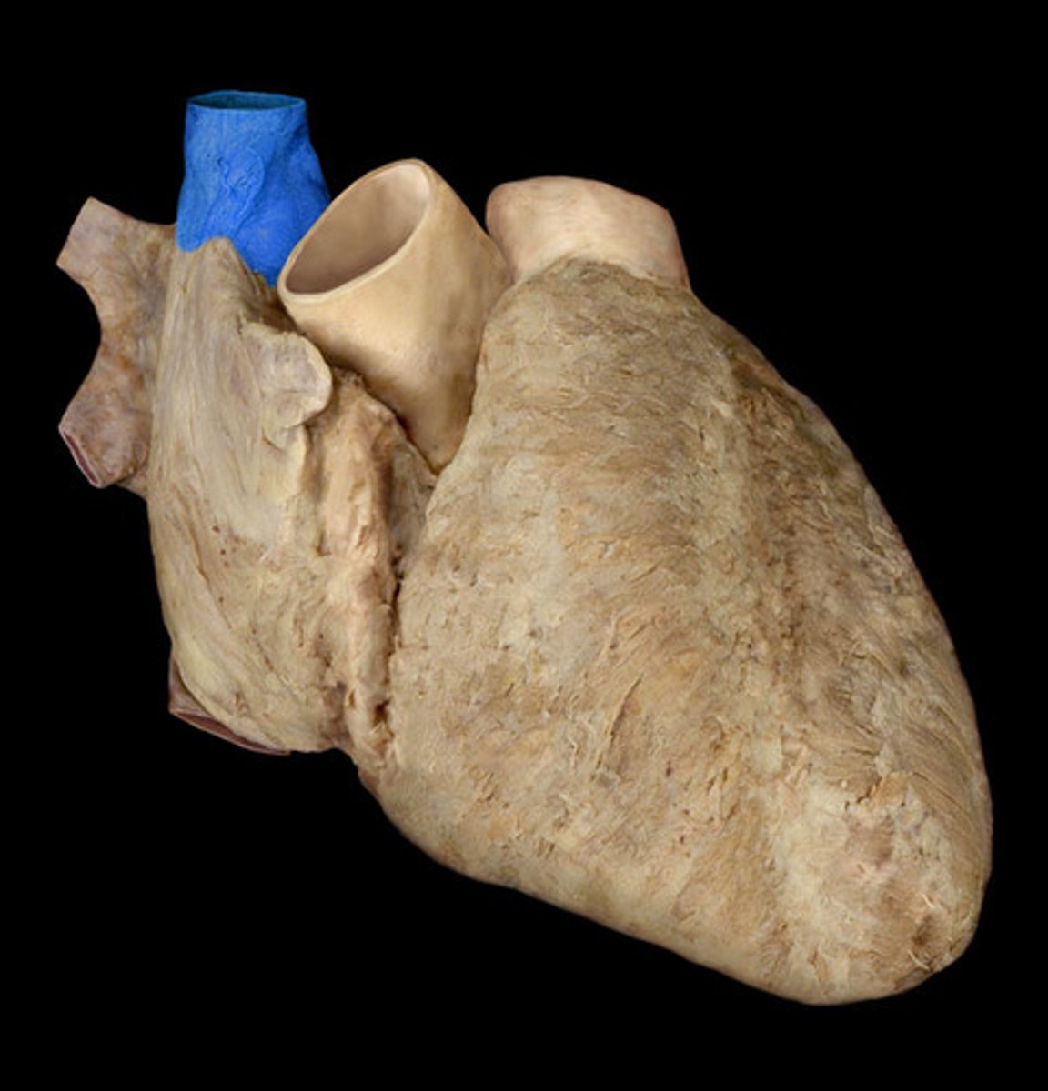
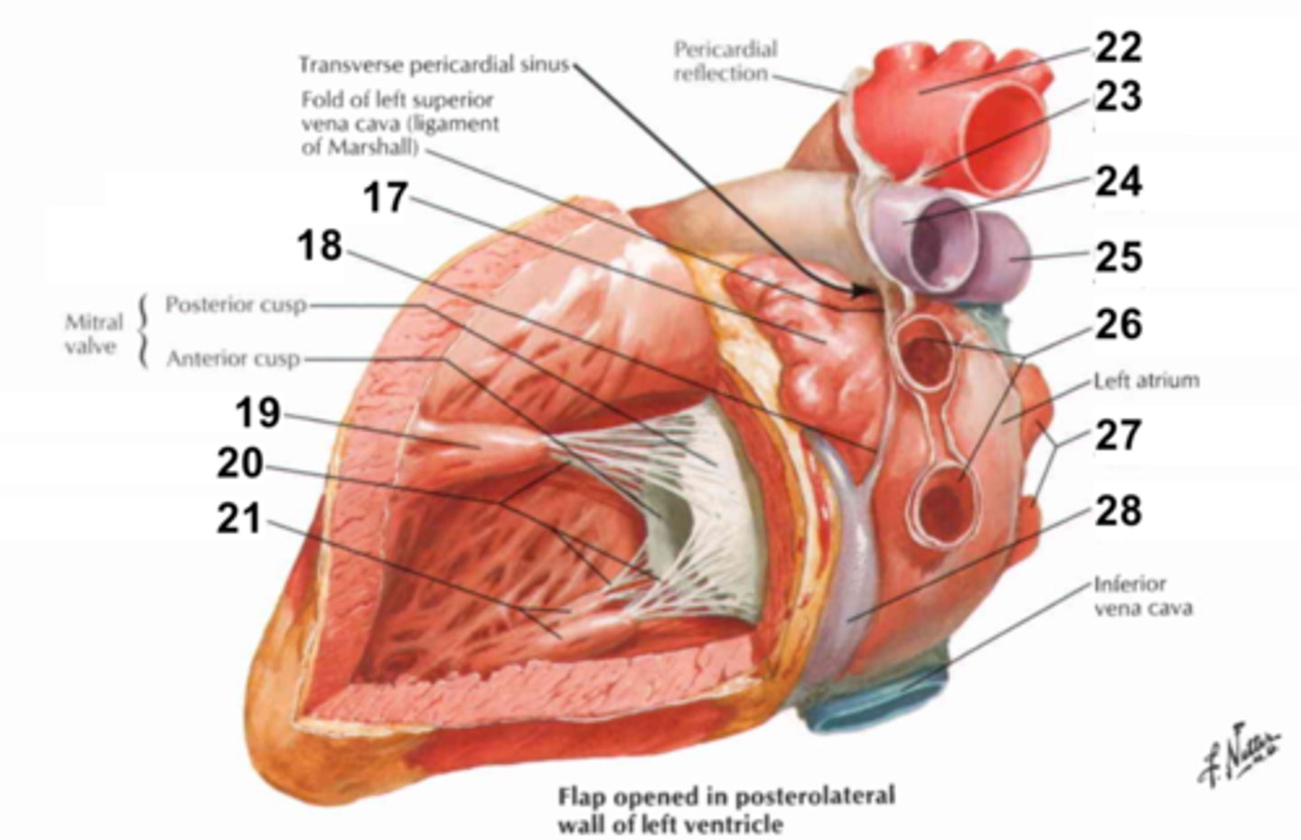
Left Auricle
Name the Structure of the Left Atrium
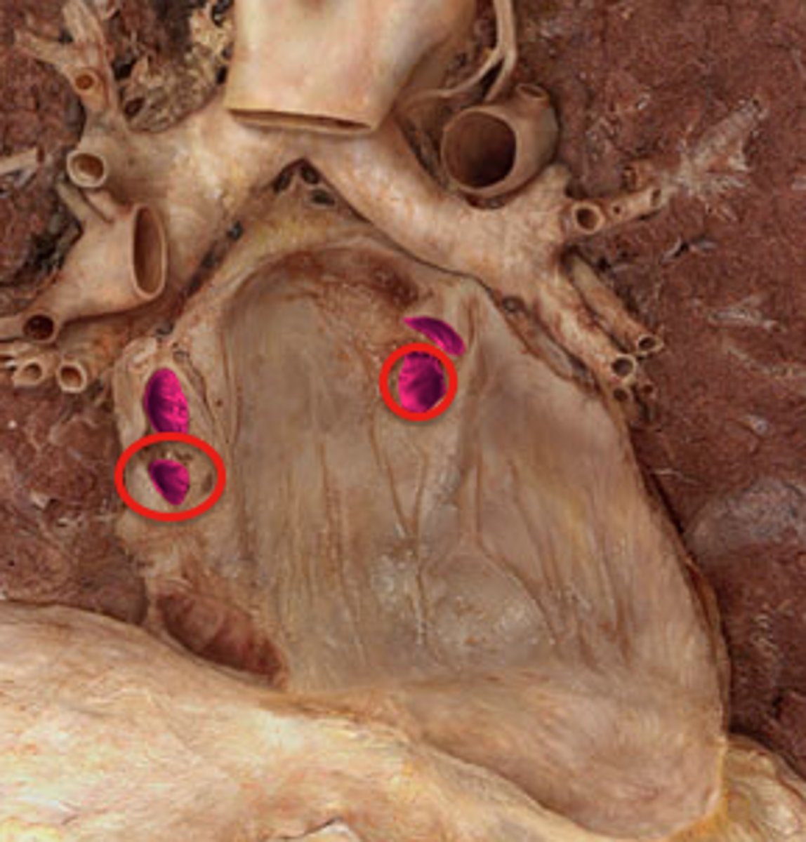
Superior Pulmonary Veins
Name the Structure of the Left Atrium
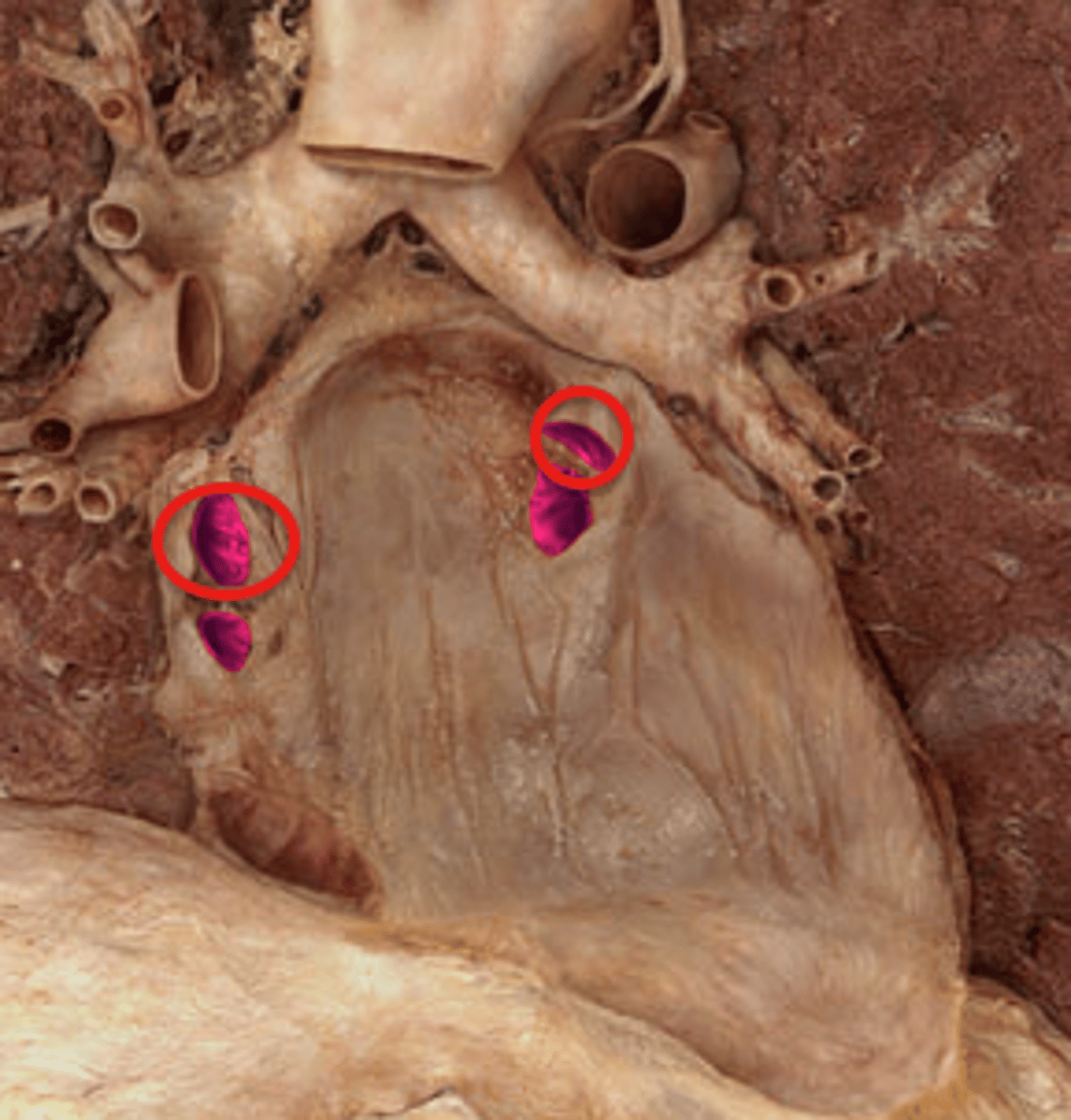
Inferior Pulmonary Veins
Name the Structure of the Left Atrium
Crux Cordis
Name the Point Where the Horizontal and Vertical Lines Cross
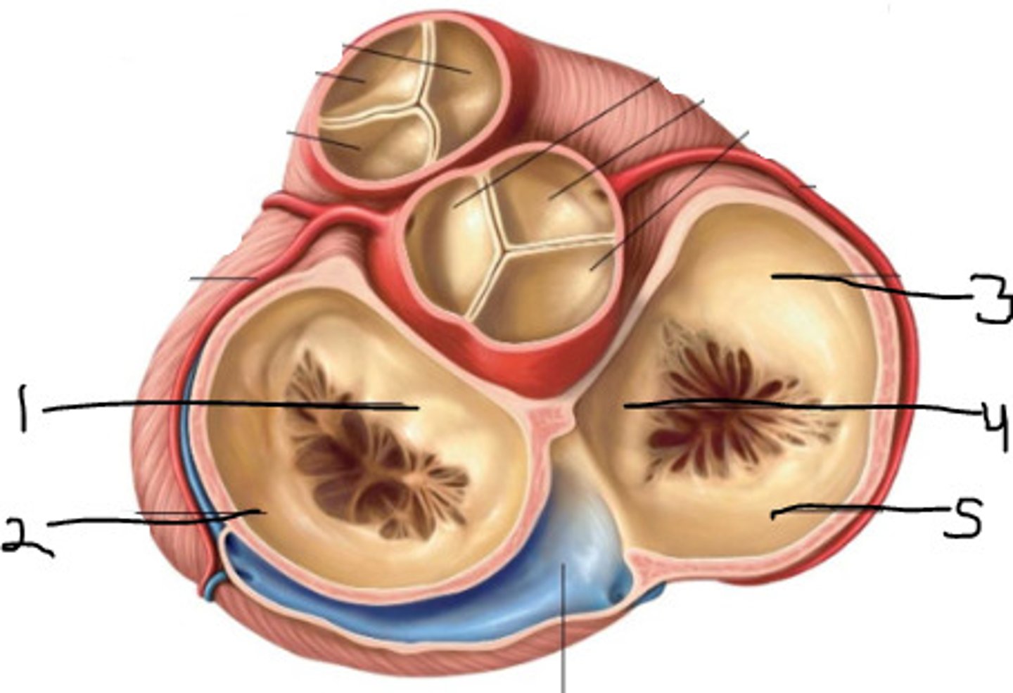
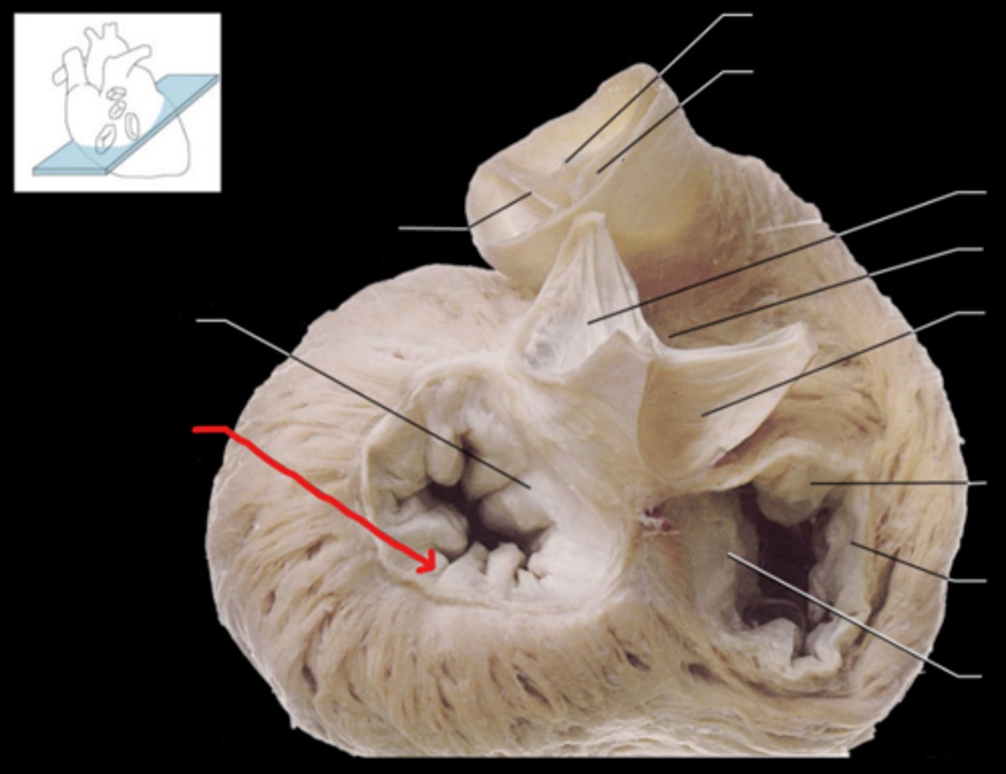
Bicuspid Valve (AKA mitral/left atrioventricular valve)
Note: contains 2 leaflets - anterior & posterior, which connect to papillary muscles via chordae tendineae
Name the Structure of the Left Ventricle
Anterior Cusp of Bicuspid
Name the Structure of the Left Ventricle
Posterior Cusp of Bicuspid
Name the Structure of the Left Ventricle
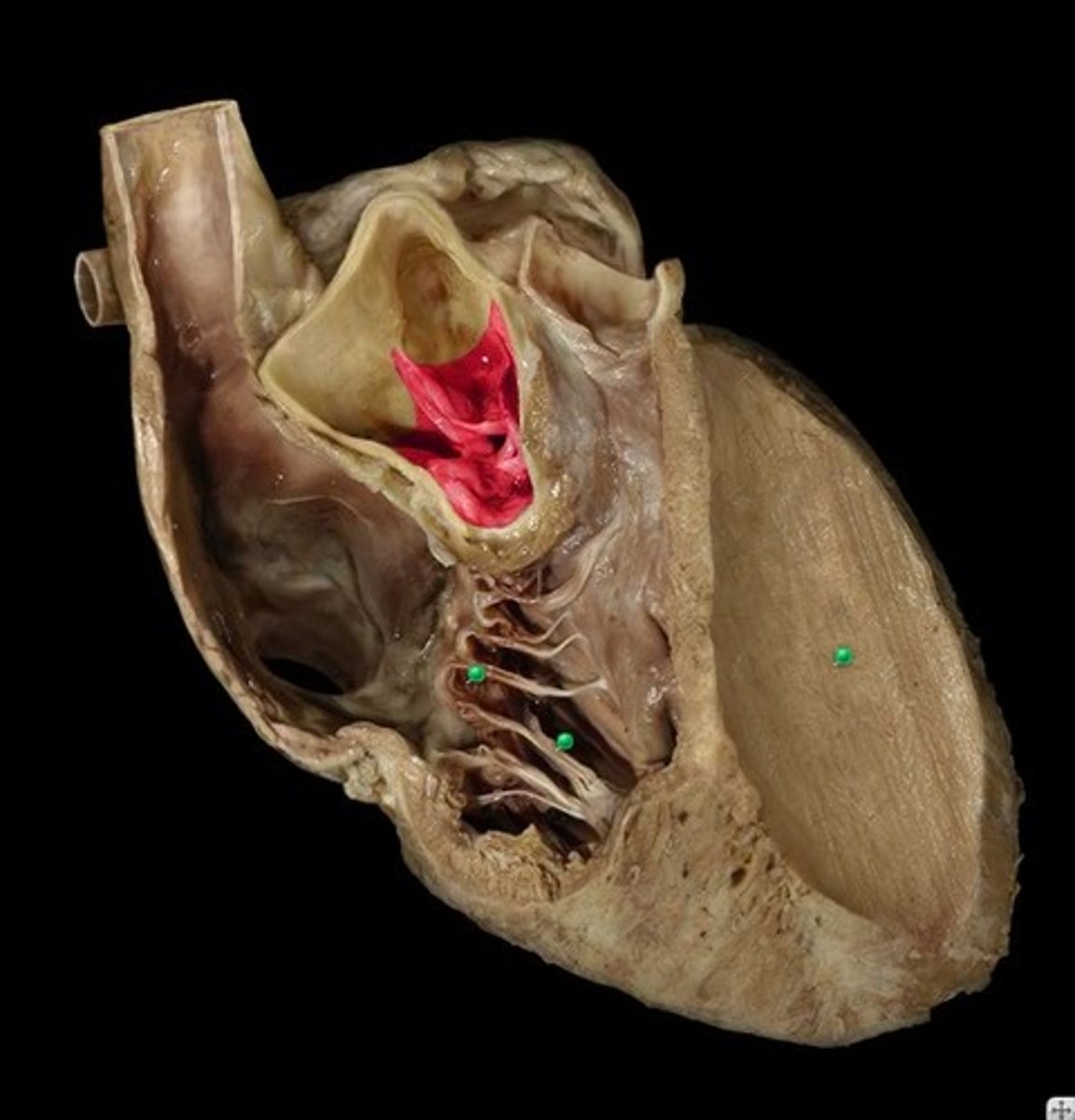
Anterior Papillary Muscle of Left Ventricle
Name Muscle #1
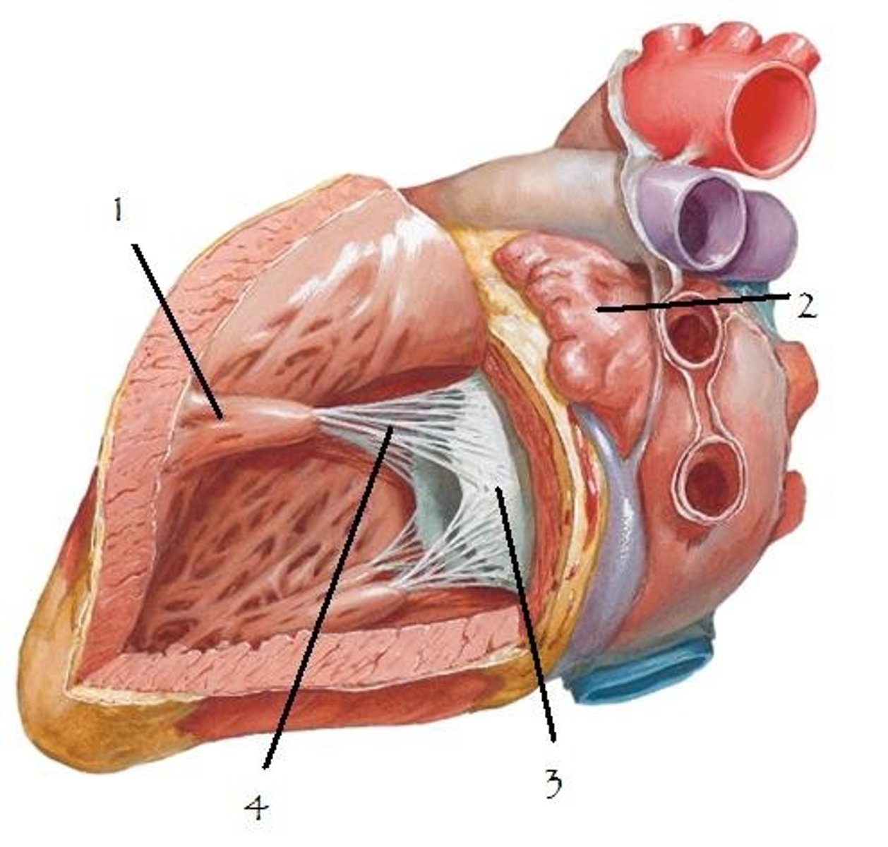
Posterior Papillary Muscle of Left Ventricle
Name Muscle #21
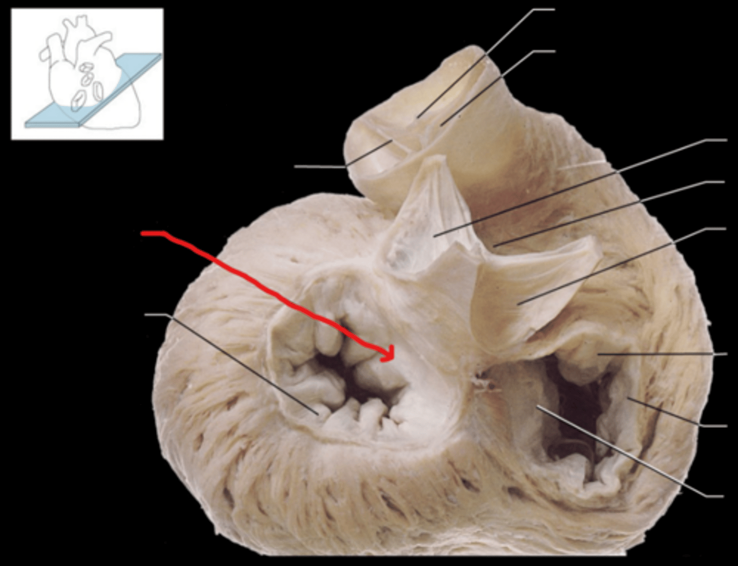
Aortic Valve
Note: contains 3 semilunar valves - right, left, posterior (non-coronary...since there is no coronary artery stemming from posterior valve)
Name the Structure of the Left Ventricle
Posterior Cusp of Aortic Valve
Name the Structure of the Left Ventricle
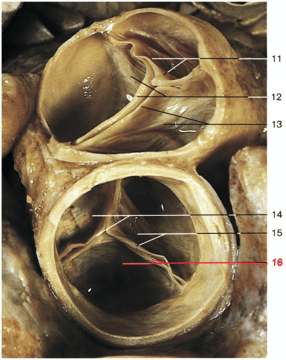
Right Cusp of Aortic Valve
Name the Structure of the Left Ventricle
Left Cusp of Aortic Valve
Name the Structure of the Left Ventricle
Fossa Ovalis
Name the Structure of the Right Atrium
