Chest X-rays
1/6
There's no tags or description
Looks like no tags are added yet.
Name | Mastery | Learn | Test | Matching | Spaced | Call with Kai |
|---|
No analytics yet
Send a link to your students to track their progress
7 Terms
What is a chest X-ray?
projection of radiograph taken by radiographer of the thorax which is used to diagnose problems within that area (always taken on insp.)
provides info about conditions of lungs + chest wall
4 densities visible on CXR → bone, air, water + fat
dense structures absorb rays → appear white (bone)
Indications for CXR
when physio needs to:
detect changes in lung structure
decide on course of treatment
establish whether treatment is effective
positioning of tubes and attachments
monitor progression of lung disease
identify normal/abnormal structures
to localise pathlogy
identify precautions/contraindications
Factors influencing quality of CXR
distance
patient position
rotation
state of respiration (insp. vs exp.)
radiographic exposure
Types of CXR views
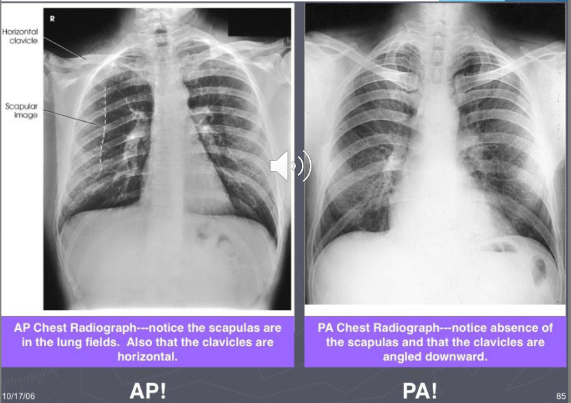
Standard = PA (prefered) or lateral
PA:
x-ray taken on insp.
beam passes back→front
person erect, arms ab.
other = AP or decubitus (lying down → pt. lies on side w x-ray beam parallel to floor)
AP:
heart magnified in this view
ant. ribs difficult to visualise
scapula in lung fields
Exposure on each x-ray view?
should be able to see vert. bodies + disc spaces to t4/T5, should only see disc spaces from T5 down
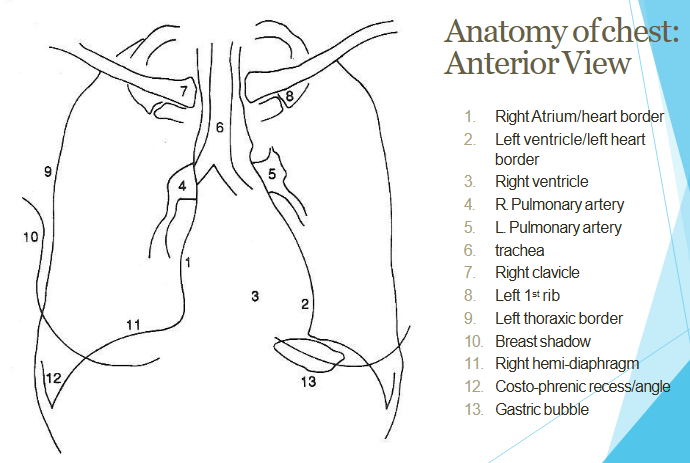
AP view anatomy rev.
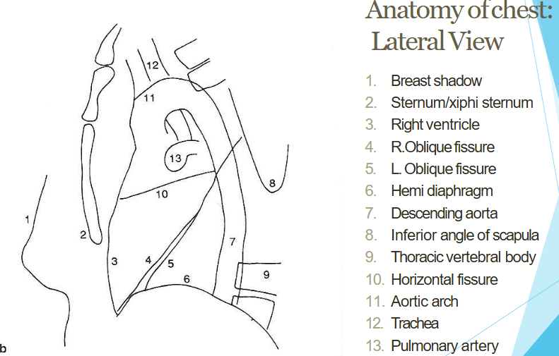
Lateral view anatomy rev.
Assessment of CXR
name, date, time taken
identify L & R sides
projection: PA (common), AP, lateral
position →clavicles equidistant from spinous processes
rotation: L = heart looks bigger, R = heart looks more central
bony structures → identify any #, deformities, inspiration
trachea position (midline or shifted)
heart (size, borders→ are they clear)
diaphragm (shape, costophrenic and cardiophrenic angles, R hemidiaphragm higher than left, is diaphragm clearly visualised)
lung fields
upper, middle lower zones
fizzures (must be faint → if absent, prominent or abnormal = problem)
air
silloutte sign ( +ve = dissapearing of some border of thoracic cavity)
increased interstitial markings