NEUR201
1/228
There's no tags or description
Looks like no tags are added yet.
Name | Mastery | Learn | Test | Matching | Spaced | Call with Kai |
|---|
No analytics yet
Send a link to your students to track their progress
229 Terms
Ipsilateral
Same side
Bilateral
Both sides
Contralateral
Opposite side
Proximal
Close
Distal
Far
Rostral/anterior
Towards face
Caudal/posterior
Towards tail
Dorsal/superior
Towards head
Ventral/inferior
Towards belly
Layers/nuclei
Well-defined group of cell bodies, called ganglia in CNS (structures, areas)
Tract (CNS)
Large collection of axons projecting to or away from a layer of nucleus within the CNS. Ex. corticospinal tract, optic tract
Nerves (PNS)
Fiber tracts that enter and leave the CNS. Once they enter the CNS, they’re called tracts. Ex. auditory nerve, vagus nerve
Coronal cut
Grey matter
Soma + dendrites
White matter
Axons
Ventricles (bladders)
Lateral, 3rd, 4th, and cerebral aqueduct. Empty spaces that hold cerebrospinal fluid. Blockages cause hydrocephalus
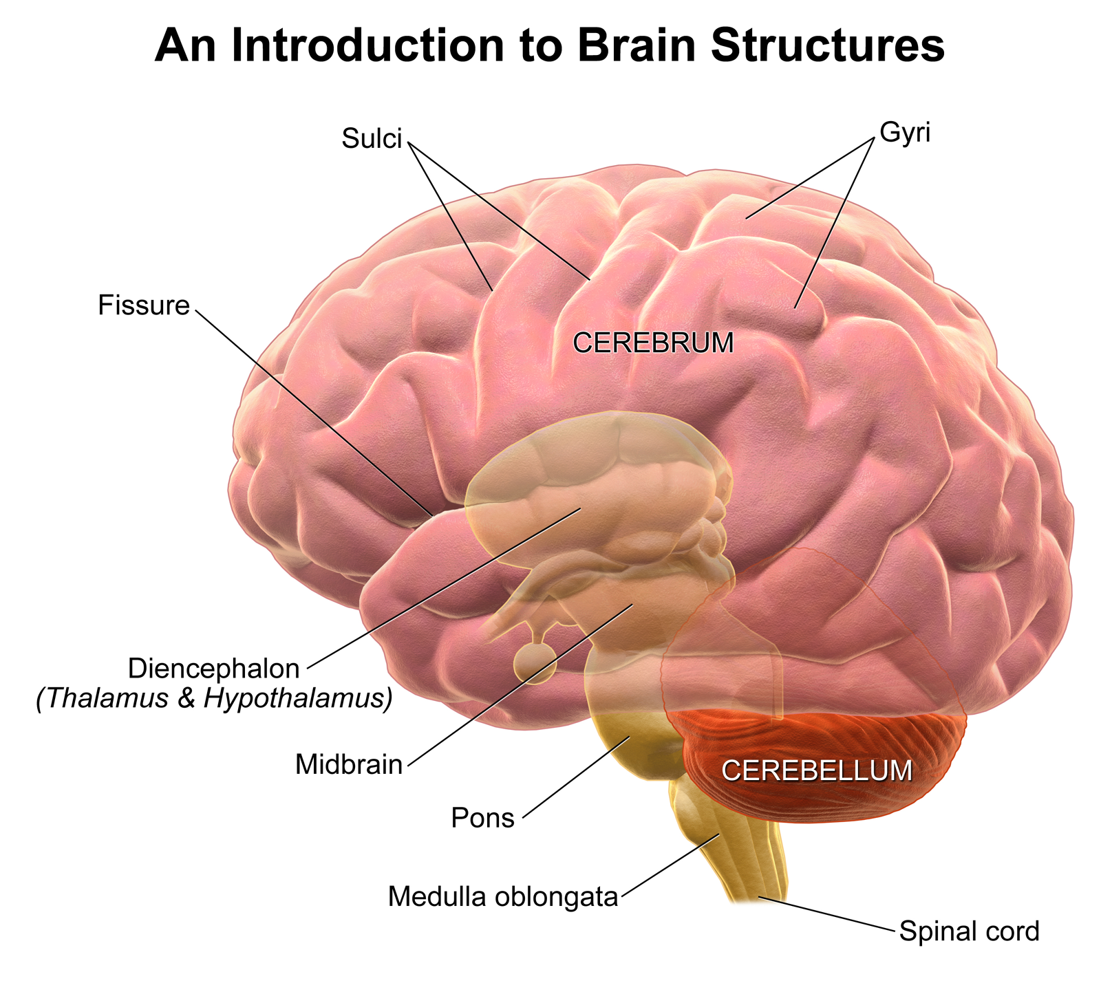
Sulcii
Grooves

Gyrii
Ridges
Sympathetic nervous system (PNS)
Innervated by chain ganglion outside of vertebrae to spinal cord. Adrenergic system (NE and Ach)
Parasympathetic nervous system (PNS)
Innervated by cranial nerve 10 (vagus nerve, CNS). Primarily cholinergic system (Ach)
Cranial nerves
Special class of nerves that project afferently or efferently. 12 pairs; convey sensory and motor signals to and from the head. 2 sets, left and right. They all connect to places outside of the brain which makes them part of the PNS
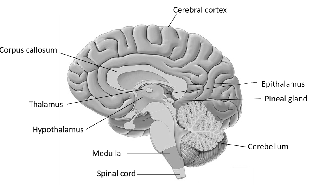
Forebrain (prosencephalon in embryo)
Telencephalon (cerebral cortex, basal ganglia), diencephalon (thalamus, hypothalamus)
Midbrain (mesencephalon in embryo)
Tectum, tegmentum
Hindbrain (rhombencephalon in embryo)
Pons, medulla oblongata, cerebellum, brain stem
Triune brain theory
Overly simple but still helpful. Not backed up by science. Popularized in 60s by Paul McLean. Reptilian brain (brainstem), mammalian brain (limbic system), neo-mammalian brain (cerebral cortex)
Somatic motor system
Motor nerves
Visceral motor system
Sympathetic/parasympathetic divisions, automatic ganglia and nerves. Automatic NS
Sylvian fissure (lateral fissure)
Separates temporal lobe from parietal + frontal lobes

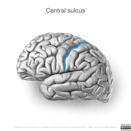
Central sulcus
Separates parietal lobe from frontal lobe
Vagal tone
The ability of the vagus nerve to influence the body. Measured using heart rate variability (higher usually better)
Dermatomes
Correspondences between skin (sensory and motor) and spinal cord. You can tell where a paralyzed person has their spinal cord injury with this
Dorsal root
Sensory, afferent, posterior, back side of spinal cord, nerves

Ventral root
Motor, efferent, anterior, front side of spinal cord, nerves
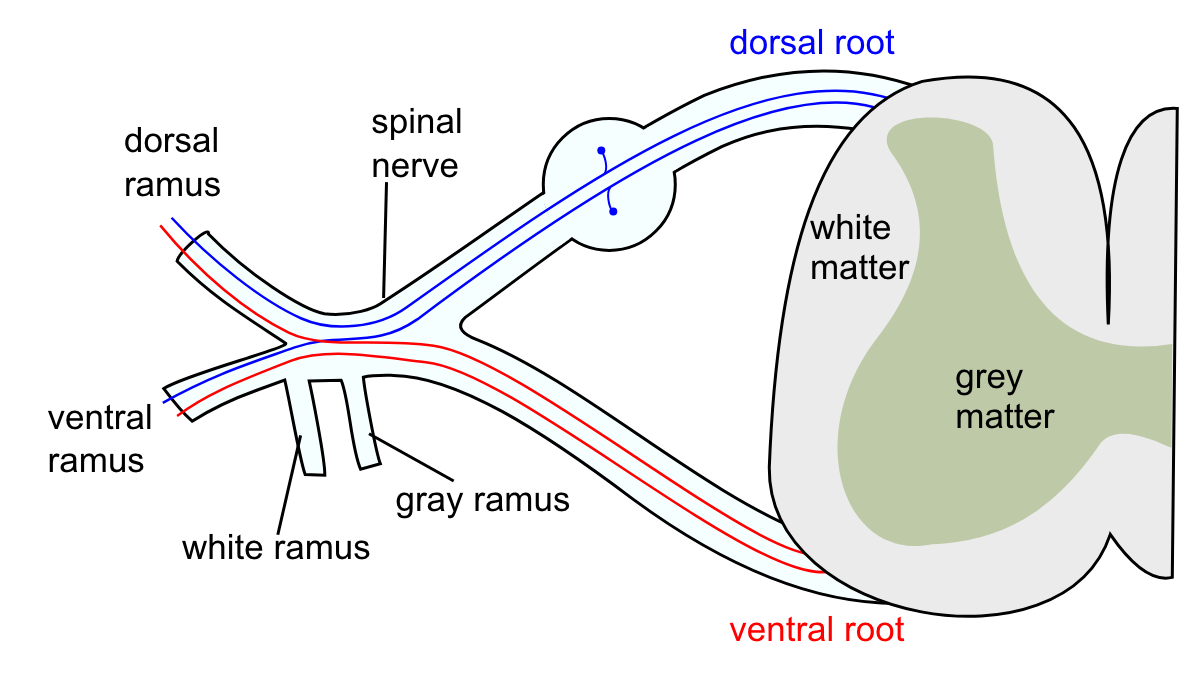
Inter-neuron
Neuron that passes messages between 2 neurons. Lateral horn and central zone
Spinal cord divisions
Dorsal horn (sections 1-6), lateral horn (section 7), ventral horn (section 8-9), central zone (section 10)
Upper motor neurons
Motor neurons in brain and brain stem
Lower motor neurons
Come out of spinal cord and communicate with skeletal muscles to tell them to contract using Ach
Myotatic reflex
Knee jerk reflex. Happens at level of spinal cord. Extensor (extend) vs. flexor (reflex) muscles. Afferent nerve → excitatory extensor → inhibitory interneuron → inhibitory flexor
Flexion/crossed extension reflex
Afferent nerve → excitatory interneuron → inhibitory interneuron → motor neuron. Stimulated leg flexes to withdraw, opposite leg extends to support. Some reflexes can be controlled, this is called gating. Some reflexes are easier to override than others.
Cranial nerve 1
Olfactory nerve (sensory)

Cranial nerve 2
Optic nerve (sensory)

Cranial nerve 3
Oculomotor nerve (motor, pupillary movement)

Cranial nerve 4
Trochlear nerve (motor, eye movement)

Cranial nerve 5
Trigeminal nerve (sensory and motor, mastication)

Cranial nerve 6
Abducens nerve (motor, also eye movement)

Cranial nerve 7
Facial nerve (sensory and motor, facial, taste)
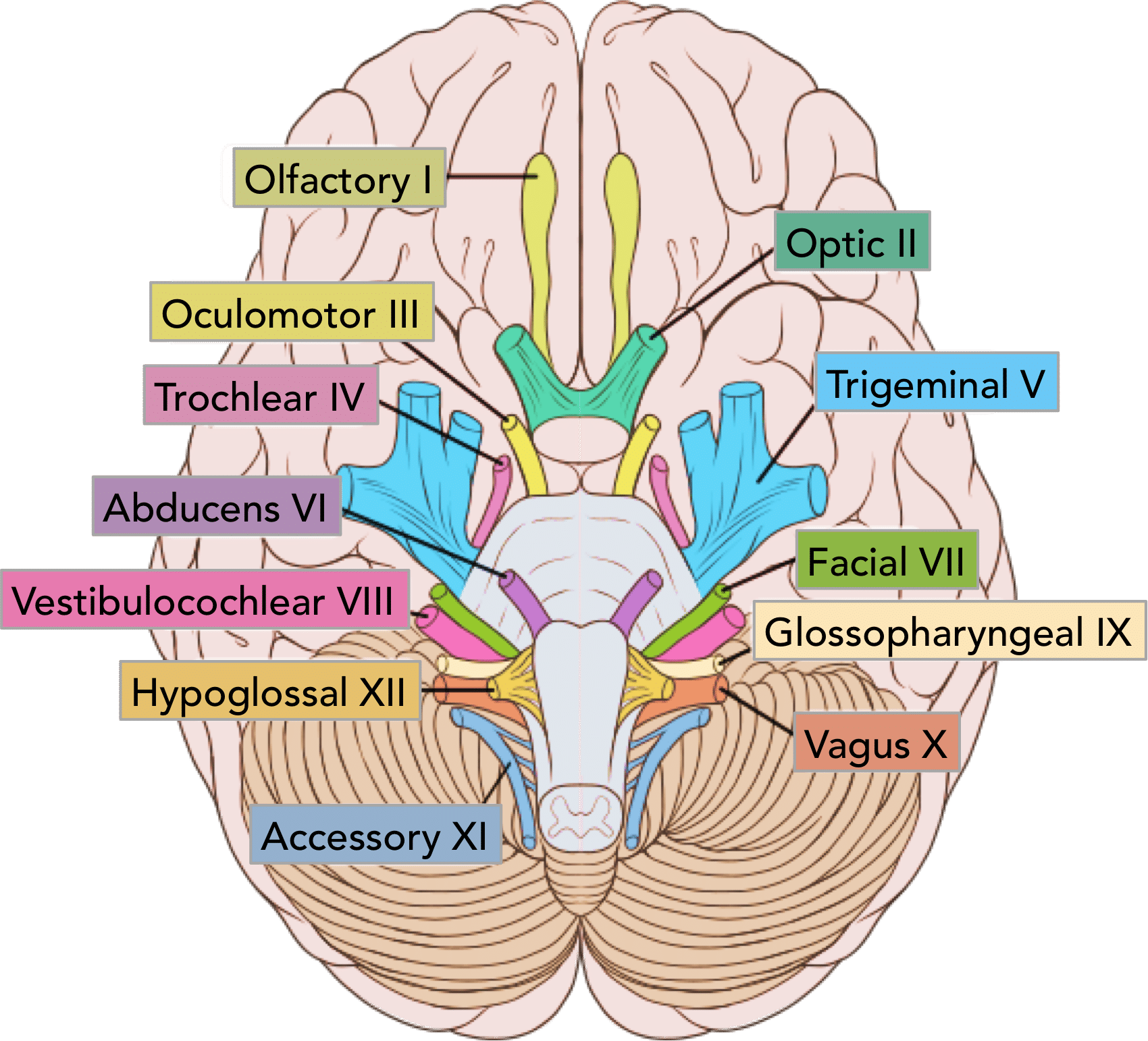
Cranial nerve 8
Vestibulocochlear nerve (sensory, hearing/sense of balance)

Cranial nerve 9
Glossopharyngeal nerve (sensory and motor, swallowing, taste, salivation, somatic from ear)

Cranial nerve 10
Vagus nerve (sensory and motor, automatic + parasympathetic)

Cranial nerve 11
Spinal accessory nerve (motor, shoulder and neck muscles)

Cranial nerve 12
Hypoglossal nerve (motor, movements of tongue)

Midbrain
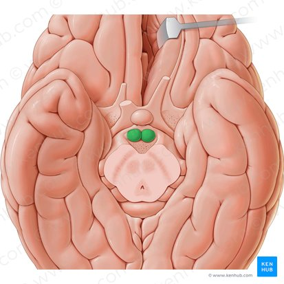
Tectum (dorsal). Superior colliculus (vision), inferior colliculus (auditory). Substantia nigra (dopamine/motor. Death of this can cause Parkinson’s). Cranial nerves 3, 4, 5, and 7. Most superior part of brain stem

Pons
Superior olivary nucleus (auditory), locus coeruleus (attention). Cranial nerves 5, 6, 7, 8, 9, 10. Attention isn’t just a higher order function
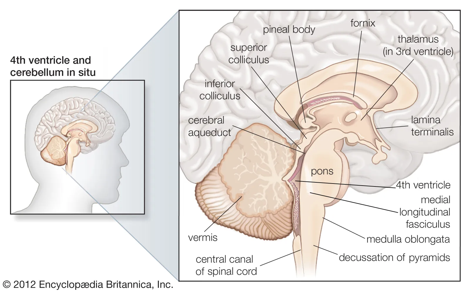
Medulla
Inferior olivary nucleus (motor and learning), reticular formation (activating system, on/off switch), nucleus ambiguus. Cranial nerves 5, 6, 8, 9, 10, 11. Most inferior part of brain stem. Nerve 11 is connected to spinal accessory nucleus

Cerebellum
Little brain. Important for motor control, balance, and motor learning. May also play a role in cognition. Treelike structure
Parts of the cerebellum
Cerebrocerebellum (input from cerebral cortex and output to thalamus to coordinate motor signals), spinocerebellum (proprioceptive info, regulating movement and error correction), vestibulocerebellum (balance and visual fixation, vestibular info from inner ear)
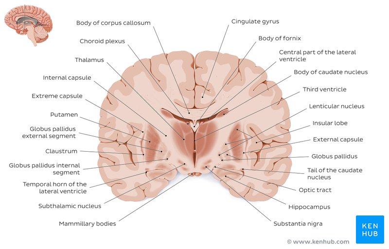
Diencephalon
Interior, medial structures that develop from the prosencephalon. Thalamus ((dorsal thalamus, epithalamus, hypothalamus, pituitary gland). Thalamus is inside basal ganglia + limbic system. Basal ganglia covers the thalamus, it’s anterior of the thalamus
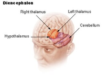
Thalamus
Sensory relay. Receives information from spinal cord and cranial nerves. Olfactory info is an exception. Medial, lateral and anterior sections divided by interior medullary lamina
Thalamus subsections
Pulvinar (visual attention top down, in the back), medial geniculate nucleus (below pulvinar, auditory), lateral geniculate nucleus (below pulvinar, visual), ventral posterior (somatosensory), ventral anterior (motor)
Connections in the thalamus
6 layers of cerebral cortex. Receive info from thalamus particularly related to motor (ventral anterior) and somatosensory (ventral lateral). Topdown and bottom up control/communication. This is good for synchronizing brain activity but can also contribute to seizures
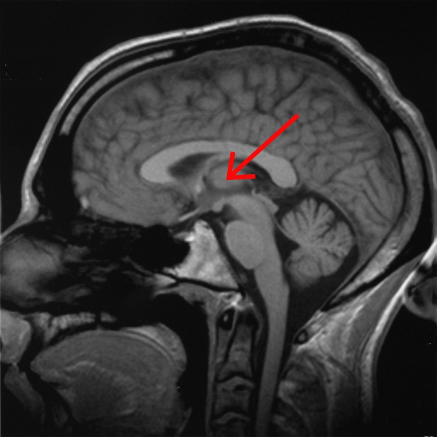
Epithalamus and pineal gland
Posterior to thalamus, midline structure (not paired), regulates endocrine function including melatonin.
Subthalamic nucleus
Integrates thalamus and basal ganglia, important for motor control, target for deep brain stimulation in Parkinson’s
Hypothalamus
Regulates blood flow, metabolism, reproduction, and threat response. Receives info from sensory and higher order areas and behavioral response. Causes changes in the body by regulating hormones

Connections in hypothalamus
Contextual info (cerebral cortex, amygdala, hippocampal formation) → hypothalamus (compares info to biological set points) ← sensory inputs (visceral and somatosensory pathways, chemosensory and humoral signals)
Mammillary bodies
Posterior part of hypothalamus. Memory and reward processing. Damaged by thiamine deficiency, esp. with alcoholism, which can lead to Wernicke-Korsakoff syndrome
Paraventricular nucleus
More superior part of hypothalamus. Aka PVN. Corticotropin-releasing hormone (CRH). Vasopressin, oxytocin mediation. Overall, nuclei in hypothalamus contribute to homeostasis and behavioral responses
HPA axis
Hypothalamic pituitary adrenal axis. PVN hypothalamus: corticotropin releasing hormone binds to anterior pituitary which releases adrenocorticotropic hormone (ACTH). When this reaches the kidneys, they bind to the adrenal glands there to release cortisol
Cortisol effects
Released when your body is under stress (different, slower system than the sympathetic, but they work together often. Chronic stress). Causes increased sugar availability, immune system suppression, reproductive system suppression, etc.
Lateral ventricles
Located in each cerebral hemisphere

3rd ventricle
On midline, situated in the diencephalon between the thalamus and hypothalamus

Cerebral aqueduct
A narrow channel connecting the 3rd and 4th ventricle
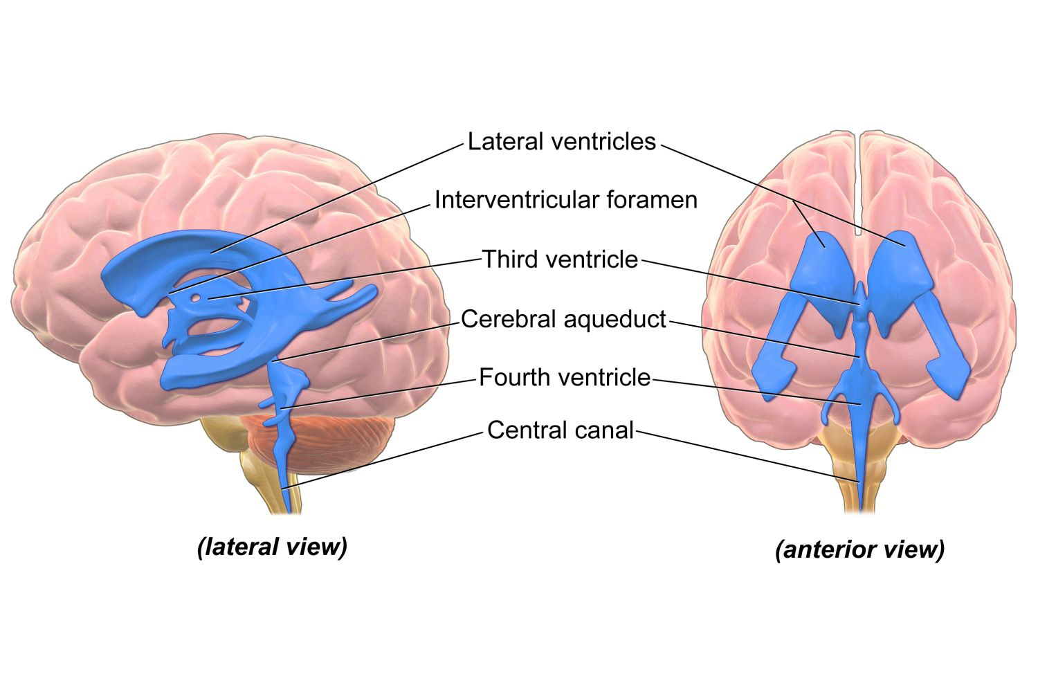
4th ventricle
On midline, located in the hindbrain between cerebellum and pons
Basal ganglia
Ventral striatum (nucleus accumbens), dorsal striatum (caudate and putamen), globus pallidus, ventral pallidum
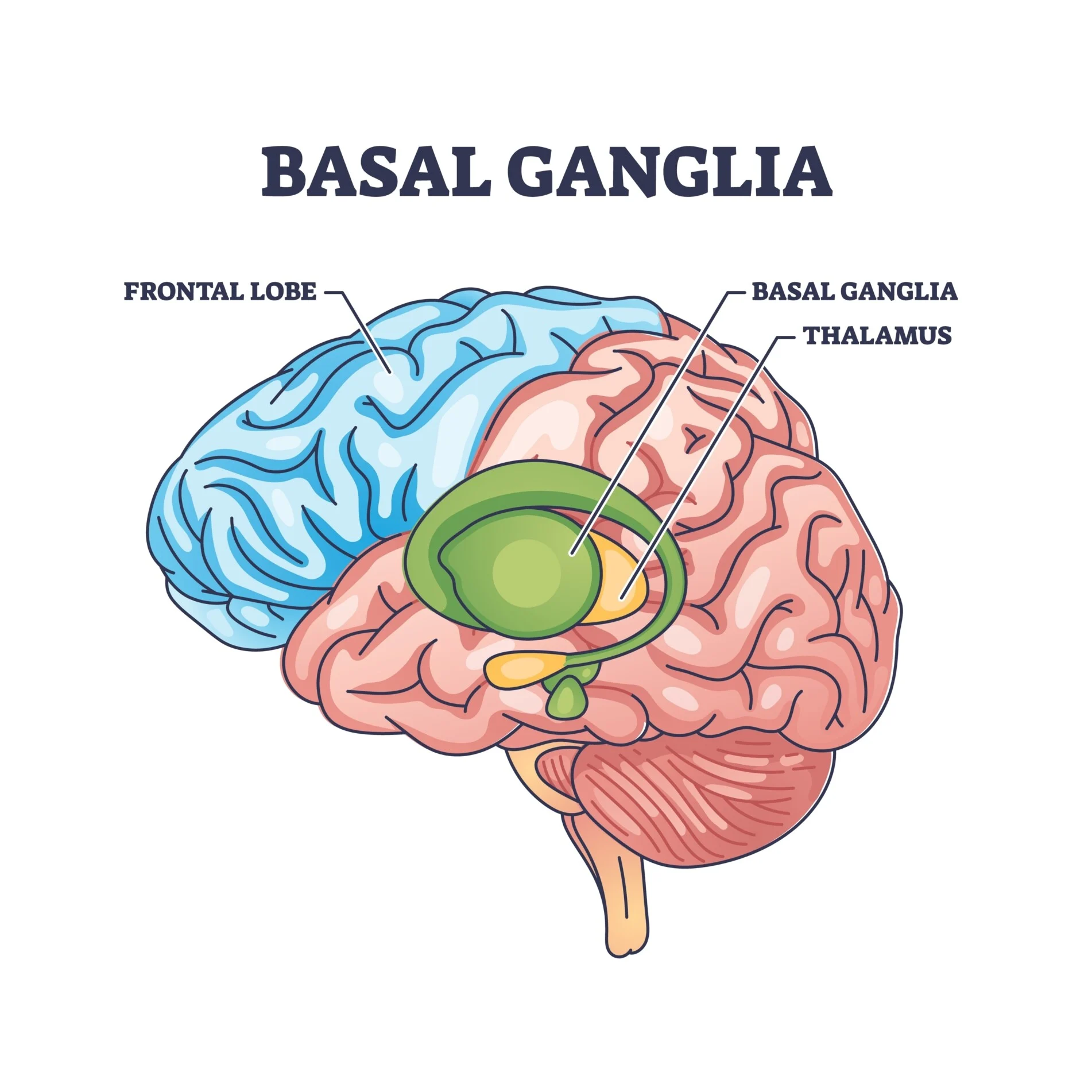
Motor functions of basal ganglia
Caudate and putamen made of medium that receives input from cortex and then creates a feedback loop (body movement loop and oculomotor loop) back to cortex. Caudate = eye movements (cranial nerves 3, 4, and 6), putamen = skeletal muscle. Receives input from substantia nigra (dopamine)
Basal ganglia circuits
Tonic (slow but consistent) vs. phasic (only happens in phases) firing. Main output from the basal ganglia is tonic inhibitory (GABAergic). Inhibit an inhibitory neuron = excitatory. Disinhibitory circuit via a chain of neurons. Reminder of IPSP and EPSP
Parkinson’s disease
Hypokinetic. Loss of dopamine-producing neurons in substantia nigra and ventral tegmentum (midbrain) → loss of nigrostriatal dopaminergic pathway → loss of transient inhibition from direct pathway to the GP internal segment → decreasing thalamic excitation of the motor cortex. Treatment: L-DOPA, deep brain stimulation
Huntington’s disease
Hyperkinetic. Autosomally dominant inheritance. Atrophy of primarily the caudate and putamen and secondarily, frontal and temporal cortices. Loss of D2-expressing medium spiny neurons → higher excitability of globus pallidus external segment → decreases excitation of subthalamic nucleus to the GP internal segment → overall decrease in inhibitory output from the basal ganglia. Can be mimicked in monkeys using GABA agonist
Reward functions
Nucleus accumbens and ventral pallidum act in reward circuits. Communicate to frontal regions to elicit reward. Dopaminergic
Limbic system
Amygala, hippocampus. Extends off of basal ganglia
Cortex
Frontal/temporal/parietal/occipital lobes. medial longitudinal fissure, central sulcus, sylvian/lateral fissure, gyrus
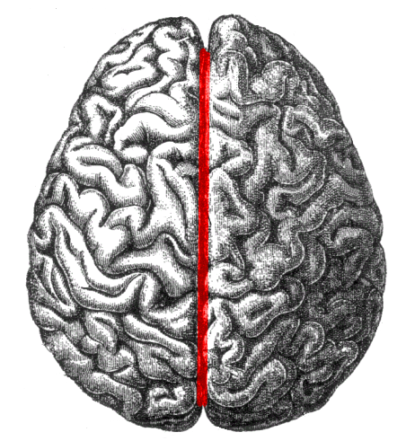
Medial longitudinal fissure
Separates hemispheres. Not the same as corpus callosum (CC is medial/interior)
Brain development
As the fetal brain develops, neurons create layers with similar types of cells that synapse within and outside of cortex. Neurons migrate. Leads to idea of cytoarchitecture. Brodmann’s areas. Generally, major features are consistent between humans and similar across species. Bigger body = bigger brain, but humans have bigger brains than expected given their size. About 86 billion neurons for humans. Counts of number of neurons are inconsistent
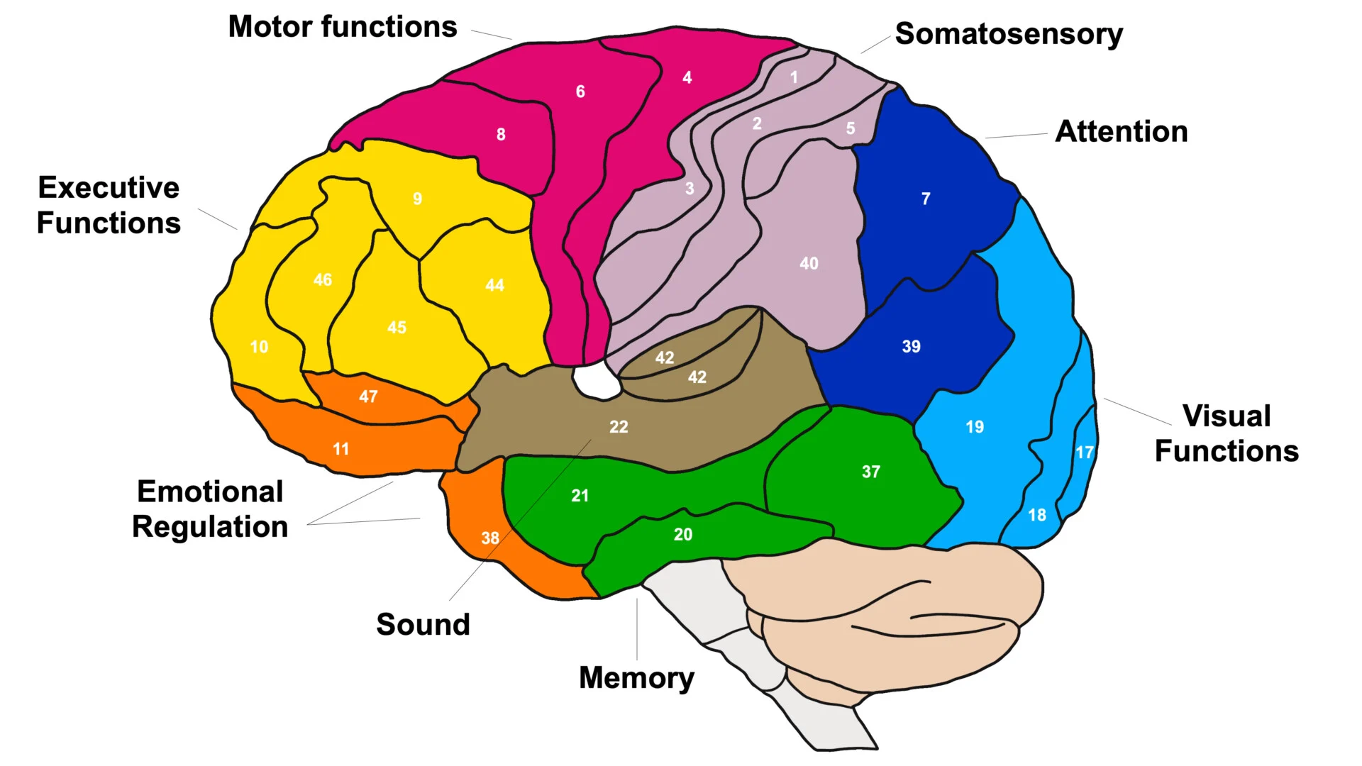
Function of the occipital cortex
Primarily processing of visual stimuli. Hierarchical processing, increased complexity from primary visual cortex (V1, edges and lines) → secondary visual cortex (V2, shapes) → V3/V4 (objects) → inferotemporal cortex (V5, faces/object recognition). At level of V1, single cells respond to a specific orientation of light in a specific location of the visual field. Each cortex has a primary part
Function of the parietal cortex
Primary somatosensory cortex (S1, postcentral gyrus. Responsive to body senses (afferent), organized as a homunculus), posterior parietal cortex (visual spatial functions, association cortex (part of brain where different sensory info is being combined))
Function of the temporal cortex
Primary auditory cortex (A1, superior temporal gyrus), language (Wernicke’s area in superior temporal gyrus; middle temporal gyrus), memory (entornihal and parahippocampal gyrus), limbic cortex, recognition (inferior and fusiform gyrus)
Function of the frontal cortex
Top-down control. Primary motor cortex (M1, precentral gyrus), supplementary motor cortex/premotor cortex and frontal eye fields, prefrontal cortex (frontal pole, orbital gyrus, parts of superior and middle frontal gyrii), language (Broca’s area in left inferior frontal gyrus)
Glial cells
Support cells in nervous system
Reticular theory
All neurons connected in net. This assumes that synapses don’t exist but they do
Convergence in neuron
More dendrites
Divergence in neuron
More axon terminals
Astrocytes
CNS glial cell. Help with blood-brain barrier, buffer ions and neurotransmitters, and secrete chemicals for synaptogenesis
Oligodendrocytes
CNS glial cell. Myelinate neuronal axons
Microglia
CNS glial cell. Macrophage activity and secrete cytokines. Important for immune system
Schwann cells
Myelinate neuronal axons and participate in recovery of function resulting from damage. Not to be confused with oligodendrocytes
Golgi stain
Uses silver to stain 1% of cells for visualization
Myelin stains
Trace glial cells for visualization
Fluorescent dye
Label specific amino acids for visualization
Nissl stain
DNA and RNA turn blue
Afferent neural circuit
Towards the CNS. SENSORY (PNS → CNS)
Efferent neural circuit
Away from the CNS. MOTOR (CNS → PNS)