Dr. KOKA
How is the heart a pump
atria receives blood returning to heart
ventricles pump blood out
septum
divides left and right halves
what do blood vessels consist of
veins arteries and capillaires
pulmonary and systemic circulation
portal system joins two capillary beds in series
blood
cells and plasma
transport of material
gases, nutrients, waste, communication
defense against pathogens and temperature homeostasis
the heart is mostly composed of ___________
myocardium
what are the twos sets of heart valves
atrioventricular and semilunar
atrioventricular valves
between atria and ventricles
tricuspid and bicuspid/mitral valve
where is the tricuspid valve located
on the right side
where is the bicuspid/mitral valve located
on the left side
Semilunar valves
between ventricles and arteries
aortic valve
pulmonary valve
ventricular contraction
the AV valves remain closed to prevent backward blood flow
ventricular relaxation
the SL valves prevent blood that has entered the arteries from flowing back into the ventricles
contractile cells
striated fibers organized into sarcomeres
autorhythmic cells (pacemaker)
signal for contraction
smaller and fewer contractile fibers compared to contractile cells
do not have organized sarcomeres
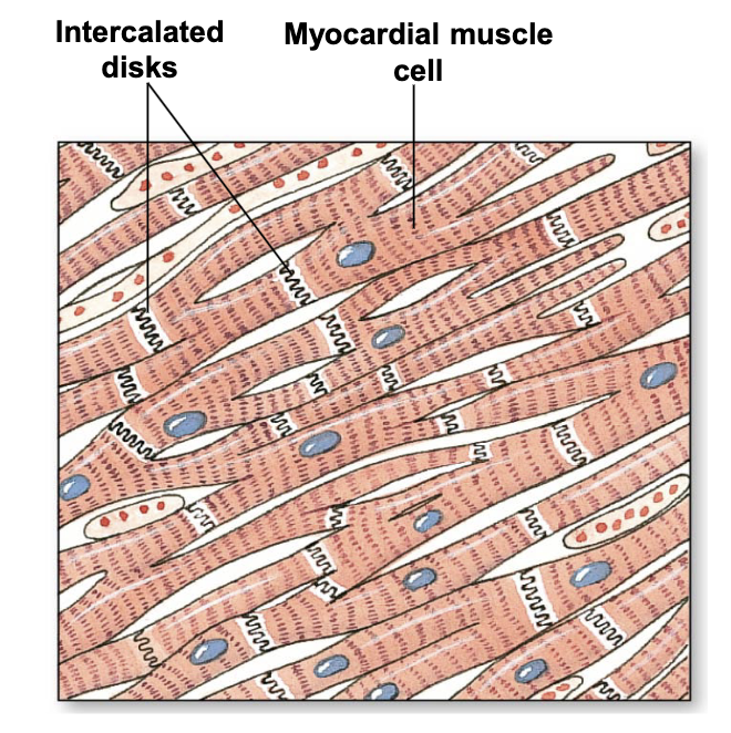
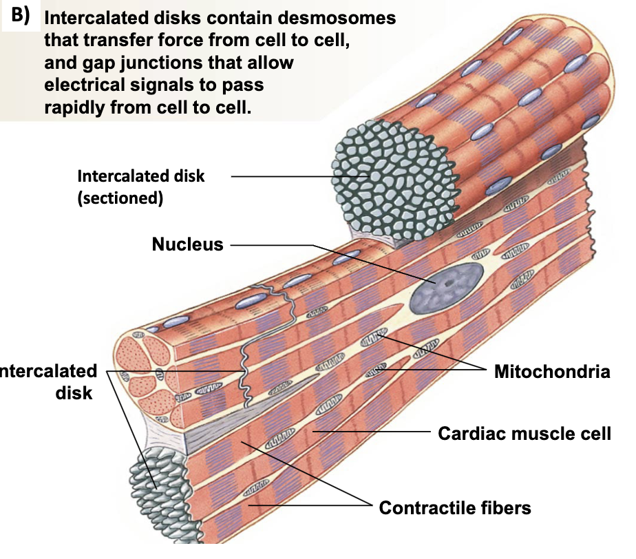
myocardial muscle cells
branched, single nucleus, connected to each other by intercalated disks
intercalated disks
contains desmosomes that transfer force from cell to cell
gap junctions that allow electrical signals to pass rapidly from cell to cell
cardiac vs skeletal muscle
smaller
single nucleus
branched
ic disks
t tubules are larger and branched
sarcoplasmic reticulum is smaller
mitochondria takes 1/3 of volume
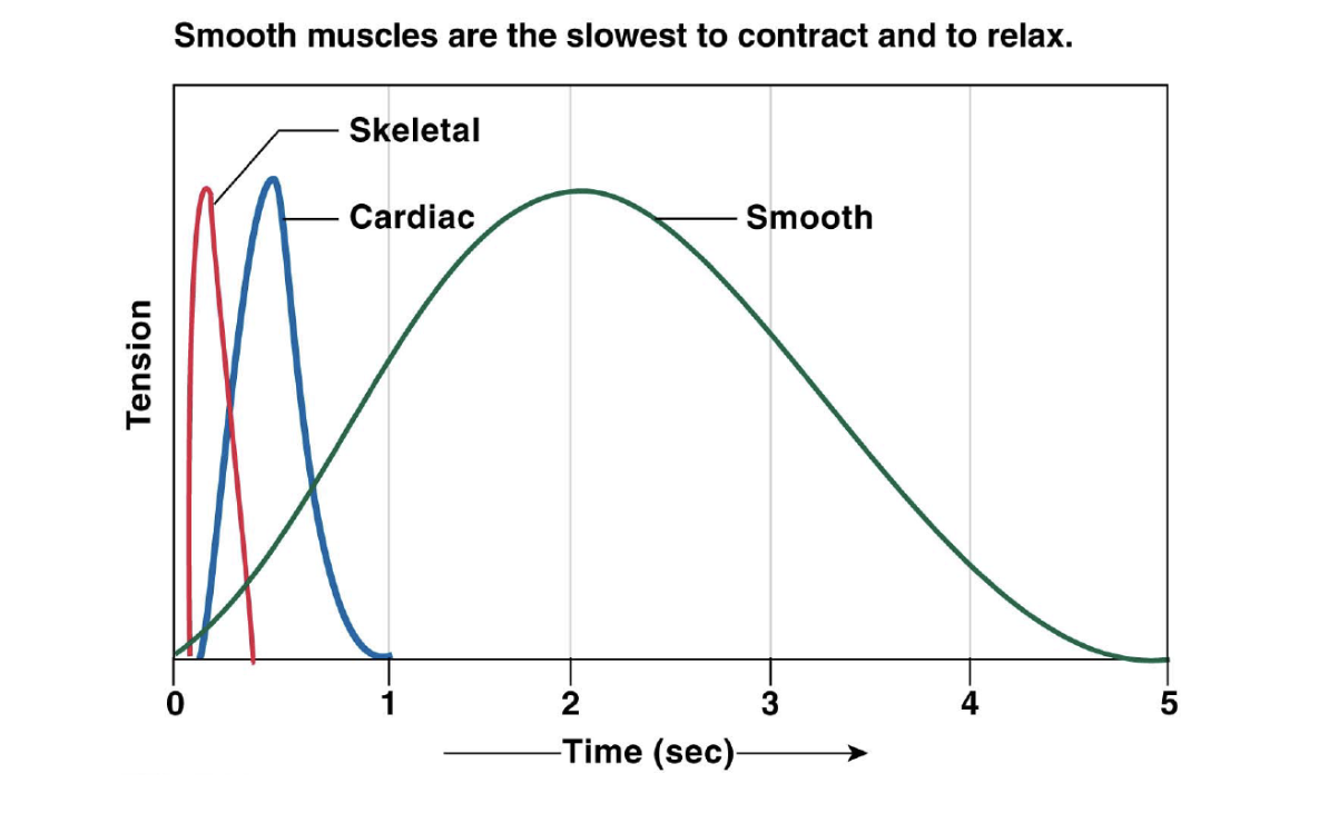
muscle twitch in diff types of muscle
myocardial autorrhythmic cells
unstable membrane potential (pacemaker potential)
depolarization due to CA2+ channels opening
Myocardial contractile cells
depolarization due to Na+ entry
Repolarization due to K+ exit
long AP (plateau) due to Ca2+ entry in the cell prevents tetanus
AP of cardiac contractile cell
Na+ channels open
Na+ channels close
Ca2+ channels open; fast K+ channels close
Ca2+ channels close; slow K+ channels open
resting potential
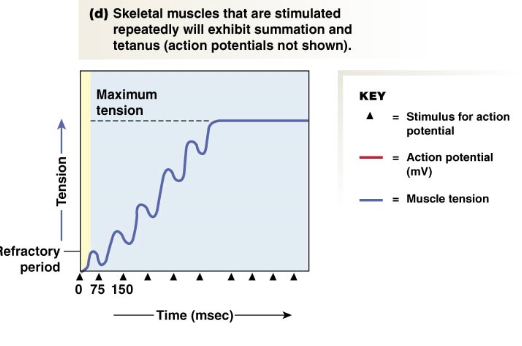
tetanus
sustained contraction
the ____ myocardial action potential helps prevent tetanus
longer
importance of preventing tetanus
heart musckles must relax between contractions so the ventricles can fill with blood
relaxation
calcium removed from cytoplasm: back into the SR with Ca2+ ATPase and out of the cell through Na+ - Ca2+ exchanger
force generated is ___________ to the number of active cross bridges
Proportional
(determined by how much calcium is bound to troponin)
sarcomere length affects force of ____________
contraction
Cardiac Muscle AP
voltage-gated L-type Ca2+ channels int the cell membrane (extracellular calcium contributes 10%)
ryanodine receptors open in the SR
calcium binds to troponin
cross-bridge cycle as in skeletal muscle
extracellular coupling in cardiac muscle
AP enters from adjacent cell
Voltage-gated Ca2+ channels open. Ca2+ enters the cell
Ca2+ induces Ca2+ release through ryanodine receptor channels (RyR)
local release causes Ca2+ spark
Summed Ca2+ sparks create a Ca2+ signal
Ca2+ ions bind to troponin to initiate contraction
relaxation occurs when Ca2+ unbinds from troponin
Ca2+ is pumped back into SR for storage
Ca2+ is exchanged with Na+ by the NCX antiporter
Na+ gradient is maintained ny the Na+-K+-ATPase
conducting system of heart steps
sa node depolarizes
electrical activity goes rapidly to AV node via internodal path
depolarization moves more slowly across atria. conduction slows through AV
depolarization moves rapidly through ventricular conducting system to apex of heart
depolarization wave spreads upward from apex
three waves
P, QRS, T
P wave
depolarization of the atria
QRS complex
wave of ventricular depolarization
T wave
repolarization of the ventricle
heart rate
time between two P waves or two Q waves
rhythm
regular
waves analysis
presence and shape
segment length
constant
ECG and electrical events
P wave (atrial depolarization)
P-Q/P-R segment (conduction through AV node/bundle
Q wave (depolarization of the septum
r wave (atrial repolarization)
s wave (ventricle contraction)
s-t segment (
t wave (ventricular repolarization
qrs
normal ECG
third degree block
missing T, relaxation is missing
atrial fibrillation
contracting more, depolarize more
ventricular fibrillation
do not have the qrst
diastole
cardiac muscle relaxes
systole
cardiac muscle contracts
beginning of cycle
heart at rest
mechanical events if cardiac cycle
late diastole: both sets chambers and ventricles are relaxed and fill passively (end diastolic volume)
atrial systole: first contraction; atrial contraction forces a small amount of additional blood into ventricles
isovolumic ventricular contraction: first phase of ventricular contraction pushes AV valves closed but does not create enough pressure to open semilunar valves
ventricular ejection: as ventricular pressure rises and exceeds pressure in the arteries, the semilunar valves open and blood is ejected (end systolic volume)
Isovolumic ventricular relaxation: ventricles relax, pressure in ventricles fall. blood flows back into cusps of semilunar valves and snaps them closed
first heart sound, “lub”
vibration following closure of the AV valves
second heart sound, “dub”
vibrations created by closing of semilunar valve
auscultation
listening to the heart through chest wall using stethoscope
driving pressure is created by…
ventricles
if blood vessels dilate, blood pressure ________
decreases
if blood vessels constrict, blood pressure ________
increases
flow through a tube is inversely proportional to resistance
1/R
if resistance ________, flow decreases
increases
if resistance ________, flow increases
decreases
Resistance is proportional to length of tube
resistance increases as length increases; vice versa
resistance is proportional to viscosity
resistance increases as viscosity increases; vice versa
resistance is inversely proportional to tube radius to the 4th power
resistance decreases as radius increases; vice versa
vasoconstriction
decrease in blood vessel diameter/radius and decreases blood flow
vasodilation
increase in blood vessel diameter/radius and increases blood flow
flow of blood is
directly proportional to pressure gradient
inversely proportional to resistance to flow
flow rate = flow rate
the volume of blood thats passes a given point per unit time
velocity of flow
distance a fixed volume of blood travels in a given period of tiem
end diastolic volume
volume of blood present in ventricle at the end of diastole
end systolic volume
voluem of blood in the ventricle at the end of systolic ejection phase
stroke volume
amount of blood pumped by one ventricle during contraction
volume before contraction - volume after contraction
EDV-ESV
average - 70 mL
cardiac output
volume of blood pumped by one ventricle in a given period of time
CO=HRxSV
average is 5L/min
ejection fraction
percentage of EDV ejected with one contraction
average is 52%
Left ventricle fractional shortening
the fraction the left ventricle shortens during a cardiac cycle
average= 30-45%
Preload
force load acting to stretch the LV fibers at the end of diastole
amount of blood returning to heart
afterload
the force that must generate in order to overcome vascular resistance and eject blood out of the left ventricle
combined load of EDV and arterial resistance during ventricular contraction
force of contraction is affected by:
length of muscle fiber (dependent on volume of blood)
contractility of heart
as stretch of the ventricular wall increases, so does stroke volume
preload is the degree of myocardial stretch before contraction
chemical that affects contractility
inotropic agent
chemicals that have positive inotropic effects
epinephrine, norepinephrine, and digitalis
chemicals with negative inotropic effects ________ contractility
decrease
frank-starling law of heart
SV increases as EDV increases
increases the volume of blood in the ventricles
EDV is determined by what?
venous return
venous return is affected by
skeletal muscle pump
respiratory pump
sympathetic innervation of veins
parasympathetic innervation causes
lower rate of depolarization in autorhythmic cells
sympathetic innervation and epinephrine causes
higher rate of depolarization in autorhythmic cells