lecture 5, chronic diseases
1/26
There's no tags or description
Looks like no tags are added yet.
Name | Mastery | Learn | Test | Matching | Spaced | Call with Kai |
|---|
No analytics yet
Send a link to your students to track their progress
27 Terms
Structure of the granuloma
Inside, there are macrophages, T cells, and a fibrous layer.
m1 and what affects it
m1→ is very proinflammatory.
in TB the bacteria shift from M1 to M2
TNF alpha → a very important factor M1
KLF4
wants more M2 and less M1
NO
more M1 and less M2, radical oxygens killing M1
what type of necrosis do we have in TB?
Caseous necrosis = a type of cell death where tissue turns into a soft, white, “cheese-like” material
Immune system tries to kill TB
TB resists → chronic inflammation
Cells die in the center
👉 So:
Outside = immune cells
Inside = dead material (caseous necrosis)
two types of giant cells
1) Foreign body giant cells: Nuclei are scattered randomly
2) langhans : the nuclei are moslty periphery (seen mostly in TB) in a horse shoe formation
what causes the most industrial plagues?
virus
explain the extracellular state of viurs
A virion = complete virus particle outside a cell
Envelope (sometimes present)
Lipid layer from host cell
Has spike proteins
👉 Used for:
entering host cells
recognition
intracellular state
when capsid is removes and it exits in the cell, ( some nucleic acid)
three big groups of viruses?
helical
polyhedral
complex
what is the subunit of capsids?
capsomere
Helical symmetry (rod-shaped)
Structure:
Capsid proteins wrap around genome like a spiral (helix)
Looks like a rod / tube
🧠 Key rules (from your slide):
👉 Length = genome length
More RNA/DNA → longer virus
👉 Width = protein size
Determined by capsid subunits
Icosahedral symmetry (spherical)
20 triangular faces
Looks like a sphere (but actually geometric)
🧠 Key idea:
👉 “Most efficient way to build a closed shell”
💡 Why?
Uses few proteins
Very stable
💡 Example:
Human papillomavirus (HPV)
🔥 Easy way to remember:
👉 “Icosahedral = soccer ball shape”
Where can viruses replicate, and how are different types of viruses grown/studied?
BACK (Answer):
👉 Replication:
Viruses replicate only in specific host cells or organisms
→ (host specificity / tropism)
👉 Bacterial viruses (bacteriophages):
Easiest to grow
Use bacteria as hosts
Common model systems
👉 Animal viruses:
Grown in:
Cell cultures
Tissue cultures
👉 Plant viruses:
Hardest to study
Often require whole plant growth
What are the phases of viral replication?
Attachment (adsorption) of the virus to a susceptible host cell
Entry (penetration) of the virion or its nucleic acid
Synthesis of virus nucleic acid and protein by cell metabolism as redirected by virus
Assembly of capsids and packaging of viral genomes into new virions (maturation)
Release of mature virions from host cell
example of an early stage protein?
NEF
example of late stage protein
VPU
What are the three mechanisms of entry of animal viruses?
Direct penetration
→ Viral genome enters directly through the membraneMembrane fusion
→ Viral envelope fuses with host cell membraneEndocytosis
→ Cell engulfs virus into vesicle → uncoating releases genome
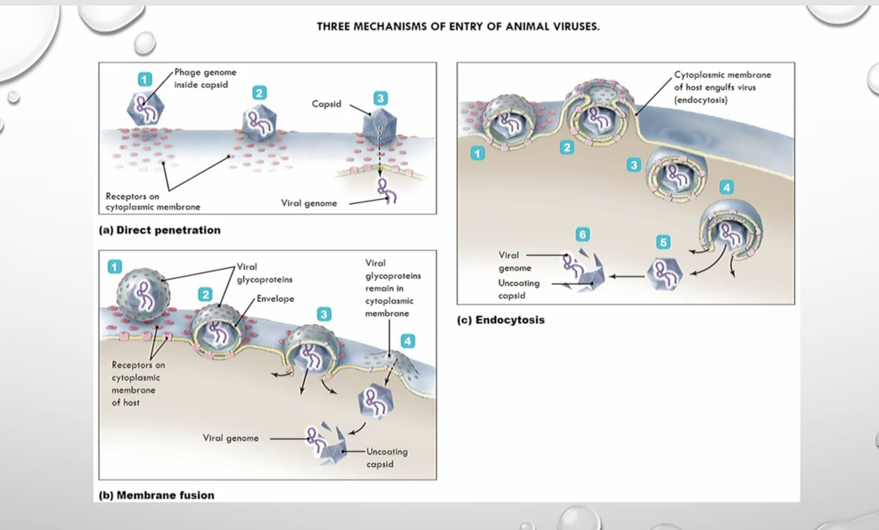
HIV is an exception, and it keeps the capsid unti it gets to the nucleas.
What is the eclipse phase in viral replication?
Eclipse phase = period after entry where NO infectious virus particles are detectable
Virus has entered cell
Capsid is removed (uncoating)
Virus exists only as nucleic acid
Viral components (proteins + genome) are being made
❗ No complete virions yet → cannot detect virus
What IS happening during eclipse?
Inside the cell:
Early enzymes made
Viral genome replicated
Viral proteins made
💥 Then what happens?
👉 After eclipse:
Assembly starts
New virions form
👉 Then graph goes 📈 UP FAST
Virulence factors
Antiphagocytic factors:
Factors prevent phagocytosis by the host’s phagocytic cells
👉 Bacterial capsule:
Composed of chemicals not recognized as foreign
Slippery; difficult for phagocytes to engulf bacteria
👉 Antiphagocytic chemicals:
Prevent fusion of lysosome and phagocytic vesicles
👉 Leukocidins:
Directly destroy phagocytic white blood cells
What are the stages of infectious disease and what happens in each?
Incubation period:
No signs or symptoms; pathogen is multiplyingProdromal period:
Vague, general symptoms (e.g., mild fever, fatigue)Illness:
Most severe signs and symptoms; peak pathogen levelsDecline:
Signs and symptoms decrease as immune system gains controlConvalescence:
No signs or symptoms; recovery phase
explain listeria
non spore,gram positive
no enzymes or toxins,
uses lysom o to destroy the fusion of lysosome and phagosom,
survives in our cells,uses the cell actin to infect the cells next to it,
What is tuberculosis and what are the three types of TB?
Tuberculosis (TB):
Respiratory disease caused by Mycobacterium tuberculosis
Increasing in Canada and the United States
Pandemic in other parts of the world
👉 Three types of tuberculosis:
Primary TB:
Results from the initial infection with M. tuberculosisSecondary TB:
Reestablishment of active infection after period of dormancyDisseminated TB:
Results when infection spreads throughout the body
What are the key features of Mycobacterium cell wall and what do they cause?
Cell wall contains a waxy lipid called mycolic acid
👉 Results in unique characteristics:
Slow growth
Protection from lysis after phagocytosis
Capacity for intracellular growth
Resistance to Gram staining, detergents, many antimicrobial drugs, and desiccation
What is cord factor and what does it do in Mycobacterium tuberculosis?
Cord factor:
Glycolipid produced by virulent strains of M. tuberculosis
👉 Functions:
Cells remain attached end-to-end → form “cords”
Inhibits migration of neutrophils and is cytotoxic
Prevents fusion of endosomes (phagosomes) and lysosomes
Stimulates granuloma formation via cytokine production
What is granulomatous inflammation and what is the structure of a granuloma?
Granulomatous inflammation:
A distinctive pattern of chronic inflammation evoked by agents like Mycobacterium tuberculosis
Characteristic feature = granuloma
👉 Granuloma structure:
Small (0.5–2 mm) collections of modified macrophages (epithelioid cells)
Surrounded by a rim of lymphocytes → nodular appearance
👉 Additional components:
Vascular elements
Fibroblasts and collagen (it can calsify and become harder)
PMNs and plasma cells