Cranium and Facial Bone Projections
1/6
There's no tags or description
Looks like no tags are added yet.
Name | Mastery | Learn | Test | Matching | Spaced |
|---|
No study sessions yet.
7 Terms
Lateral Facial Bones
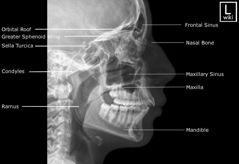
Part Position: Head in true lateral; IPL perpendicular; IOML parallel to bottom of IR.
Central Ray: Perpendicular to zygoma (midway between outer canthus and EAM).
Collimation: 6×10 L/W
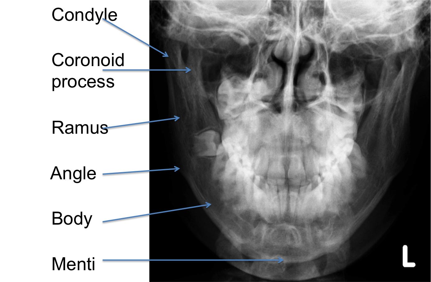
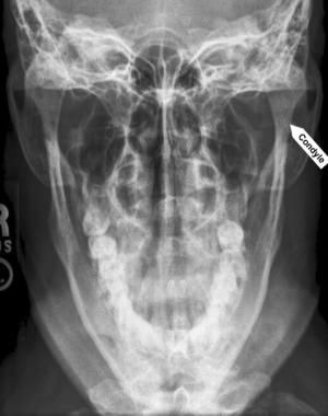
Axiolateral Mandible
Part Position: Head in true lateral; IPL perpendicular; IOML parallel to bottom of IR.
Central Ray: 20-25* cephalic midway between outer canthus and EAM.
Collimation: 6×10 L/W
Parietoacanthial (Waters)
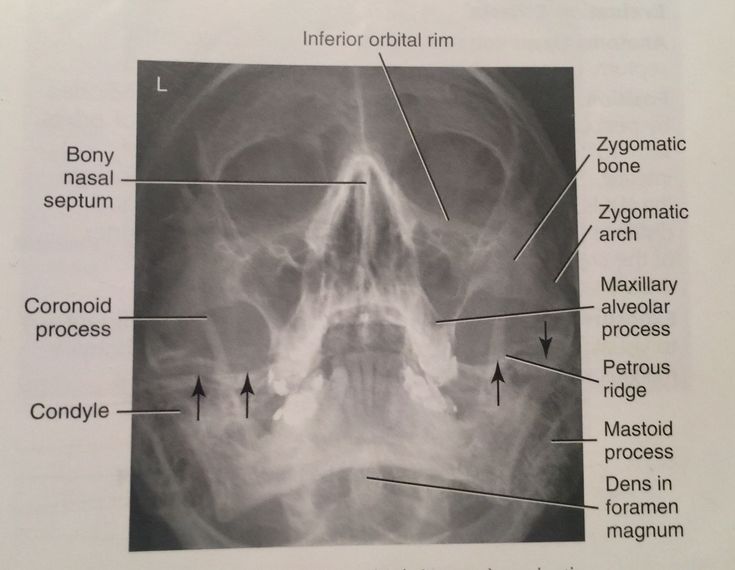
Part Position: Chin on IR; MML perpendicular; OML forms 37° angle with IR.
Central Ray: Perpendicular to exit at acanthion.
Collimation: 10×10
SMV for Zygomatic Arches
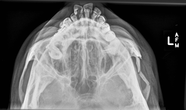
Part Position: Vertex on IR; IOML parallel to IR.
Central Ray: Perpendicular to IOML, entering at mid-throat, 1 inch posterior to outer canthi.
Collimation: 8×6 c/w
AP Axial (Modified Towne) for Zygomatic Arches
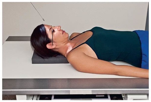
Part Position: Back of head on IR; OML or IOML perpendicular.
Central Ray: 30° caudad to OML (or 37° to IOML), entering 1 inch above nasion.
Collimation: 3×8 c/w
PA for Mandible
Part Position: Forehead and nose on IR; OML perpendicular to IR.
Central Ray: Perpendicular to exit the acanthion.
Collimation: 6×8 l/w
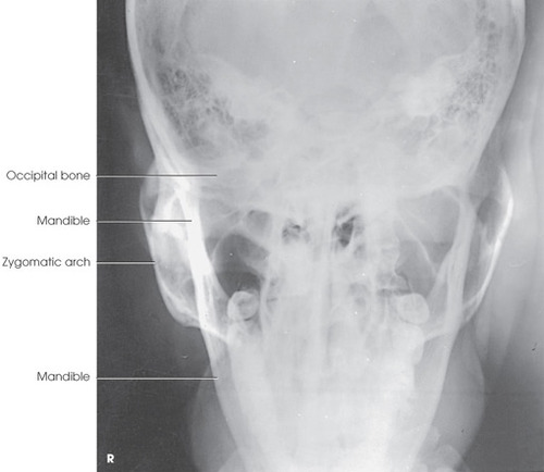
PA Axial for Mandible
Part Position: Forehead and nose on IR; OML perpendicular to IR.
Central Ray: 20-25° cephalad, exiting at acanthion.
Collimation: 6×8 l/w