BSC2011 Exam 4 Study Guide
1/78
There's no tags or description
Looks like no tags are added yet.
Name | Mastery | Learn | Test | Matching | Spaced | Call with Kai |
|---|
No analytics yet
Send a link to your students to track their progress
79 Terms
which animal groups exhibit the different types of symmetry?
bilateral = most animals
radial = cnidarians, ctenophores
asymmetry = sponges
how do unicellular and multicellular organisms differ in regards to getting nutrients to the cells?
unicellular organisms = get nutrients through diffusion
multicellular organism = must provide nutrients and oxygen to all cells w/ specialized cells, organs, and organ systems
how does the BMR differ between animals of different sizes
smaller endothermic organisms have higher BMR than larger ones per gram of body weight
how does homeostasis maintain internal conditions?
if conditions stray from set point, homeostasis mechanisms kick in
what are the general differences between negative and positive feedback + examples of each?
negative feedback = counteracts internal changes. ex. change in glucose
positive feedback = strengthens response to stimulus. ex. birthgiving
4 mechanisms of heat transfer
radiation, conduction, convection, evaporation
radiation
the transfer of heat by electromagnetic (infrared) radiation. ex. sun shining on lizard
conduction
direct transfer of heat between two objects. energy transferred from hotter objects to colder ones. ex. lizard sitting on hot sidewalk
convection
transfer of heat brought about by the movement of a gas or liquid. ex. atmospheric air warming lizard
evaporation
heat of vaporization or the amount of energy needed to change them from a liquid to a gas phase… always heat loss. ex. lizard skin water loss
ectotherms
regulate temperature using behaviors. ex. snakes, lizards
endotherms
create internal metabolic heat. conservation or dissipation heat transfer controlled by amount of blood flow to animal surface. ex. mouse
4 tissue types
epithelia, connective, muscles, neurons
epithelia funciton
line cavities, open spaces, and surfaces
connective tissue function
connect tissues together, provide support
muscle function
generate movement
neurons
generate and send electrical signals
simple squamous epithelium
flat, irregular round shape. single layer.
location: lung alveoli, capillaries
stratified squamous epithelium
flat, irregular round shape. multiple layers.
location: skin, mouth, vagina
simple cuboidal epithelium
cube shaped, central nucleus. simple layer.
location: glands, kidney (renal) tubules.
some gland ducts are stratified
simple columnar epithelium
tall, narrow, nucleus toward base. single layer.
location: digestive tract (unciliated), bronchi (ciliated), intestinal lining w/ microvilli
pseudostratified columnar epithelium
tall, narrow, nucleus along cell. appear to be stratified by isnt.
location: upper respiratory tract
different types of connective tissue
blood, adipose tissue, loose connective tissue, dense connective tissue, bone, cartilage
blood
has RBCs (erythrocytes) and WBCs (leukocytes). fluid portion = matrix, the plasma. transports material through the body to bring nutrients to cells and remove waster material form them
adipose tissue
made up of adipocytes that collect and store fat in the form of triglycerides for energy metabolism. also serve as insulation to help maintain temperatures and act as cushioning. ex. fat under skin, visceral fat (around organs). no fibroblasts or real matrix
loose connective tissue
has sampling of all components of matrix: fibroblasts, macrophages, some lymphocytes, some neutrophils. fibers: collagen, elastic, reticular. found in every blood vessel, helps keep vessel in place. found around and between most body organs. tough, yet flexible. anchors epithelia
dense connective tissue
cells: fibroblasts, macrophages. large amounts of collagen fibers, few cells or matrix material. fibers arranged regularly or irregularly.
irregular arrangement = areas where body stress occurs in all directions, like skin dermis
regular arrangement - tendons (muscles to bone) and ligaments (bone to bone_
bone
cells: osteoblasts, osteocytes, osteoclasts. dual matrix: organic (collagen/elastic → flexibility) + inorganic (calcium salts → hardness)
types: compact and spongy
provide structural support + tendon attachment points.
location: vertebrate skeletons
cartilage
large amount of matrix. cells, chondrocytes, make matrix and fibers of the tissue. few collagen and elastic fibers = hyaline cartilage.
fibrocartilage = large amount of collagen
location: shark skeleton, fetal bones, human ears, invertebral discs
different types of muscle tissues
smooth, skeletal, cardiac
smooth muscle
no striations + single, centrally located nucleus. involuntary
location: visceral organs, walls of blood vessels, tubes of digestive system, tubes of reproductive systems
skeletal muscle
has striations. relatively long w/ multiple nuclei along the edge.voluntary
cardiac muscle
has striations + single, centrally located nucleus. involuntary but can be influenced by the autonomic nervous system to speed up or slow down. intercalated disc = assists in passing electrical impulse from one cell to the next and maintains strong connection between nearby cardiac cells.
location: heart
what are the different cellular components of nerve tissue?
neurons, neuroglia
neurons
generate and transmit electrical impulses
neuroglia
support and insulate neurons and eliminate foreign materials in and around neurons
what are the different parts of a neuron?
cell body (soma) = contains nucleus
dendrites = highly branched extensions. conduct electrical impulses toward cell body
axon = sign cytoplasmic extension. conducts impulses away from cell body
what is the function of the myelin sheath of neurons?
insulate the axon so the electrical impulse is transferred more efficiently
what are the main organs of the vertebrate digestive system and their general functions?
mouth and pharynx = entry
esophagus = delivers food to stomach
stomach = preliminary digestion
small intestine = digestion and absorption
large intestine/colon = absorption of water or minerals
cloaca or rectum = expel waste
how do the digestive systems of humans and rabbits differ (both with monogastric stomachs)?
rabbits small intestine and cecum are enlarged to allow more time to digest plant material + more surface area for nutrient absorption
rabbits digest food 2x: food passes through system and passes as cecotrophs. rabbit re-ingests cecotropes
what are the primary accessory organs of the vertebrate digestive system and their general functions?
liver = produces bile
gallbladder = stores and concentrates bile
pancreas = produce “pancreatic juice.” digestive enzymes and bicarbonate buffer
how does the avian digestive system differ from other vertebrates?
have crop in esophagus, which stores good
2 stomachs: (1) proventriculus contain digestive juices to break down good. (2) gizzard grinds food
wastes are excreted from cloaca - dont have separate opening for urine and feces
in general, how does the ruminant digestive system differ from other mammals? give examples of ruminants
ruminant examples: cows, goats, sheep, deer
multi-gastric = have 4 stomachs - rumen, reticulum, omasum, and abomasum
what is the function of each chamber of the ruminant stomach?
rumen and reticulum = contain prokaryotes that are able to digest cellulose fiber
omasum = remove water from cud
abomasum = where cud is digested by enzymes produced by the ruminant
follow the digestive process of a piece of food (bolus) through the digestive system. least each organ and its energy function, in order, from mouth to anus
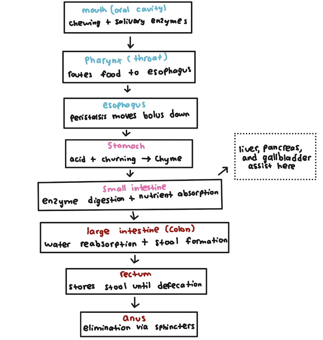
how does peristalsis work to move a bolus of food through the alimentary canal?
alternating contractions of circular and longitudinal smooth muscle bands pushes bolus along the digestive track
how does the oblique muscle layer of the stomach help mechanical digestion?
it helps mix food with gastric juices
what are the functions of the different cells lining the stomach?
mucus-secreting cells = lubricates food bouls
parietal cells = secrete HCI and intrinsic factor (for Vitamin B 12 absorption)
chief cells = secrete pepsinogen (inactive form of pepsin) for protein digestion
what are the functions of the villi and microvilli
both have functions in the small intestine
villi = folds on the small intestine lining that increase the surface area to facilitate the absorption of nutrients
microvilli = projections of the epithelial lining to further increase surface area for absorption
in a balanced diet, what different food groups should be included?
fruits, vegetables, grains, and protein
how are carbohydrates digested and absorbed?
starch and glycogen are broken down into glucose by amylase and maltase
sucrose and lactose are broken down by sucrase and lactase, respectively
monosaccharides are absorbed by Na+ - cotransporters or facilitated diffusion across the brush boarder
how are proteins digested and absorbed?
digestion begins in the stomach and continues through the small intestines where amino acids are absorbed
pepsin in stomach breaks down proteins → peptides
various peptidases from pancreas and small intestine → amino acids in small intestine
these are transported to the liver via the hepatic portal vein
liver regulates distribution of amino acids to the rest of the body
how are lipids digested and absorbed?
digested in the mouth, stomach, and mostly the small intestine
bile emulsifies lipids to breakup globules for more effective lipase action in small intestine
gastric and pancreatic lipases breakdown lipids → fatty acids and monoglycerides
packaged as micelles to move through microvilli
reformed triglycerides w/ cholesterol, coated w/ proteins → chylomicrons → lymph vessels → circulatory system
what are the four essential nutrients + examples?
vitamins (e.g. vitamin C and D)
amino acids (e.g. valine, glutamine, glycine)
long-chain unsaturated fatty acids (e.g. omega-3s, bs, and qs)
minerals (e.g. calcium, iron, iodine)
what are the three general functions of circulating blood and a circulatory system?
transportation, regulation, protection
what are the components of blood plasma? what is the hematocrit?
nutrients, wastes, hormones, ions, proteins (albumin and fibrogen), 92% water. hematocrit = fraction of total blood volume occupied by red blood cells (RBCs)
what is the function of hemoglobin?
binds and transports oxygen from the lungs to the body’s tissues and organs. carries oxygen to cells and carbon dioxide to lungs
describe the steps of how a blood clot forms
stimulated by chemicals released from damaged tissue
vessels constrict near damage, reducing blood loss
injury site releases chemicals to activate and attract other platelets → become spiked and stick to each other and wound → platelet plug
coagulation → circulating fibrinogen converted to fibrin, forming mesh to trap more platelets and RBCs → clot
where do blood cells form? from what cells do they arise?
they come from the bone marrow from pluripotent stem cells
what are examples of the most basic types of circulation systems in animals?
sponges = circulate water using incurrent pores and ine excurrent pore (osculum)
hydra circulate water through a gastrovascular cavity
nematodes are thin enough that the digestive tract can also be used as a circulatory system
open circulatory system
hemolymph (blood mixes with interestial fluid), no capillaries, animal movement → hemolymph back to heart through ostia. decreased energy, decreased O2 to organs. ex. arthropods, mollusks
closed circulatory system
blood separate from interestial fluid, unidirectional flow (enclosed in vessels- arteries → capillaries → veins), blood moved by heart. increased energy, increased O2 to organs. ex. annelids, vertebrates. more efficient at delivering oxygen to tissues
difference between hemolymph and blood
hemolymph = blood mixed with interestial fluid
blood = separate from interestial fluid
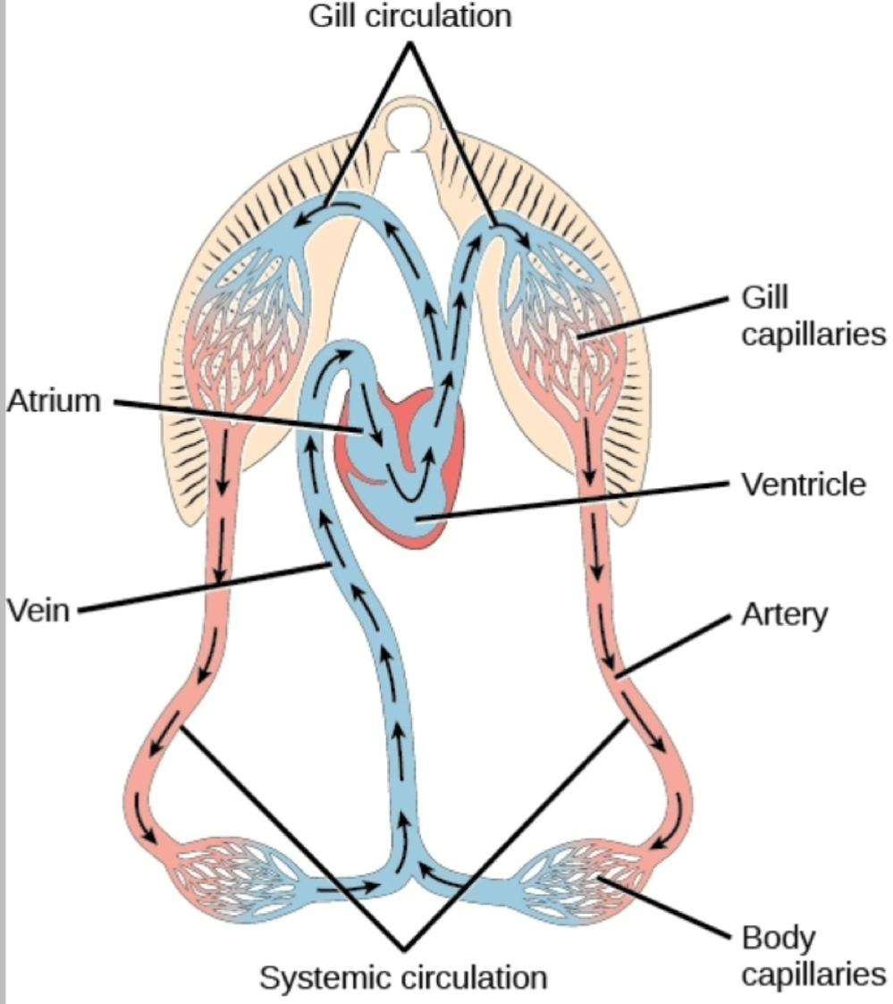
circulatory system of a bony or cartilaginous fish with a 2-chambered heart.
circulatory system of a lungfish or amphibian with a 3-chambered heart
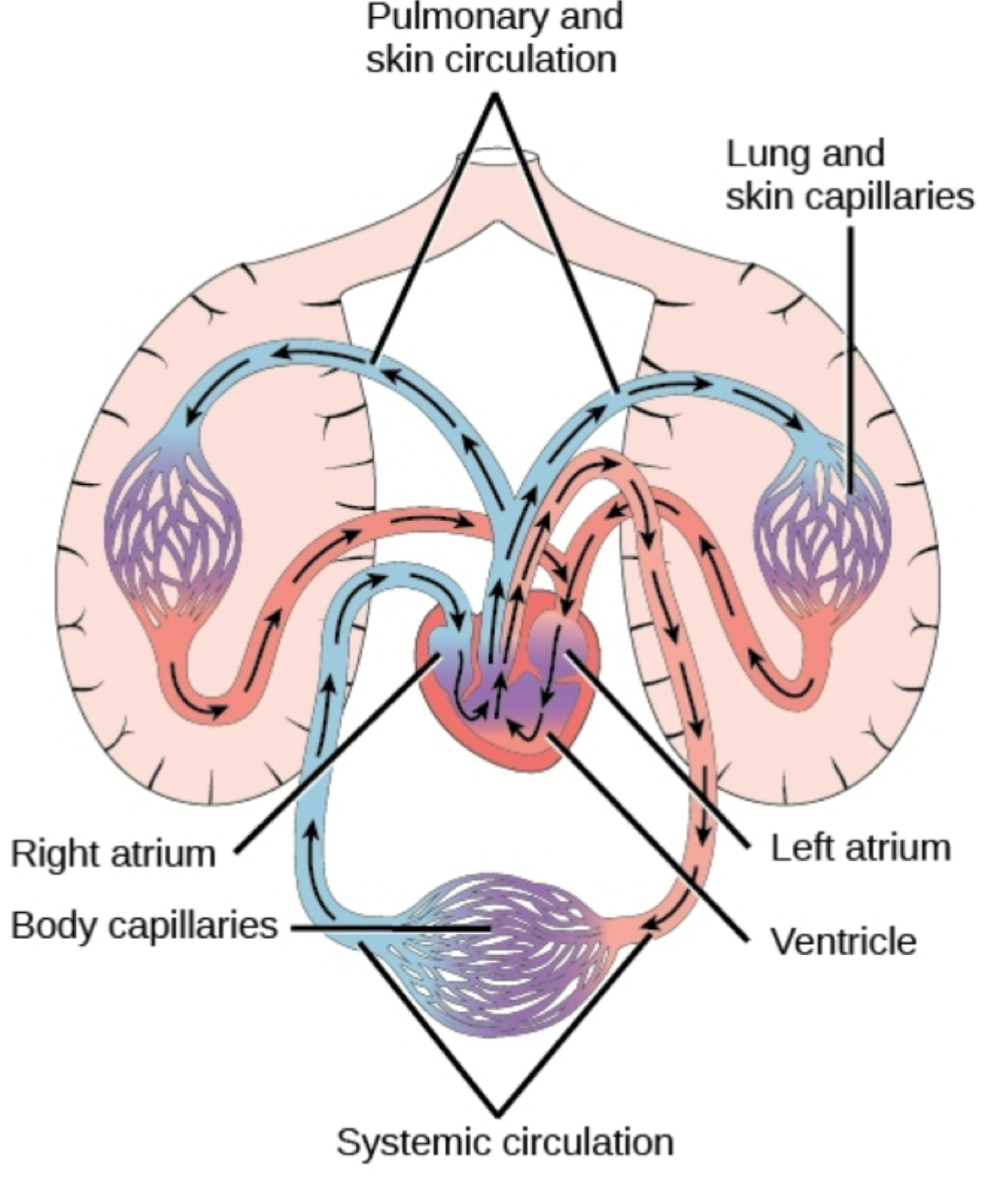
how does a typical, non-crocodilian heart differ from that of amphibians and crocodilians?
they have a septum, which partially subdivides the ventricle, further reducing mixing of blood
path of blood flow through the 4-chambered human heart
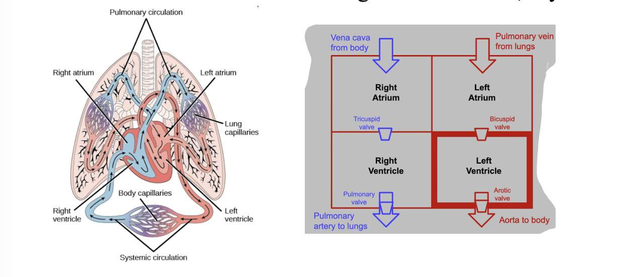
describe the cardiac cycle (from diastole to systole back to diastole), including the diff components of the cycle, and how they function
the cycle begins with diastole - the ventricles relax and fill with blood as the AV valves (tricuspid on the right, mitral on the left) open
next, SA node (pacemaker in right atrium) fires electrical signal before sending it through the Bundle of His → Purkinje fibers, causing both ventricles to contrast simultaneously - this is systole. the AV valves close (“lub”) and the semilunar valves open, ejecting blood to the lungs and body
finally, ventricles relax, the semilunar valves close (“dub”), and the cycle returns to diastole
what are the “heart sounds” and what do they indicate is happening within a beating heart?
“lub” = AV valves closing
“dub” = semilunar valves
murmurs = defective valves
arteries structure
4 tissue layers: endothelium, elastic fibers, smooth muscle, and connective tissue. walls thick → no material exchange across wall. more elastic fibers in walls
veins structure
4 tissue layers: endothelium, elastic fibers, smooth muscle, and connective tissue. wall thick → no material exchange across wall. thinner layer of smooth muscle
capillaries structure
single layer of endothelial cells. greatest cross-sectional surface area → lowers blood pressure. about diameter of single RBC to enhance gas exchange
arteries function
recoil each time they receive blood from heart (pulse). helps to “smooth” out blood flow
veins
venous pumps helps blood return to heart. skeletal muscle contractions (body movements) and one-way venous valves. keeps one-way flow toward heart
how does blood and interstitial fluid flow at the level of the capillaries?
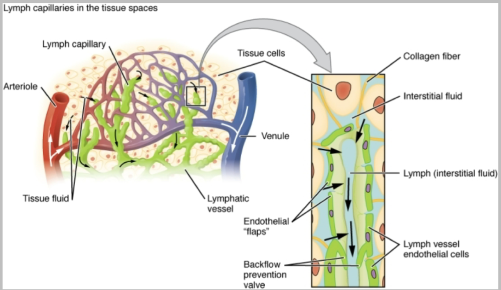
most fluid leaves at the arteriole end (higher blood pressure) of capillary and returns at venule end (lower pressure). fluid that does not return to capillaries is returned to circulation by the lymphatic system
major components of the lymphatic system and their functions
lymph = continues into progressively larger vessels with one way valves (like veins, very low pressure)
lymphatic vessels = drain into subclavian veins through thoracic duct
lymph nodes = sites for immune system cells to filter and “sample” lymph for pathogens
lymph organs = thymus and spleen
path of lymphatic fluid from capillary beds back to circulatory system
atherosclerosis
accumulation of fatty material within arteries - plaque. impedes blood flow
arterioscelosis
arterial hardening due to calcium deposition