Chapter 8, Lesson 3: The Vertebral Column and Thoracic Cage
1/25
Earn XP
Description and Tags
Flashcards from Chapter 8, Lesson 3 of McGraw Hill Anatomy and Physiology, Ninth Edition, by Kenneth S. Saladin.
Name | Mastery | Learn | Test | Matching | Spaced |
|---|
No study sessions yet.
26 Terms
Spine functions
Supporting and protecting the skull, trunk, and spinal cord by absorbing stresses
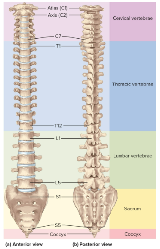
Vertebral regions
7 cervical vertebrae
12 thoracic vertebrae
5 lumbar vertebrae
5 sacral vertebrae
4 vertebrae in the coccyx
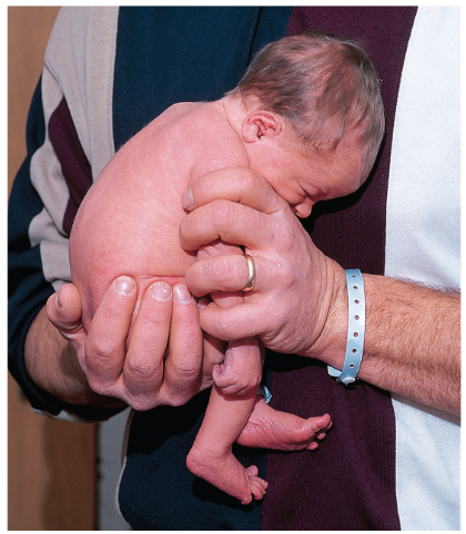
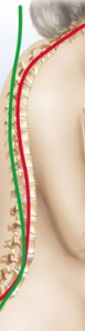
Primary curvature
The first C-shaped convex curve at birth; persists as the thoracic and pelvic spine
Secondary curvatures
Develop with crawling and walking in childhood; creates the cervical and lumbar areas concave curves
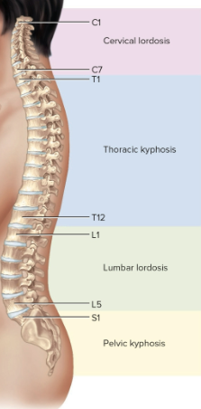
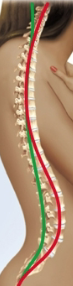
Final curvatures
Spine turns into an S shape: cervical lordosis, thoracic kyphosis, lumbar lordosis, and pelvic kyphosis
Lordoses curve in, kypohses curve out
Abnormal curvatures
Can result from disease, paralysis, posture, or congenital defects like scoliosis (sideways)
Hyperkyphosis
An exaggerated thoracic curvature usually from osteoporosis, the spine curve goes overly out
Hyperlordosis
An exaggerated lumbar curvature usually caused by pregnancy or obesity, the spine curve goes overly in
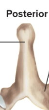
Spinous process
Projection upward from one vertebra to meet another articular process above
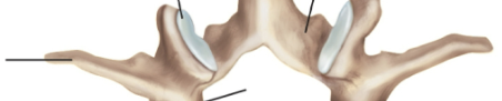
Transverse process
Lateral extension from a vertebtra
Intevertebral foramen
Opening between pedicles of two adjoining vertebrae
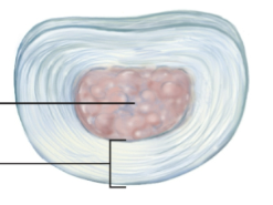
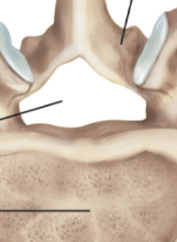
Intervertebral discs
Pads between the vertebrae that bind them together to support the weight of the body
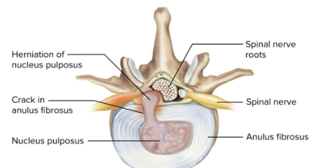
Herniated disc
The slipping or rupturing of a disc; can put painful pressure on the spinal nerve or cord
Cervical vertebrae
Notated as C1 to C7; C2 to 6 have forked spinous process while C1 and C2 are the atlas and axis
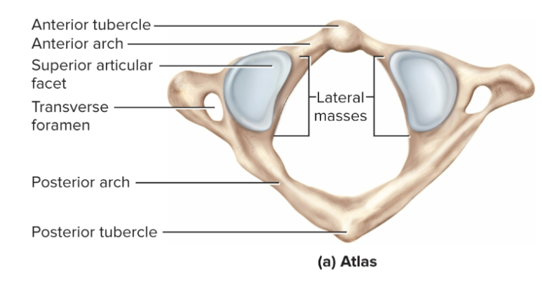
Atlas
The first cervical vertebrae (C1), supports the head and allows nodding “yes” (pitch) with anterior and posterior arches
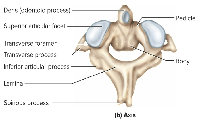
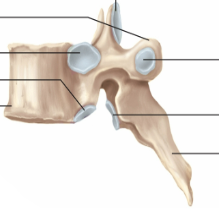
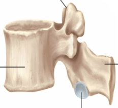
Axis
The second cervical vertebrae (C2), allows nodding “no” (yaw)
Thoracic vertebrae
Notated as T1 to T12; has downward angled spinous processes that correspond to the 12 pairs of ribs they are attached to
Lumbar vertebrae
Notated as L1 to L5; have thick, stout bodies and blunt, squarish spinous processes
Sacrum
Bony plate forming the posterior wall of the pelvic girdle, notated as S1 to S5 that begin fusing around age 16
Anterior surface is smooth and concave while posterior surface is very rough
Coccyx
Consists of four smaller vertebrae notated as Co1 to Co4, fuses into single, triangular bone by age 20 to 30
Can be fractured during childbirth or hard fall and provides pelvic muscular attachment
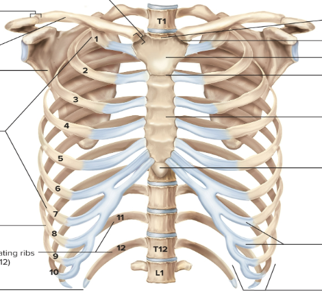
Thoracic cage
Consists of the thoracic vertebrae, sternum, and ribs to enclose the lungs and heart
Sternum
The bony plate anterior to the heart, divided into the manubrium (superior portion), body (long mdidle portion), and xiphoid (inferior point)
Ribs
12 pairs with ends attached to vertebral column and sternum; costal cartilages attach ribs to sternum
True ribs
Ribs 1 to 7, each directly connected to the sternum
False ribs
Ribs 8 to 12, lacking independent connections to the sternum
Floating ribs
Ribs 11 and 12 (also false), no connection at all to sternum or cartilages