INTRODUCTION TO ENTEROBACTERIACEAE
1/55
There's no tags or description
Looks like no tags are added yet.
Name | Mastery | Learn | Test | Matching | Spaced | Call with Kai |
|---|
No analytics yet
Send a link to your students to track their progress
56 Terms
Enterobactaeriacaea morphology characteristics:
gram-negative bacilli
They have flagella for motility
Most have fimbriae or pili for attachment to other bacteria or host cells
They have a complex cell wall
Outer membrane:
Lipopolysaccharide portion which is responsible for the endotoxin and porin protein and lipoprotein.
Peptidoglycan
gram-negative bacilli peptidoglycan is thicker compared to that of gram-positive microorganisms.
Capsular antigen (K antigen)
found in the capsule
Somatic antigen (O antigen)
found in the cell wall
Flagellar antigen (H antigen)
which is found in the flagella
Determinants of Pathogenicity: WHY THERE IS FEVER FOR A GI TRACT BACTERIA?
If an organism is engulfed by a macrophage, this organism will release endotoxin and will stimulate production of Interleukin-1 and this goes into the blood stream and into the Hypothalamus of the brain stimulating the pituitary gland to produce prostaglandins which will cause fever and this is why fever is present if you have a gram-negative infection.
Endotoxin
Integral part of the cell, Heat labile, toxoid cannot be produced, produced by gram - negative organisms only.
Subunit B (Enterotoxin)
attaches to the plasma membrane of epithelial cells lining the small intestine.
Subunit A (Enterotoxin)
induces the formation of Cyclic AMP from ATP in the cytoplasm
As a result, epithelial cells discharges large amounts of fluids and electrolytes such as chlorides, potassium, sodium, and bicarbonates. Normal contractions are disturbed leading to severe diarrhe
Subunit B (Shiga toxin)
binds to the cellular receptor (GB3 or Globotriaosylceramide) found on the endothelial cells. It allows entry of Subunit A and modifies RNA component of the ribosome to inactivate it (inhibits protein synthesis).
Verotoxin/Shiga-like toxin
similar to Shiga toxin
Colonization Factors
Capsule
Vi Antigen
Fimbriae
O Antigen
Intracellular survival
Capsule
Inhibits phagocytosis
Vi Antigen
found in Salmonella, protects the organism from complement-mediated killing
Fimbriae
attachment
O Antigen
adherence of the organism to the host cell
Intracellular survival
characteristics that makes them survive intracellularly
Mode of transmission
From the feces (from humans or animals) containing pathogenic microbes or their toxins, they can be transmitted through the foods, fluids (water and milk), or fingers. The organisms/toxins are ingested by humans, it goes into the gut or the intestinal tract of humans. The organisms multiply in the gastrointestinal tract and toxins may be produced but infection remains localized in the gastrointestinal tract. If they are in the gastrointestinal tract, they produce diarrhea and will be excreted out also through the feces.
Some organisms invade/the toxins are absorbed so they become disseminated they go to other parts of the body and causes symptoms of systemic infection like fever but can also be excreted out through the feces.
Sites of Infection
Central Nervous System
Lower Respiratory tract
Bloodstream
Gastrointestinal tract
Urinary tract
Central Nervous System
Escherichia
Lower Respiratory tract
Klebsiella, Enterobacter, Escherichi
Bloodstream
Escherichia, Klebsiella, Enterobacter
Gastrointestinal tract
Salmonella, Shigella, Escherichia, Yersinia
Urinary tract
Escherichia, Proteus, Klebsiella, Morganella
other specimens used for diagnosis:
Cerebrospinal fluid
sputum
blood
stool
urine
General specimen:
Generally, stool specimens are used for the diagnosis of the gram-negative bacilli because they are usually found in the gastrointestinal tract.
Stool specimen is processed within 1 hour and no preservative is added
rotavirus, direct wet mount, ELISA, and assays
Specimen cannot be processed within 1 hour then a transport medium is needed
Carry Blair, Glycerol M Phosphate (Shigella), Stuarts medium (Shigella)
Enrichment medium
Tetrathionate broth
Bile salts- Salmonella and Shigella; Brilliant green- Salmonella
Selenite F broth
Hajna GN broth
If growth, inoculate the plating medium. In some specimens like CSF, it can be inoculated directly to the plating medium
Slightly selective
allows growth of all gram (-) organisms; Eosin Methylene Blue and McConkey
Moderately selective
allows growth of Salmonella and Shigella only; Salmonella-Shigella Agar, Hektoen Enteric Agar, and XLD (Xylose Lysine Deoxycholate)
Highly selective
growth of Salmonella typhi and other salmonella; BGA (Brilliant Green Agar), BSA (Bismuth Sulfide Agar)
Eosin Methylene Blue Agar
Sugar: Lactose and Sucrose
Indicator: Eosin Y and Methylene Blue
Lactose Fermenters: Dark-colored colonies, Escherichia coli- Greenish-metallic sheen
Non-Lactose Fermenters: Colorless colonies
This medium contains aniline dyes which is inhibits other bacteria other than gram-negative bacill
MacConkey Agar
Sugar: Lactose
Indicator: Neutral Red
Lactose Fermenters: Pink Color
Non-Lactose Fermenters: Colorless
It inhibits gram-positive organisms because it contains bile salts and Crystal Violet.
Salmonella Shigella Agar
Sugar: Lactose
Indicator: Neutral Red
Lactose Fermenters: Pink Color
Non-Lactose Fermenters: Colorless
High concentrations of bile salts and sodium citrate to only allow growth of Salmonella and Shigella.
Hektoen Enteric Agar
Sugar: Lactose, Sucrose, and Salicin
Indicator: Bromothymol Blue
Lactose Fermenters: Bright Orange to Salmon colonies
Non-Lactose Fermenters: Green-Colored colonies
If you have an organism which produces H2S, you will have Blue green colonies with black centers.
Xylose Lysine Desoxycholate
Sugar: Lactose, Sucrose, and Xylose
Indicator: Phenol Red
Lactose Fermenters: Yellow Colored colonies
Non-Lactose Fermenters: Red Colored colonies or Pink
Brilliant Green Agar
Sugar: Lactose and Sucrose
Indicator: Phenol Red
Lactose Fermenters: Yellow to Greenish Yellow colonies
Non-Lactose Fermenters: Pink with an Intense Red background
Bismuth Sulfite Agar (BSA)
Sugar: Dextrose
Indicator: Bismuth Sulphite
If there is Salmonella Typhi, it produces jet-black colonies with metallic sheens.
If there are other colonies Dull-Green, Brown or sometimes Colorless colonies
Once the specimen has been inoculate in the plated medium, usually it would produce a typical colonies in the plating medium.
Confirm these with Biochemical Tests
Triple Sugar Ion
Sugars: Lactose, Sucrose, and Fructose (Glucose or Dextrose 0.1%)
Indicator: Phenol Red
Acid Production: Yellow Color
There is a Slant and a Butt
The lactose fermenter will utilize first Glucose, since only have 0.1%, this will be utilized immediately.
The organism will also use Lactose
As a result of the utilization of these sugars, it will have mixed acid production
Since the indicator present is phenol red, when there is acid production, it will produce yellow color.
Peptides can also be used to convert it to Amines, but this can only be achieved in the presence of Oxygen.
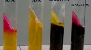
Lactose Fermenter with an Acid slant and Acid butt
If the organism produces Gas it would produce Cracks or Bubbles
If you have Non-Lactose Fermenters the initial reaction would be to use Glucose. Since all of the Enterobacteriaceae utilizes Glucose.
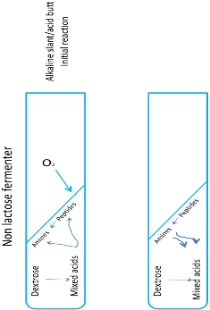
When glucose is consumed, lactose can no longer be used.
As a result of glucose fermentation, it will also have acid production in the butt the organism will then utilize proteins or peptides, Peptide Degradation requires oxygen and will have only utilization of peptides in the slant, since this is an alkaline reaction, your slant would be alkaline (Red Color) since your indicator is phenol red.
When all amines have been utilized, the reaction will go down, but it cannot go down deep into the butt.
If gas is produced, bubbles and cracks would be noticed.
Non-Lactose Fermenter with Hydrogen Sulfide (H2S)
Blackening of medium due to the reaction of H2S with Iron, producing Iron Sulfide or FeS producing a black color.
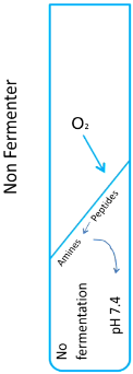
Alkaline slant and Alkaline butt but contains H2S.
Does not use any of the sugar, only uses Proteins thus, have an Alkaline reaction.
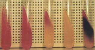
Different reactions
Alkaline Slant and Acid Butt
Acid Slant and Acid Butt
Acid Slant and Acid Butt with H2S (Fermenters with H2S)
Alkaline Slant and Acid Butt with H2S (Fermenters with H2S)
Alkaline Slant and Alkaline Butt (Non-Fermenters)
Alkaline Slant and Acid Butt (Non-Lactose Fermenters)
Acid Slant and Acid Butt (Lactose Fermenters)
Alkaline Slant and Acid Butt (H2S)
Indole Test
Tryptophan, the bacteria will ferment it to tryptophanase to Indole + Pyruvic acid + Ammonia
With the addition of Aldehyde Indicator (Kovac’s or Ehrich’s reagent), it will result into a purple ring
Negative will be yellowish or greenish ring
Methyl Red Test & Voges-Proskauer Test
Taken together since they use the same medium
Methyl Red Test
Glucose is converted into pyruvic acid (mixed acid fermentation) with pH of less than 4.4. If methyl red is added, produces a red color (negative: yellow)
Voges-Proskauer Test
Follows a different pathway of Glucose utilization.
Glucose is broken down to acetoin. When KOH is added and exposed to air, it will be converted to diacetyl.
When α-naphthol is added, it will produce a pink-red complex (negative: yellow)
Citrate Utilization Test
Determines whether the organism utilizes citrate as a source of Carbon
Indicator: Bromothymol Blue
Positive: Blue
Negative: Green or retains its original color
Indole, Methyl Red, Voges-Proskauer, and Citrate Utilization are generally called as IMViC tests
Urease Test
A rapid identification test for Proteus
Converts urea to NH3 and CO2, NH3 is alkaline
Indicator: Phenol Red
Positive if Alkaline: Dark pink color
Negative: Yellowish or retains its color.
Nitrate Reduction Test
Reduced Nitrate to Nitrite using the enzyme, Nitrate reductase. Adding sulfanilic acid will produce diazonium salt
When α-naphthylamine is added to diazonium salt, it produces a red water-soluble azo dye
Positive: Red Color (but will not stay long)
Negative: Orange
Sulfite Indole Motility Test
Medium is Semi Solid and contains Iron so that if the organism produces H2S, you have blackening of the medium
Non-motile organisms usually grows only in the line of stabbing.
If it is a motile organism, it causes haziness of the medium, which is conclusive that the growth is beyond the line of stabbing.
Phenylalanine Deaminase Test
If the organis has this certain enzyme, it is converted into phenylpyruvic acid
Adding 10% FeCl3 produces green color
Identifies Proteus group
Orthonitrophenyl-β-D-galactopyranoside (ONPG)
Broken down by β-galactosidase into orthonitrophenol which is yellow in color.
Determines whether if the organism is a Late or Slow Lactose Fermenter
If the organism is slow or a late lactose fermenter, they have beta-galactosidase.
Positive: Yellow Color
Carbohydrate Fermentation Test
Some organism can be identified only from the other species by using carbohydrate
Indicator: Phenol Red
Positive: Yellow; Negative: Red
Rapid Diagnostic Tests
For faster diagnosis (API)
Enterotube test (same principle with API)
If done in biochemical test, some organisms still can’t be differentiated with biochemical tests, so we need to use Serologic Tests like in aSalmonell.
With a Serologic Test, we can now differentiate different species.
Treatment
Penicillin Derivatives, since gram negative bacilli are resistant to Penicillin
Ampicillin
Carbenicillin
Mezclocillin
Piperacillin
Ticarcillin
Cephalosporins
Aminoglycosides
Amikacin
Gentamicin
Kanamycin
Tobramycin
Netilmicin
Polymyxin
Colistin
Polymyxin B
Quinolones
Sulfonamides
Tetracyclines
Other antimicrobials
Chloramphenicol
Nalidixic Acid
Nitrofurantoin
Trimethoprim-sulfamethoxazole
Imipenem