Oct 31 (MT) Microbiology and Immunology - Mid-Term
1/193
Earn XP
Description and Tags
Name | Mastery | Learn | Test | Matching | Spaced | Call with Kai |
|---|
No analytics yet
Send a link to your students to track their progress
194 Terms
Microbiology
Study of microorganisms, including bacteria, viruses, fungi, and parasites.
It explores their structure, function, behavior, and interactions with the environment.
It’s crucial in various fields, such as medicine, agriculture, and industry, helping to prevent and control diseases, develop vaccines, and improve food production.
Microorganisms
Small living organisms that are invisible to the naked eye.
They include bacteria, viruses, fungi, and protozoa.
They play important roles in various ecosystems, such as decomposing organic matter, aiding digestion in animals, and producing food and medicine.
They can be both beneficial and harmful to humans.
Prokaryotes
Single-celled organisms without a nucleus or membrane-bound organelles.
They have a simple structure and are found in domains Bacteria and Archaea.
They include bacteria and cyanobacteria, and are crucial for nutrient cycling and decomposition.
They have circular DNA and reproduce through binary fission.
Are diverse in shape, size, and metabolism, and can be found in various environments.
Eukaryotes
Organisms with cells that have a nucleus and membrane-bound organelles.
They include animals, plants, fungi, and protists.
Examples: humans, trees, mushrooms, and amoebas.
How Humans Use Microbes
They help in food production, such as fermentation of yogurt and cheese.
Used in the production of antibiotics, insulin, and other medicines.
They play a role in wastewater treatment and bioremediation.
They’re used in the production of biofuels and in agriculture for nitrogen fixation and pest control.
How Microbes Benefit Human Health
They help you digest food, protect against infection and even maintain your reproductive health.
We tend to focus on destroying bad microbes.
But taking care of good ones may be even more important.
You might be surprised to learn that your actually outnumber your own cells by 10 to 1.
Opportunistic Pathogen
They take advantage of weakened immune systems or compromised barriers to cause infection.
It typically does not cause disease in healthy individuals.
Typically characterized in the medical literature as organisms that can become pathogenic following a perturbation to their host
E.g., disease, wound, medication, prior infection, immunodeficiency, and ageing
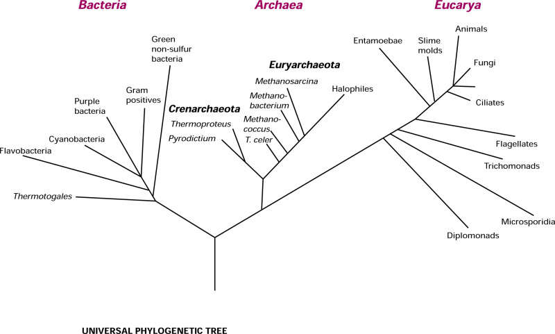
Microbes Tree of Life
Bacteria, Archaea, and Eukarya.
The lifeforms contained within each domain are further subdivided according to kingdom, phylum, class, order, family, genus, and species.
The domain Eukarya, commonly referred to as eukaryotes, is the most recognizable of the three.
Bacteria and Archaea, together the prokaryotes, outnumber the Eukarya.
In the evolutionary history of our planet, prokaryotes were the first and, for a long time, only life forms.
Bacteria
Tiny single-celled organisms
Found everywhere: air, water, soil, and living organisms
Some bacteria are beneficial, aiding digestion and producing vitamins
Others can cause diseases like pneumonia and food poisoning
Used in various industries like food production, medicine, and biotechnology
Archaea
They are single-celled organisms that can survive in extreme environments, such as hot springs and deep-sea vents.
They have a unique cell structure and genetic makeup, making them distinct from bacteria and eukaryotes.
They play important roles in various ecological processes, including nutrient cycling and decomposition.
Eukarya
These include protists, fungi, and some algae.
They are single-celled or multicellular organisms with a true nucleus and membrane-bound organelles.
Examples include amoebas, yeast, and diatoms.
Robert Hooke (1600s)
English scientist who coined the term "cell" to describe the basic unit of life.
He also discovered Hooke's Law, which explains the relationship between the force applied to an object and its resulting deformation.
Studied household objects, plants, and trees
Described cellular structures
Antonie van Leeuwenhoek (1670s)
A pioneer in microbiology who developed the first microscope and observed microorganisms, laying the foundation for the field of microbiology.
First to see bacteria.
Observed “animalcules” scraped from his teeth
Spontaneous Generation Theory
Theory that living organisms can arise from non-living matter, without the need for pre-existing life.
Disproved by Louis Pasteur's experiments, which showed that life only comes from pre-existing life.
Theory of Biogenesis
The theory that states living organisms can only arise from other living organisms through a process called reproduction.
It opposes the theory of spontaneous generation, which suggests that life can arise from non-living matter.
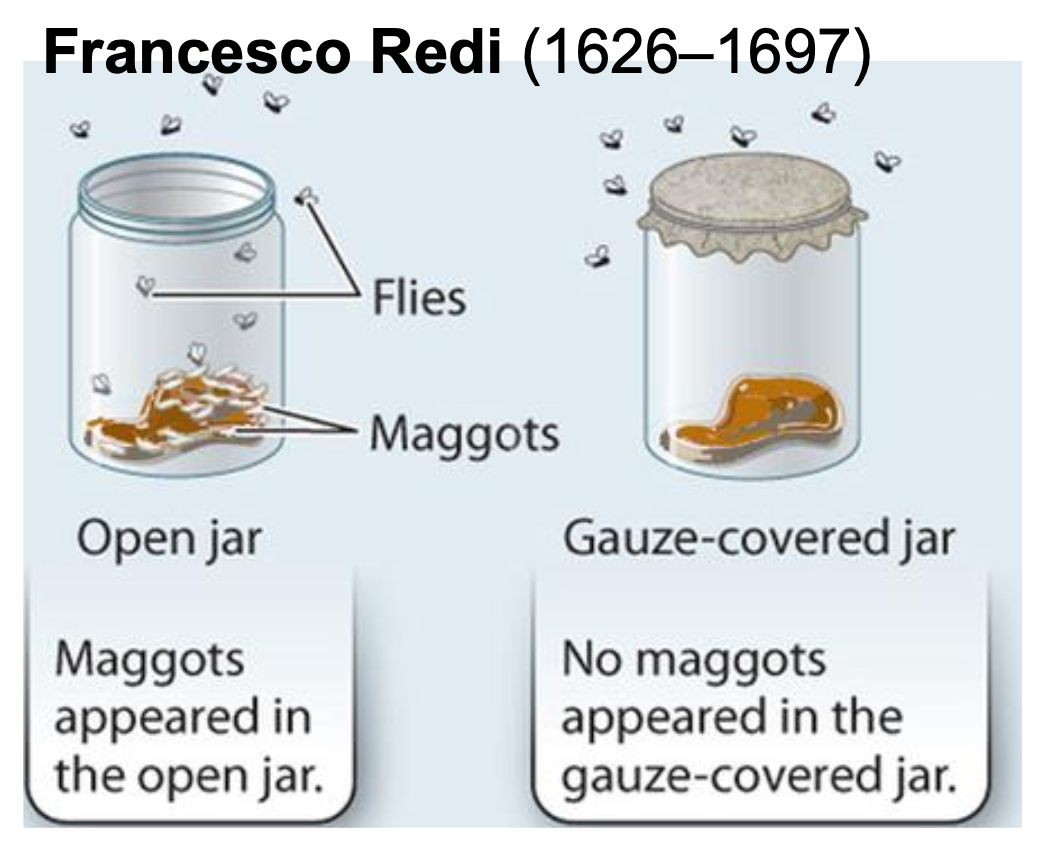
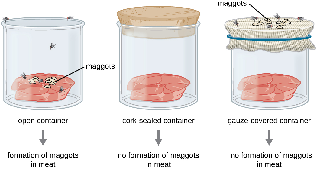
Francesco Redi (1626-1697)
Italian physician and scientist who conducted experiments to disprove spontaneous generation theory.
He demonstrated that maggots do not arise spontaneously from decaying meat, but from fly eggs.
His work laid the foundation for the concept of biogenesis.
Louis Pasteur (1822–1895)
The scientist who disproved spontaneous generation through his experiments with swan-necked flasks.
Showed that microorganisms only arise from preexisting microorganisms.
Showed that biogenesis is responsible for the
propagation of life
Hypothesized that air contained contaminating
microbes investigated his hypothesis by performing an
experiment with a specialized Swan-necked flask
Pasteurization kills off yeast and prevents stored
wine from turning bitter
Developed the first vaccine against anthrax and rabies
Robert Koch (1875)
He identified the bacterium responsible for tuberculosis and discovered the Vibrio cholerae bacterium that causes cholera.
His work laid the foundation for modern bacteriology and earned him the Nobel Prize in Physiology or Medicine in 1905.
German physician and microbiologist who developed Koch's postulates, a set of criteria to establish the causative agent of an infectious disease.
Ignaz Semmelweis (1818–1865)
Pioneering physician who advocated handwashing to reduce the spread of infectious diseases.
Introduced hand hygiene in obstetric clinics, significantly reducing maternal mortality rates.
Known as the "savior of mothers."
Joseph Lister (1827–1912)
Pioneer of antiseptic surgery.
Developed Listerine, a disinfectant. Introduced carbolic acid to sterilize surgical instruments and wounds.
Significantly reduced infection rates and improved surgical outcomes.
Florence Nightingale (1820–1910)
Pioneer of modern nursing.
Known for her work during the Crimean War
She improved sanitation and implemented evidence-based practices.
Binomial Nomenclature System
A system of naming species in biology.
It uses a two-part Latinized name; the first word is the name of the genus, and the second is the species name., to uniquely identify each species.
The genus name is capitalized and italicized, while the species name is lowercase and italicized.
For example, Homo sapiens is the binomial name for humans, with Homo representing the genus and sapiens representing the species.
Carl Linnaeus
Swedish botanist and zoologist who developed a system for classifying and naming organisms called binomial nomenclature.
Symbiotic Relationships b/w Humans & Microbes
Humans have evolved intimate symbiotic relationships with a consortium of gut microbes and individual variations in the microbiome influence host health, may be implicated in disease etiology, and affect drug metabolism, toxicity, and efficacy.
Taxonomic Hierarchy
The classification system used to categorize and organize all living organisms based on their characteristics and evolutionary relationships. It includes the following levels:
Domain, Kingdom, Phylum, Class, Order, Family, Genus, and Species.
Delightful King Philip Came Over For Great Spaghetti
Parasitism
A type of relationship where one organism benefits at the expense of another organism, often causing harm or disease.
Ex. A tiny tick attaches itself to a deer, feeding on its blood and weakening the deer over time.
Mutualism
A type of symbiotic relationship where both organisms benefit from each other's presence and actions.
They rely on each other for survival, such as pollination or cleaning.
Examples include bees and flowers, where bees get nectar and flowers get pollinated.
Commensalism
A type of symbiotic relationship where one organism benefits while the other is neither harmed nor benefited.
Ex. A bird builds its nest on a tall tree branch without causing any harm or benefit to the tree. The bird benefits by gaining a safe place to lay its eggs and raise its young, while the tree remains unaffected by the bird's presence.
Single Microbe
Definition:
A single microorganism, such as a bacterium or a fungus that exists as an individual cell.
Characteristics:
Independent and free-living.
Can move and colonize new areas.
Susceptible to antibiotics and immune responses.
They can replicate and divide to form new cells.
Limited ability to form complex structures.
Biofilm
Definition:
A community of microorganisms that adhere to each other and to surfaces, forming a protective matrix.
Characteristics:
Consists of multiple microbial species.
Attached to surfaces, such as medical devices or natural environments.
Protected by a slimy extracellular matrix.
Enhanced resistance to antibiotics and immune responses.
Can communicate and coordinate behaviour.
They can form complex structures, such as channels and towers.
Types of Media
Broths
Plates
Slants
Deeps
Aseptic Culture Techniques
Pure culture
Aseptic culturing techniques
Biological safety cabinet
Ways To View Microbes
Stains, or dyes, increase contrast
Basic dyes are some of the most commonly used stains
Dye is positively charged
Attracted to the negatively charged cell surface
Acidic dyes are used in negative staining
Dye is negatively charged
Repelled from negatively charged cell surface
Bacterial Staining Techniques
Making a smear of the specimen
Fixing the specimen by exposing it to heat (or chemical reagent)
Staining of the specimen
Simple Stains
Techniques use one dye
Used to determine size, shape, and/or cellular arrangement
Structural Staining (Capsule Staining)
Technique used to stain bacterial capsules.
Crystal violet is applied to the slide, followed by copper sulfate as a counterstain.
Capsules appear as clear halos surrounding stained cells.
Structural Staining (Flagellar Staining)
The staining method used to visualize flagella on bacteria.
Involves applying mordant, then staining with carbolfuchsin.
Flagella appear as thin, hair-like structures under a microscope.
Helps in identifying motile bacteria.
Structural Staining (Bacterial Endospore Staining)
The staining technique used to visualize bacterial endospores.
Involves the use of malachite green dye, heat, and counterstain.
Endospores appear green, while vegetative cells appear red.
Differential Stains
Techniques use more than one dye
Highlights differences in bacterial cell walls in order to discriminate between classes of cells
Most important: Gram stain and acid-fast stain
Gram Staining (Gram Positive and Gram Negative)
Consists of sequential applications of:
Crystal violet (primary stain)
Iodine (mordant)
Acetone-alcohol (decolorizing step)
Safranin (counterstain)
Acid-Fast Staining
Distinguishes between cells with and without waxy cell walls
Acid-fast bacteria
Contain waxy cell walls rich in mycolic acid
Ex. Mycobacterium tuberculosis
Light Microscopy
Uses visible light to illuminate the specimen.
This is the most common type of optical microscope.
Types of Light Microscopy
Bright Field
Dark Field
Phase Contrast
Differential Interference Contrast
Electron Microscopy
Shoots electrons at a specimen
Electrons interact with the specimen and an image is generated
Provides high-magnification and high-resolution images
Two Main Classes of Electron Microscopes
Transmission electron microscopes
Scanning electron microscopes
The Scientific Method
Make an observation.
Ask a question.
Form a hypothesis, or testable explanation.
Make a prediction based on the hypothesis.
Test the prediction.
Iterate: use the results to make new hypotheses or predictions.
Difference Between Prokaryotic Cells and Eukaryotic Cell
Prokaryotes are always unicellular, while eukaryotes are often multi-celled organisms.
Eukaryotic cells are more than 100 to 10,000 times larger than prokaryotic cells and are much more complex.
The DNA in eukaryotes is stored within the nucleus, while DNA is stored in the cytoplasm of prokaryotes.
Organisms that are Prokaryotes
Include two domains, Eubacteria and Archaea.
Examples include bacteria, archaea, and cyanobacteria (blue-green algae).
Importance of Bacteria
They play a crucial role in various ecosystems, like soil and water, by decomposing organic matter and recycling nutrients.
They also help in food production, such as fermenting yogurt and cheese.
Additionally, they contribute to human health by aiding digestion, producing vitamins, and boosting the immune system.
Importance of Eubacteria
Essential for nutrient recycling in ecosystems
Major contributors to the nitrogen cycle
Some species are beneficial to human health (e.g., probiotics)
Used in biotechnology for production of antibiotics and enzymes
Can cause diseases in plants, animals, and humans
Play a crucial role in food production and preservation
Importance of Archaea
Microorganisms that thrive in extreme environments like hot springs and deep-sea hydrothermal vents.
Crucial for maintaining ecological balance and nutrient cycling.
Play a significant role in global carbon and nitrogen cycles.
Help break down organic matter, produce methane, and fix nitrogen.
Contribute to human health by aiding in digestion and preventing harmful bacteria from colonizing the gut.
How Prokaryotic Organisms Reproduce
They reproduce through a process called binary fission. In binary fission.
The DNA of the replicates, and the cell divides into two identical daughter cells.
This allows them to rapidly multiply and increase their population.
Average Size of Prokaryotic Cells
Cells are typically smaller than eukaryotic cells, with an average size ranging from 0.2 to 2.0 micrometers in diameter.
Average Shape of Prokaryotic Cells
The average shape of the cells is typically either spherical (cocci), rod-shaped (bacilli), or spiral (spirilla).
Average Arrangements of Prokaryotic Cells
Cells can be found in various arrangements, but the most common is the single-celled arrangement, where each cell exists independently.
However, they can also form clusters, chains, or pairs depending on the species.
Cell Structures Found in Prokaryotes
Essential cell structures include:
A Cell Membrane
Cytoplasm
Ribosomes
A Nucleoid Region.
Cell Structures Found in Eukaryotes
It has several cell structures, including;
The Nucleus
Mitochondria
Endoplasmic Reticulum
Golgi Apparatus
Lysosomes
Vacuoles.
Functions of Prokaryotes
Perform various functions, including;
Nutrient recycling
Nitrogen fixation
Decomposition
Pathogen control
Crucial in the role of production of antibiotics
Used in biotechnology for genetic engineering and the production of enzymes
Functions of Eukaryotes?
Perform various functions, including;
Cell division
Energy production
Protein synthesis
Cellular communication.
They have specialized organelles like the nucleus, mitochondria, and endoplasmic reticulum, which contribute to these functions.
Mitosis
The process of cell division in which a single cell divides into two identical daughter cells, each having the same number of chromosomes as the parent cell.
Meiosis
A type of cell division that occurs in sexually reproducing organisms.
It involves two rounds of division, resulting in the formation of four haploid cells, each with half the number of chromosomes as the parent cell.
Importance of Bacterial Endospores
They allow certain bacteria to survive harsh conditions such as extreme temperatures, lack of nutrients, and exposure to chemicals or radiation.
Highly resistant structures that protect the bacterial DNA and other essential components.
This ability enables bacteria to remain dormant for extended periods, ensuring their survival until favorable conditions return.
Can be a challenge to eliminate, as they are resistant to many disinfectants and antibiotics.
Cell Wall Structure of Gram-positive Bacteria
Composed of a thick layer of peptidoglycan, which provides strength and rigidity to the cell.
It contains teichoic acids, which help in cell wall maintenance and regulation of ion movement.
The cell wall may have proteins and polysaccharides that contribute to cell adhesion and protection.
Cell Wall Structure of Gram-negative Bacteria
Composed of a thin peptidoglycan layer surrounded by an outer membrane.
The outer membrane contains lipopolysaccharides (LPS), contributing to the bacteria's pathogenicity.
This structure provides resistance to certain antibiotics and makes them more challenging to treat.
Clinical Importance of Gram-negative Bacteria
They are important due to their ability to cause a wide range of infections, including urinary tract infections, pneumonia, bloodstream infections, and gastrointestinal infections.
They possess an outer membrane that provides resistance to antibiotics and toxins, making them challenging to treat.
They can produce endotoxins, such as lipopolysaccharides, which can lead to severe sepsis and septic shock.
Understanding the clinical significance is crucial for effective diagnosis, treatment, and prevention of infections caused by these pathogens.
What is the clinical importance of Gram-positive bacteria?
They are clinically important due to their ability to cause a wide range of infections, including skin and soft tissue infections, respiratory tract infections, bloodstream infections, and urinary tract infections.
They are responsible for serious diseases such as pneumonia, meningitis, and sepsis.
They can produce toxins that contribute to the severity of infections.
They can develop resistance to antibiotics, making them challenging to treat.
Understanding the clinical importance is crucial for effective diagnosis and treatment of infections caused by these pathogens.
The Steps of Gram Staining
There are three general steps involved:
Collecting the sample.
Processing the sample.
Examining the sample.
Gram Staining Gram-Positive
Cell walls contain thick layers of peptidoglycan, a substance that forms the cell walls of many bacteria.
The peptidoglycan forms about 90% of the cell wall.
This causes them to appear blue to purple under a Gram stain.
Gram Staining Gram-Negative
Cell walls with thin layers of peptidoglycan (10% of the cell wall) and high lipid (fatty acid) content.
This causes them to appear red to pink under a Gram stain.
Function of the External Structures of Prokaryotes
Prokaryotes have external structures: cell wall, flagella, pili, and capsule.
The cell wall provides shape, support, and protection.
Flagella aid movement.
Pili helps with attachment and genetic transfer.
Capsules protect against stress and aid attachment.
These structures are essential for prokaryotes to survive, move, attach, and protect in diverse environments.
Basic Types of Membrane Transport
Simple passive diffusion
Facilitated diffusion (by channels and carriers)
Active transport
Simple Passive Diffusion
A process by which molecules move across a cell membrane from an area of higher concentration to an area of lower concentration without energy or a transport protein
Facilitated Diffusion
A passive transport process that allows the movement of specific molecules across the cell membrane with the help of channels and carriers
Active transport
The process by which cells move molecules or ions against their concentration gradient, requiring the expenditure of energy in the form of ATP
ATP (Adenosine Triphosphate)
It serves as the primary energy currency in cells, providing energy for various cellular processes.
Generated through cellular respiration, where glucose is broken down to release energy that is stored in these molecules.
This energy is then utilized by microorganisms for essential functions like metabolism, growth, and reproduction.
It’s involved in active transport, enabling the movement of molecules across cell membranes against concentration gradients.
It plays a crucial role in powering microbial activities and maintaining cellular homeostasis.
Mycoses
Fungal infections that affect humans and animals.
They can be superficial, like athlete's foot, or systemic, affecting internal organs.
Who is at Risk of Mycoses
People with:
Diabetes
Cancer
Organ transplant
Stem cell transplant
Neutropenia (low number of white blood cells)
Long-term corticosteroid use
Injection drug use
Too much iron in the body (iron overload or hemochromatosis)
Skin injury due to surgery, burns, or wounds
Prematurity and low birthweight (for neonatal gastrointestinal mucormycosis)
Are viruses alive?
No, viruses are not considered alive. They lack the characteristics of living organisms, such as the ability to reproduce on their own and carry out metabolic processes. Instead, viruses rely on host cells to replicate and survive. They are essentially genetic material (DNA or RNA) enclosed in a protein coat. While they can cause diseases and have genetic material like living organisms, viruses are classified as non-living entities.
Viruses
Viruses are not considered alive.
Viruses are not made out of cells, they can't keep themselves in a stable state, they don't grow, and they can't make their own energy.
Even though they definitely replicate and adapt to their environment, viruses are more like androids than real living organisms.
Potential Shapes of a Virus
Icosahedral: 20 triangular faces, common in adenoviruses.
Helical: Long, coiled structure, seen in tobacco mosaic virus.
Complex: Irregular shapes with additional structures, like bacteriophages.
Enveloped: Surrounded by a lipid membrane, e.g., influenza virus.
Filamentous: Long and flexible, found in Ebola virus.
Spherical: Round shape, seen in many types of viruses.
Polyhedral: Multiple flat faces, found in herpesviruses.
Bullet-shaped: Tapered ends, seen in rabies virus.
Spindle-shaped: Elongated with tapered ends, e.g., measles virus.
Toroidal: Doughnut-shaped, observed in some bacteriophages.
Potential Components of a Virus
Genetic Material: Contains the virus's genetic instructions.
Capsid: Protein coat that protects the genetic material.
Envelope (optional): Lipid layer surrounding the capsid.
Spike Proteins: Help the virus attach to host cells.
Enzymes (optional): Assist in viral replication or evasion of host defenses.
Matrix Proteins (optional): Maintain the structure of the virus.
Viral Proteins: Perform various functions during infection.
Lipid Membrane (optional): Present in enveloped viruses, derived from the host cell.
Glycoproteins (optional): Assist in viral attachment and entry into host cells.
How a Viral Envelope is Acquired by some Viruses
Some viruses acquire a viral envelope by budding through the host cell's plasma membrane or an intracellular membrane.
This process involves the viral proteins being inserted into the host cell membrane, which then surrounds the viral nucleocapsid, forming the envelope.
The envelope is derived from the host cell's membrane and contains viral glycoproteins that aid in viral attachment and entry into new host cells.
Examples of viruses with envelopes include influenza, HIV, and herpesviruses.
Function of Spikes on a Virus
Promote attachment and entry into host cells by binding to specific receptors on the cell surface.
Antigenic Drift
Gradual genetic changes in viruses result in minor variations in surface proteins.
This leads to reduced effectiveness of vaccines over time and the need for regular updates.
Antigenic Shift
A sudden change in the surface proteins of a virus, resulting in a new strain.
It occurs when two or more different strains of a virus infect the same host and exchange genetic material.
This can lead to the emergence of a new viral subtype, often causing more severe disease and making existing vaccines less effective.
Host Range
The organisms a microbe can infect.
It determines the susceptibility of different species or cell types.
Microbes vary, with some infecting closely related species (narrow range) and others infecting a wide variety of hosts (broad range).
Tropism
Refers to the directed movement or growth of microorganisms in response to a specific stimulus, such as light, gravity, or chemicals.
It allows microorganisms to adapt and optimize their survival and reproduction by moving towards or away from the stimulus.
Steps of Multiplication in a Bacteriophage
Attachment
Penetration
Replication
Assembly
Maturation
Lysis
Each step is essential for the successful replication and spread of these viral entities.
Attachment - Steps of Multiplication in a Bacteriophage
The first step in the multiplication process is the attachment of the bacteriophage to the surface of the host bacterium.
The bacteriophage recognizes specific receptors on the bacterial cell surface and binds to them.
This attachment is crucial for the subsequent steps of the multiplication cycle.
Penetration - Steps of Multiplication in a Bacteriophage
Once attached, the bacteriophage injects its genetic material, which is typically composed of DNA, into the host bacterium.
This process involves the contraction of the bacteriophage's tail sheath, allowing the viral DNA to pass through the bacterial cell wall and membrane.
Replication - Steps of Multiplication in a Bacteriophage
After the viral DNA is inside the bacterium, it takes over the host's cellular machinery to replicate its genetic material.
The viral DNA serves as a template for the synthesis of new viral components, including more copies of the viral DNA itself.
Assembly - Steps of Multiplication in a Bacteriophage
As the viral components are being synthesized, they start to come together to form new bacteriophages.
This assembly process involves the packaging of the replicated viral DNA into the newly formed viral capsids, which are protein shells that protect the genetic material.
Maturation - Steps of Multiplication in a Bacteriophage
Once the assembly is complete, the newly formed bacteriophages undergo maturation.
During this stage, the viral components mature and become fully functional, acquiring the ability to infect other bacterial cells.
Lysis - Steps of Multiplication in a Bacteriophage
The final step of multiplication, which refers to the bursting or disintegration of the host bacterium.
This process is triggered by the release of enzymes from the bacteriophage that degrade the bacterial cell wall, causing it to rupture.
As a result, the newly formed bacteriophages are released into the surrounding environment, ready to infect new host cells and continue the multiplication cycle.
Steps of Multiplication of an Animal Virus
Attachment
Penetration
Uncoating
Replication
Assembly
Release
By studying these processes, scientists can develop targeted antiviral therapies and preventive measures to combat viral diseases.
Attachment - Steps of Multiplication of an Animal Virus
In this initial step, the animal virus attaches itself to the surface of a host cell.
The virus recognizes specific molecules on the cell's surface, which act as receptors.
These receptors are like keys that fit into the virus's proteins, allowing it to bind to the cell and establish a connection.
Penetration - Steps of Multiplication of an Animal Virus
Once attached, the animal virus finds a way to enter the host cell.
There are two main mechanisms involved in this process. Some viruses enter the cell by fusing their envelope with the cell membrane, essentially merging with the host cell.
Others are engulfed by the cell through a process called endocytosis, where the cell membrane forms a vesicle around the virus, bringing it inside.
Uncoating - Steps of Multiplication of an Animal Virus
After entering the host cell, the animal virus sheds its protective protein coat.
This step is crucial for the virus to release its genetic material, which is essential for replication.
The process can occur in different ways, depending on the specific virus.
Some viruses use enzymes to break down the protein coat, while others rely on the cell's own mechanisms to remove it.
Replication - Steps of Multiplication of an Animal Virus
Once the viral genetic material is exposed, it hijacks the host cell's machinery to replicate itself.
The animal virus utilizes the cell's resources, including enzymes, ribosomes, and other cellular components, to produce multiple copies of its genetic material and viral proteins.
These replicated components will be used to assemble new virus particles.
Assembly - Steps of Multiplication of an Animal Virus
In this step, the newly synthesized viral genetic material and proteins come together to form complete virus particles.
The assembly process varies among different animal viruses.
Some viruses self-assemble, where the components spontaneously come together to form new virus particles.
Others require additional viral proteins or enzymes to facilitate the assembly process.