H.A. Integumentary System Terms
1/32
There's no tags or description
Looks like no tags are added yet.
Name | Mastery | Learn | Test | Matching | Spaced | Call with Kai |
|---|
No analytics yet
Send a link to your students to track their progress
33 Terms
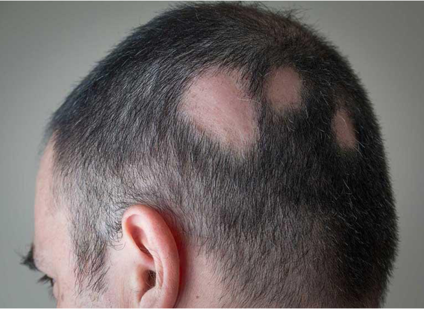
Alopecia
Baldness; hair loss
Annular
Circular shape to skin lesions
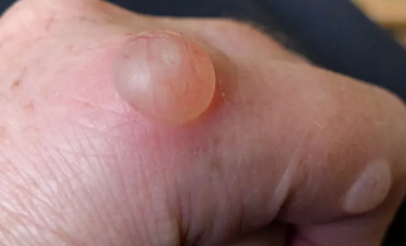
Bulla
Elevated cavity containing free fluid larger than 1 cm in diameter.
Confluent
Ski lesions that run together
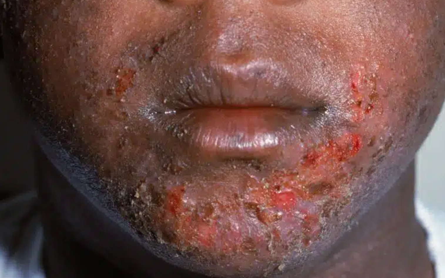
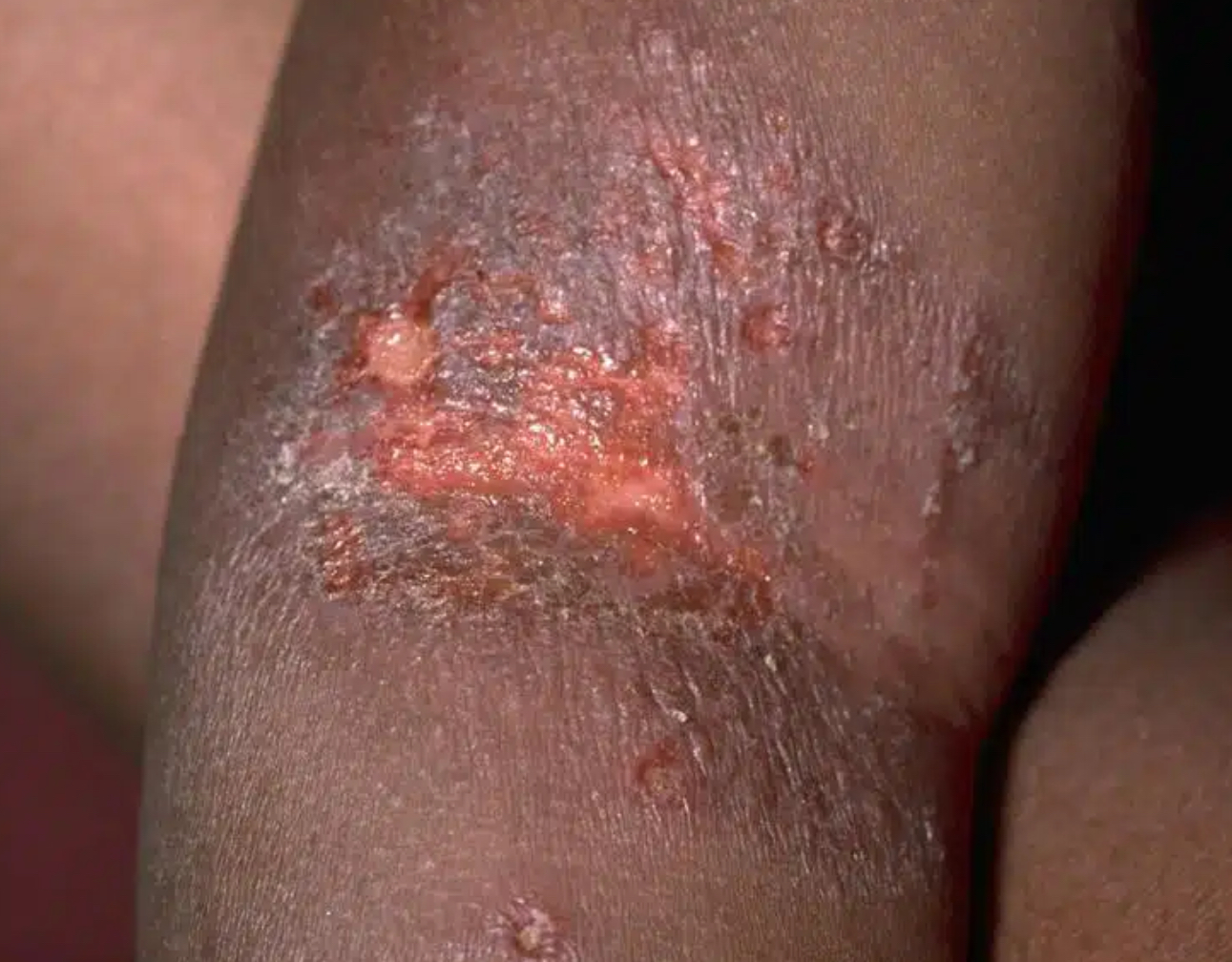
Crust
Thick, dried-out exudate left on the skin when vesicles or pustules burst or dry up.
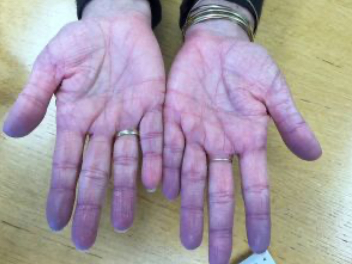
Cyanosis
Dusky blue color to skin or mucous membranes, as a result of increased amount of non oxygenated hemoglobin.
Erosion
Scooped-out, shallow depression in the skin.
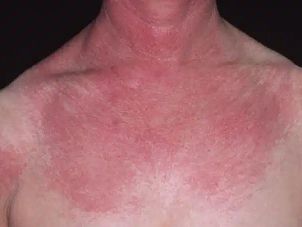
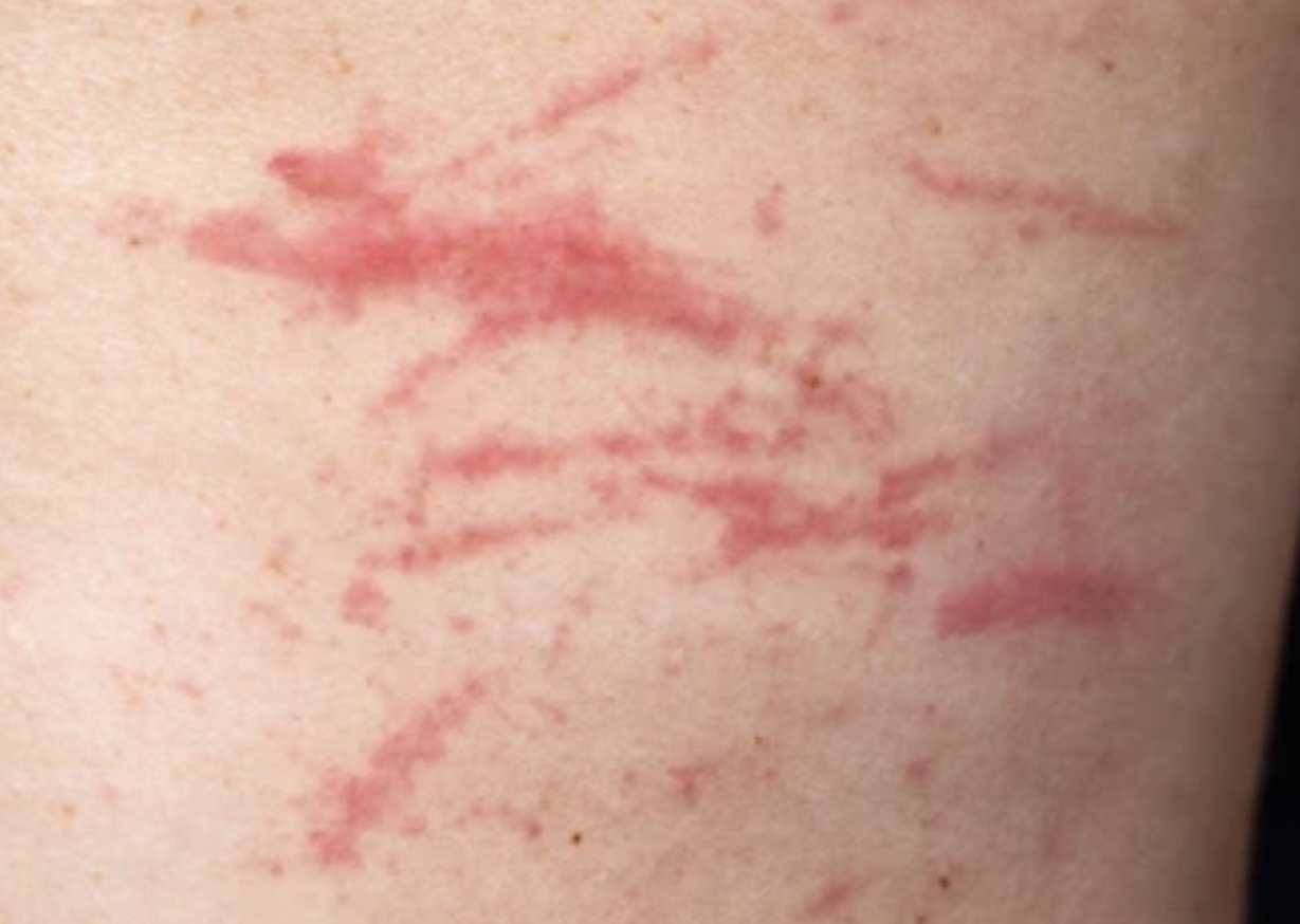
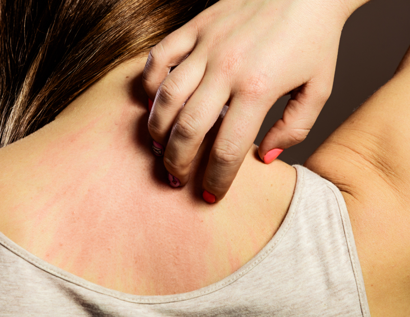
Erythema
Intense redness of the skin due to excess blood in dilated superficial capillaries, as in fever or inflammation.
Excoriation
Self-inflicted abrasion on the skin due to scratching.
Fissure
Linear crack in the skin extending to the dermis.
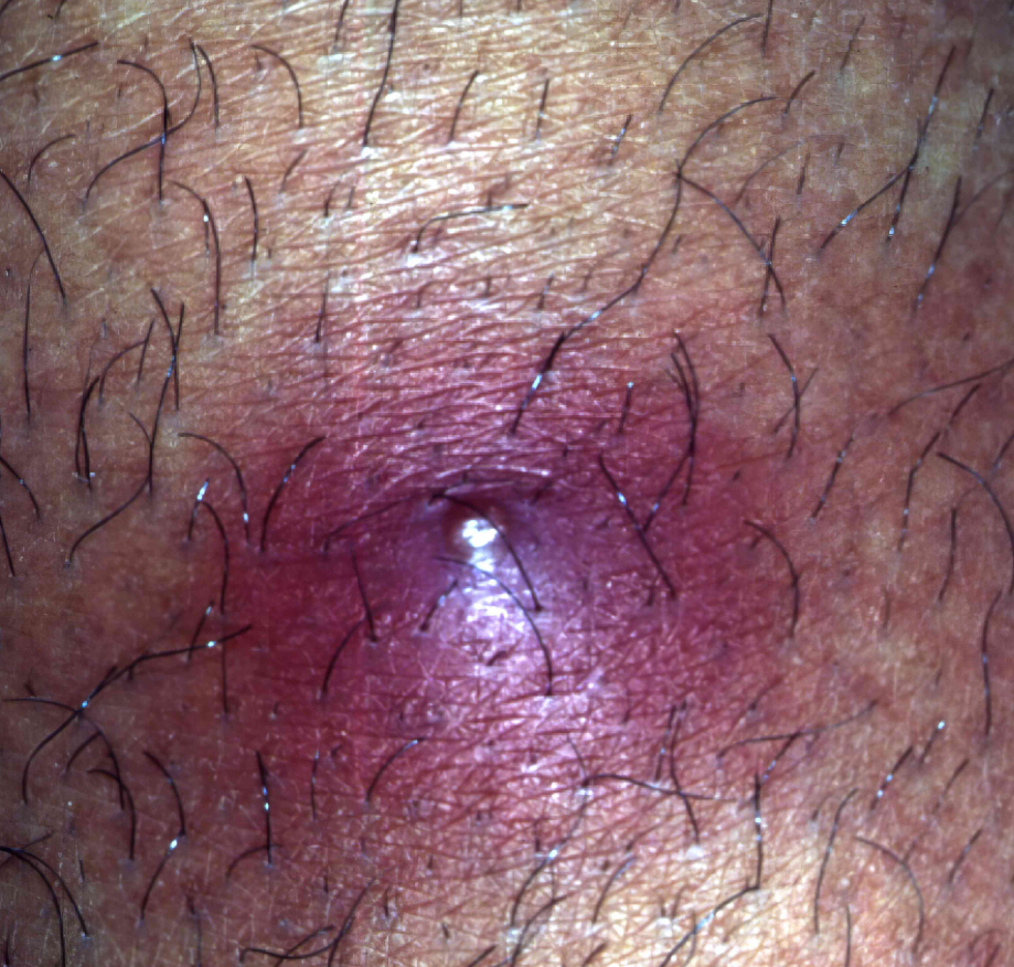
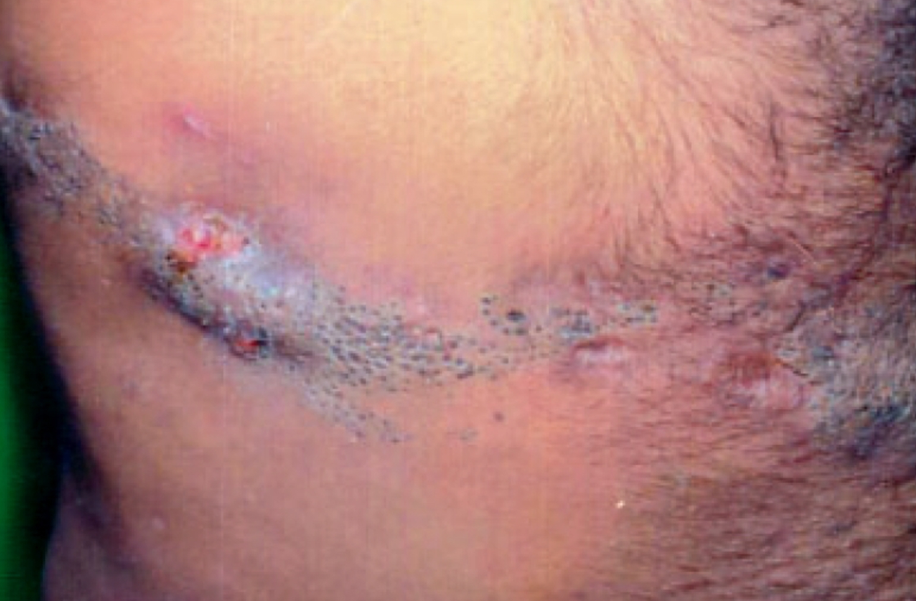
Furuncle
Boil; suppurative inflammatory skin lesion due to infected hair follicles.
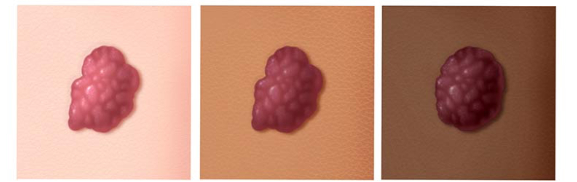
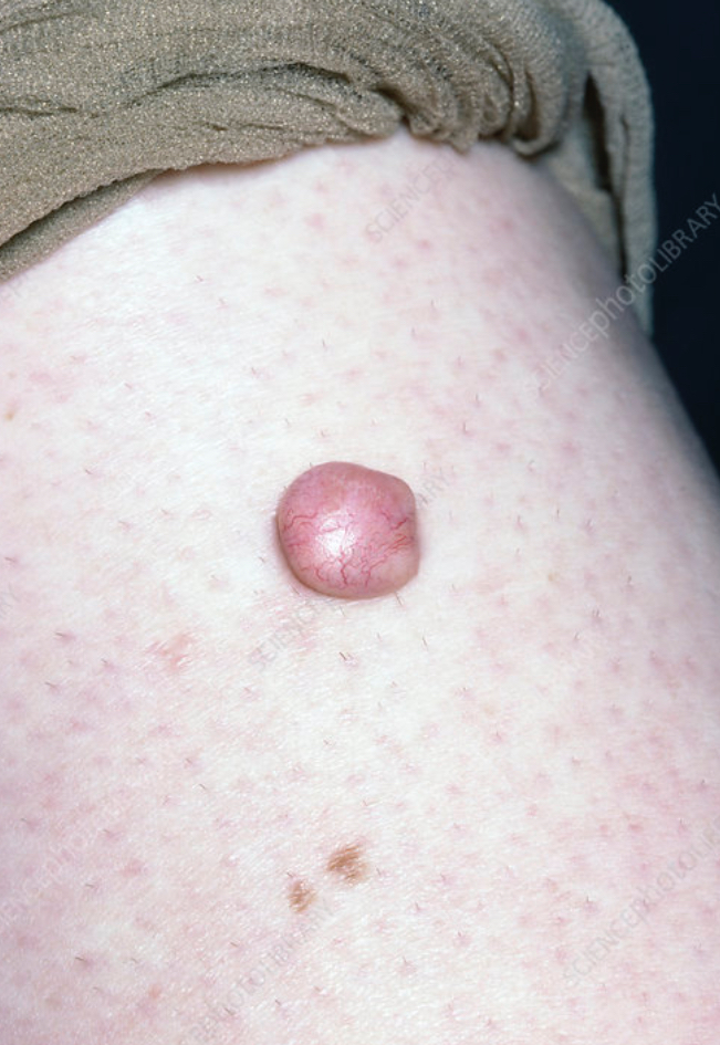
Hemangioma
Skin lesion due to benign proliferation of blood vessels in the dermis.
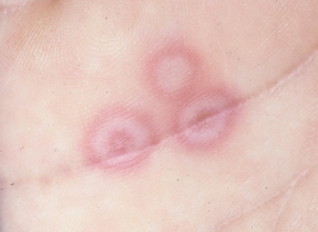
Iris
Target shape of skin lesion.
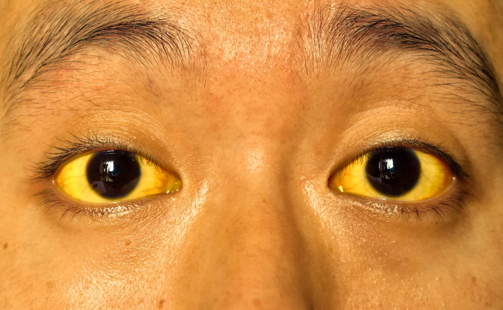
Jaundice
Yellow color to skin, palate, and sclera due to excess bilirubin in the blood.
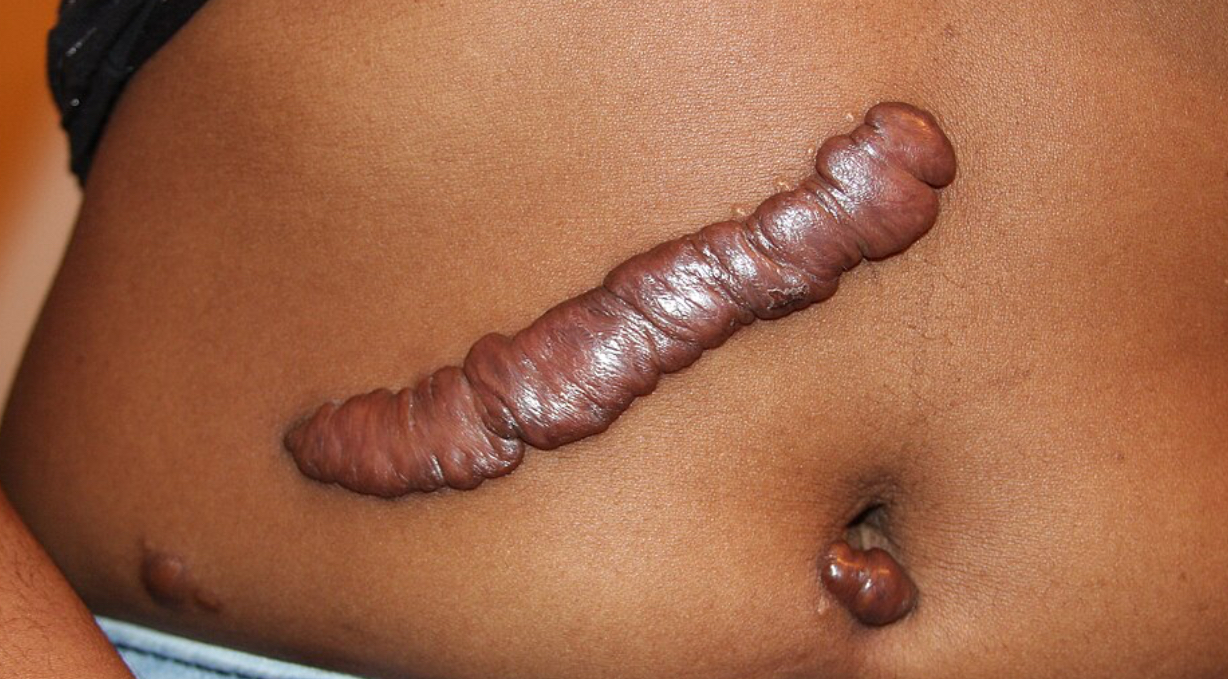
Keloid
Hypertrophic scars, elevated beyond the site of original injury.
Lichenification
Tightly packed set of papules that thickens skin; caused by prolonged, intense scratching.
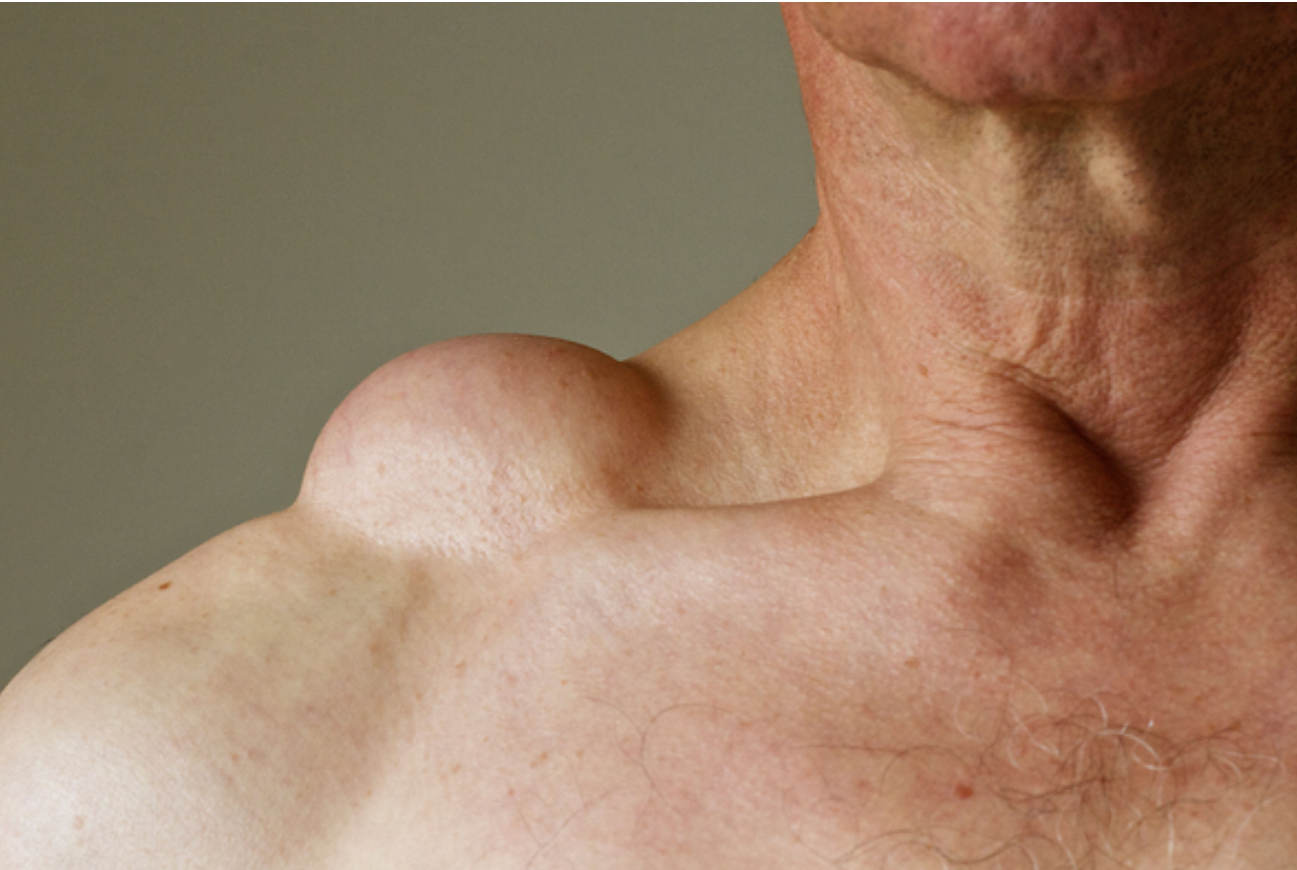
Lipoma
Benign fatty, tumor, composed of mature fat cells.
Maceration
Softening of tissue by soaking in liquid.
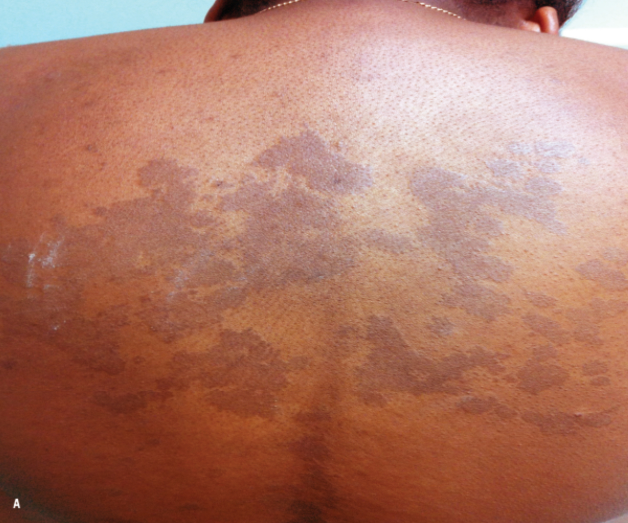
Macule
Flat skin lesion with only a color change.
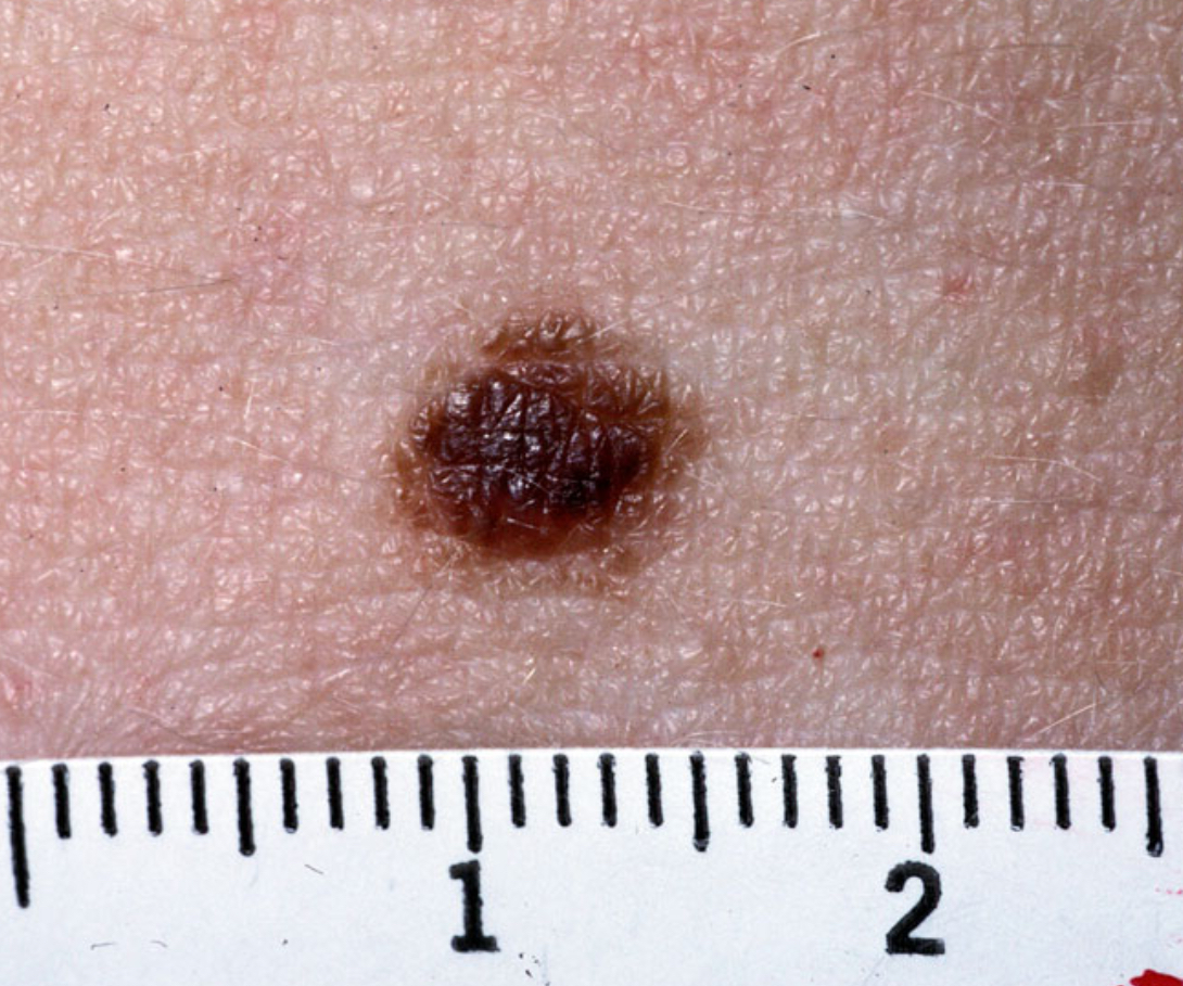
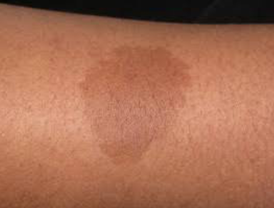
Nevus
Mole; circumscribed skin lesion due to excess melanocytes.
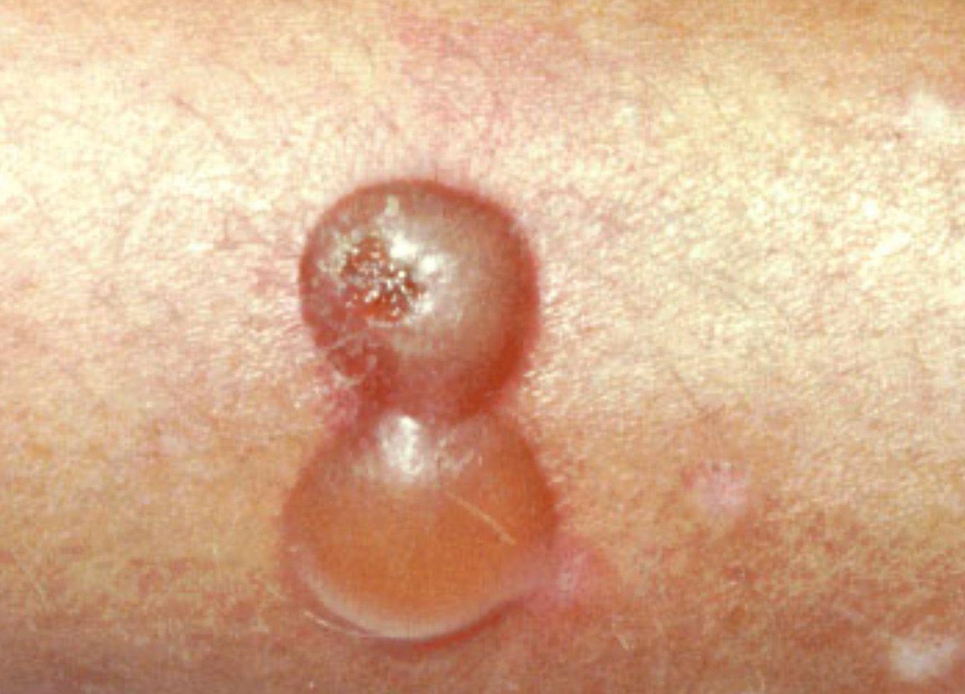
Nodule
Elevated skin lesion larger than 1 cm in diameter.
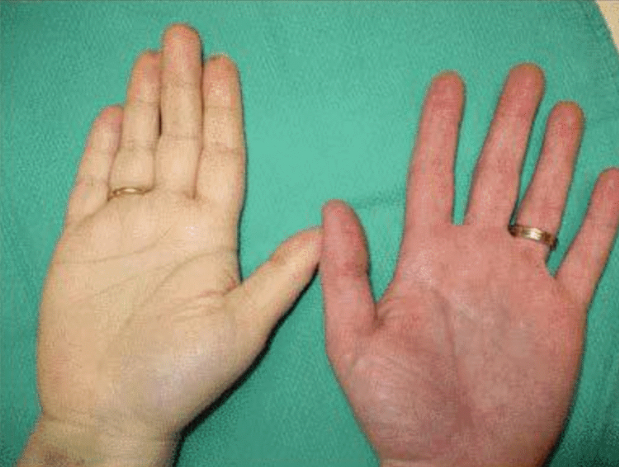
Pallor
Excessively pale, whitish-pink color to lightly pigmented skin.
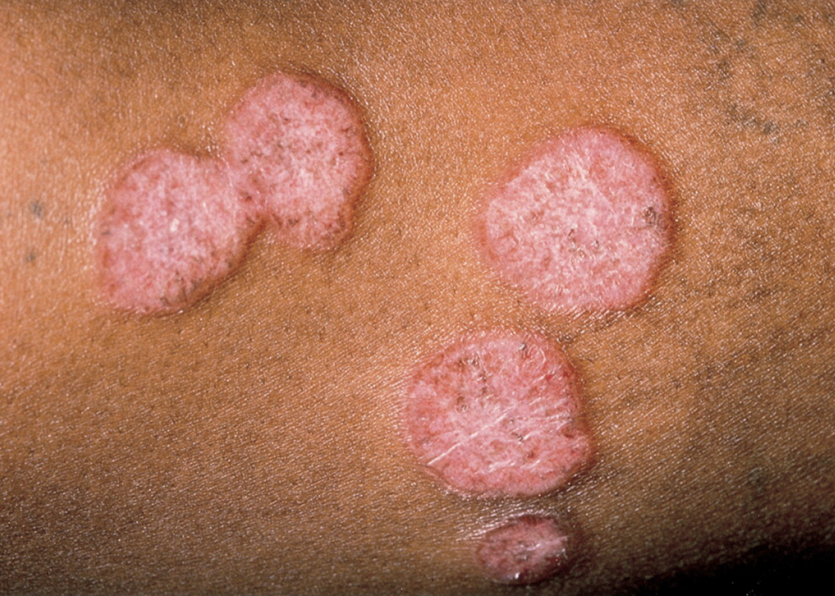
Papule
Palpable skin lesion smaller then 1 cm in diameter.
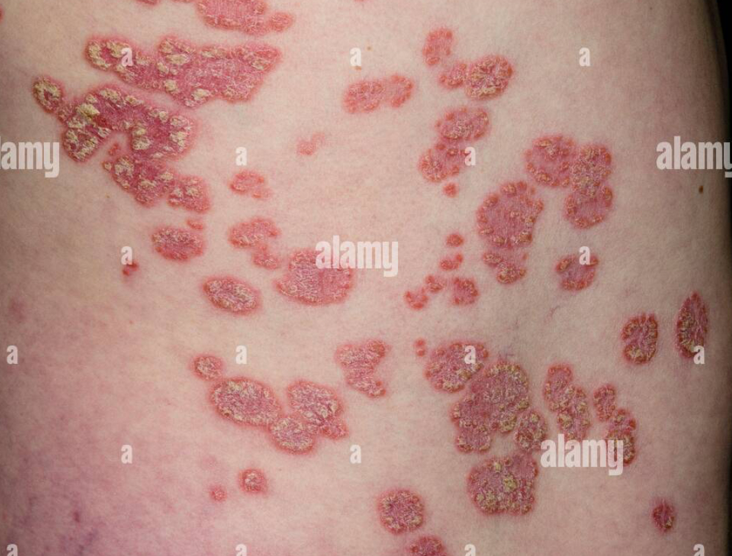
Plaque
Skin lesions in which papules coalesce or come together.
Pruritus
Itching
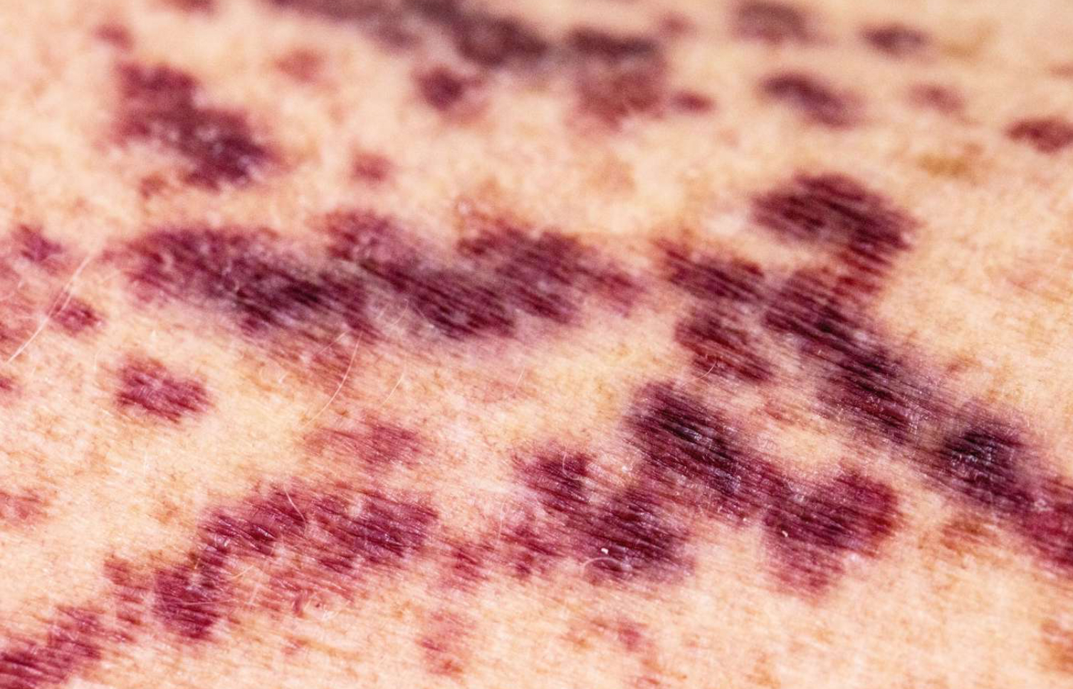
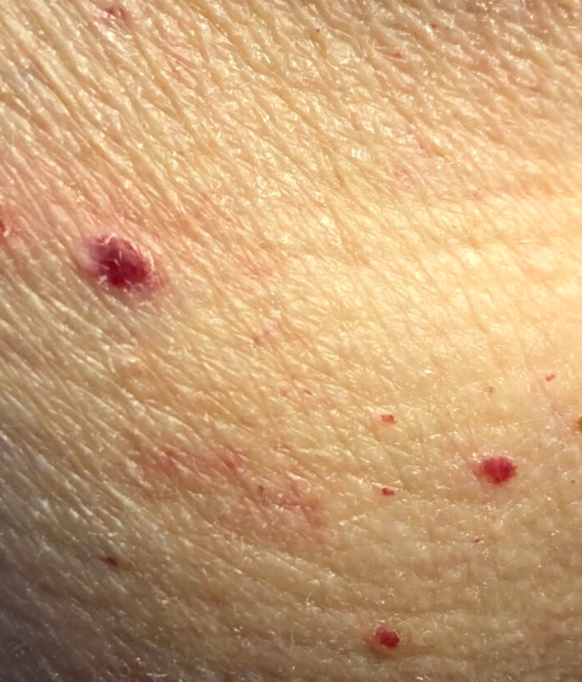
Purpura
Red-purple skin lesion due to blood in tissues from breaks in blood vessels.
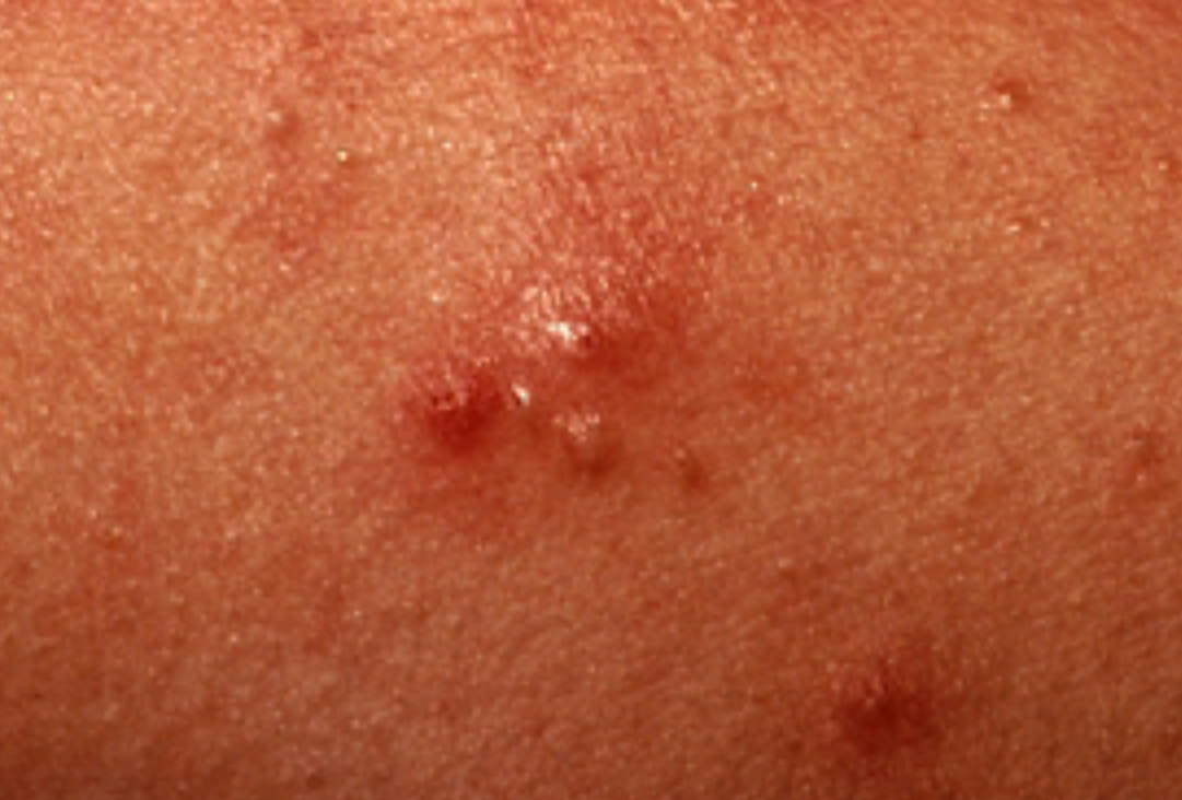
Pustule
Elevated cavity containing thick, turbid fluid.
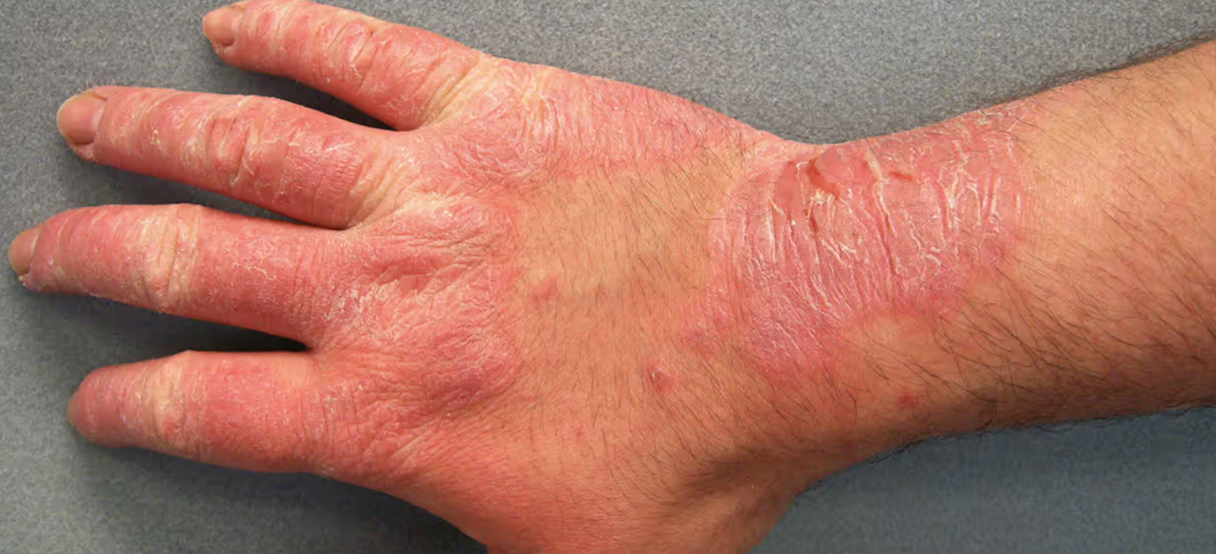
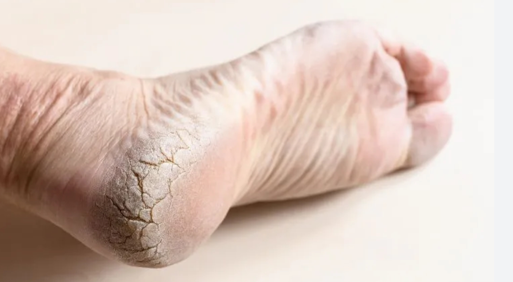
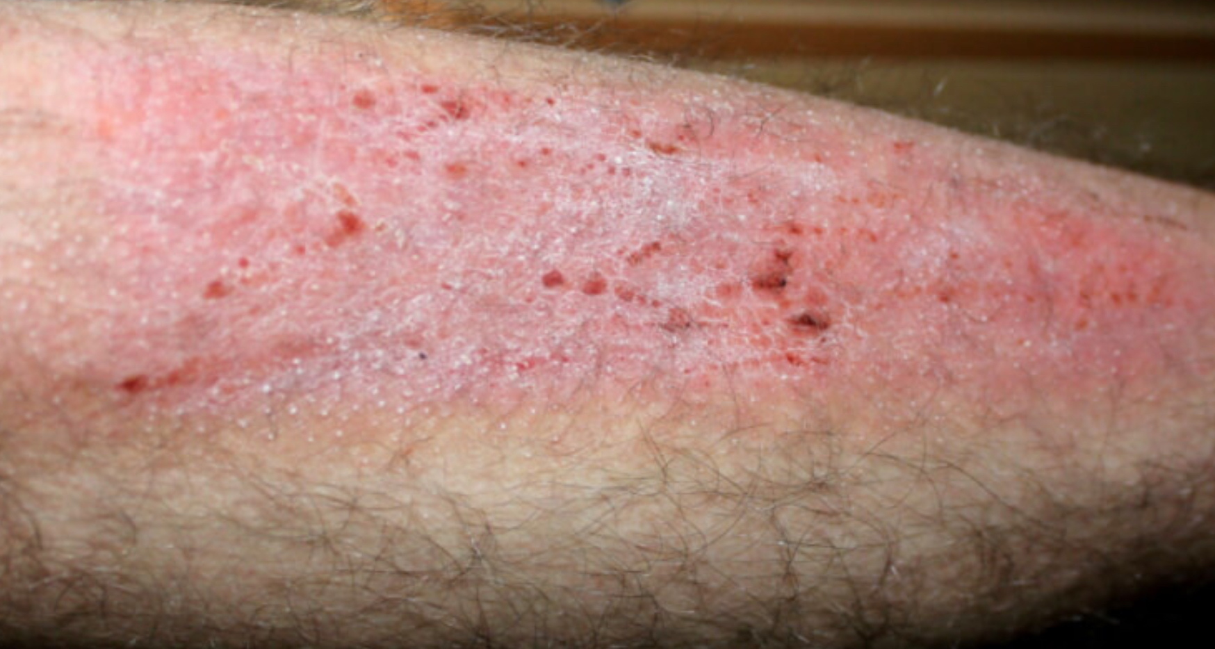
Scale
Compact desiccated flakes of skin from shedding of dead skin cells.
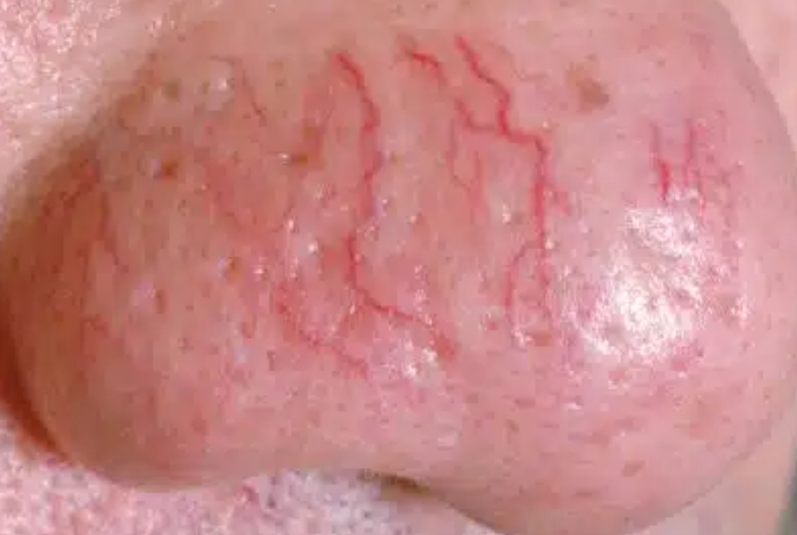
Telangiectasia
Skin lesion due to permanently enlarged and dilated blood vessels that are visible.
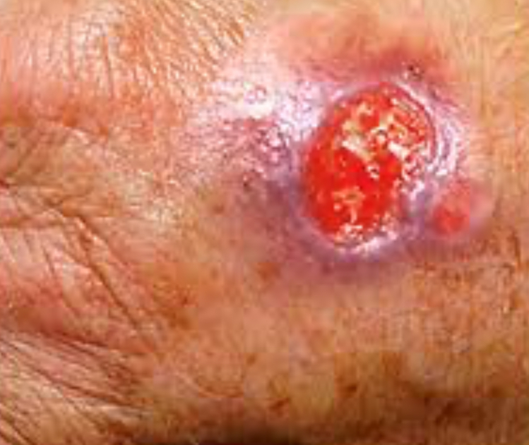
Ulcer
Sloughing of necrotic inflammatory tissue that causes a deep depression in skin, extending into dermis.
Vesicle
Elevated cavity containing free fluid up to a cm in diameter.
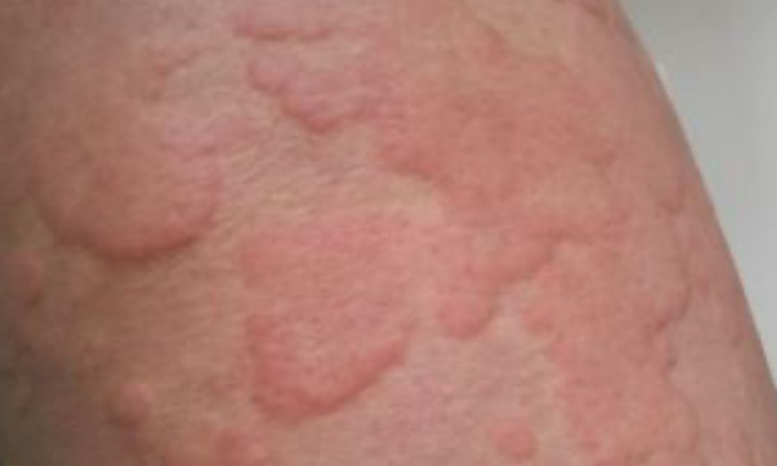
Wheal
Raised red skin lesion due to interstitial fluid.
Zosteriform
Linear shape of skin lesion along a nerve route.