HP LEC 7: The Peripheral Nervous System
1/28
There's no tags or description
Looks like no tags are added yet.
Name | Mastery | Learn | Test | Matching | Spaced | Call with Kai |
|---|
No analytics yet
Send a link to your students to track their progress
29 Terms
PNS is composed of:
Nerves: Cranial and Spinal Nerves
Ganglia
Nerves
bundles of axons in the PNS are referred to as nerves, these structures in the periphery are different than the central counterpart called a tract
Cranial Nerves
Spinal Nerves
Ganglia
group of neuron cell bodies in the periphery
can be categorized: 1) Sensory Ganglia or 2) autonomic ganglia referring to their primary functions
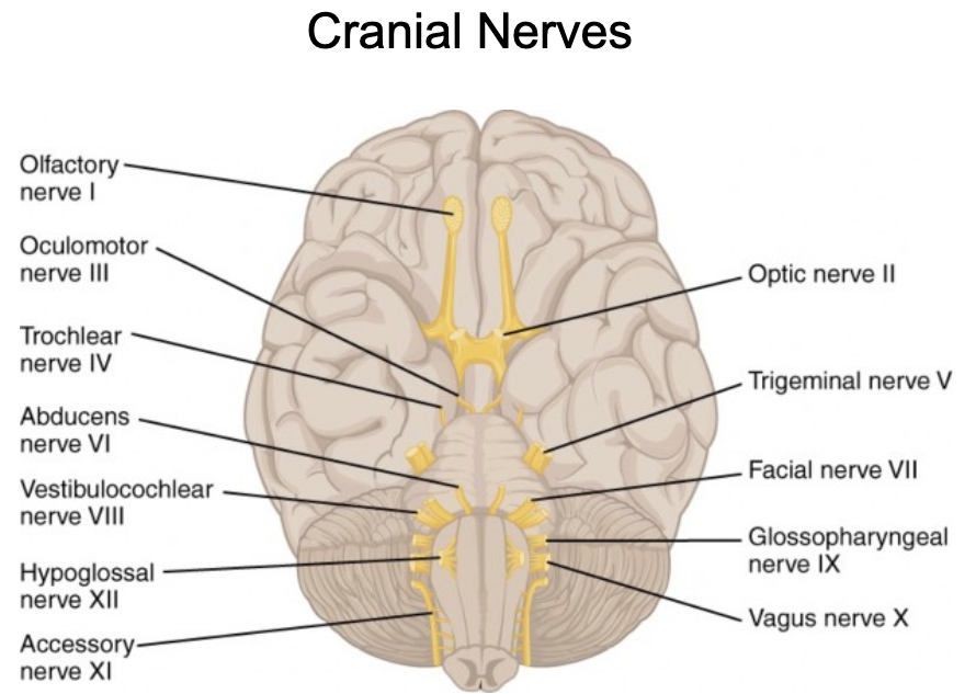
Cranial Nerves
Nerves that arise directly from the nuclei in the brain
12 pairs of Cranial Nerves
most are mixed nerves: with both sensory and motor fibers (ex: vagus nerve)
nerves associated with vision, olfactory, and hearing are sensory only
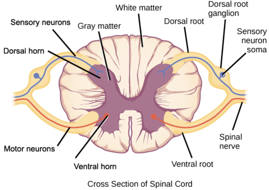
Spinal Nerves
nerves that airse directly from the spinal cord
31 Pairs: 8 Cervical, 12 thoracic, 5 lumbar, 5 sacral, 1 coccygeal
mixed nerves (can carry both motor and sensory info)
How do spinal nerves enter and motor fibers exit in the PNS?
sensory fibers enter the cord via the posterior/dorsal root, and the motor fibers exit by way of the anterior/ventral root
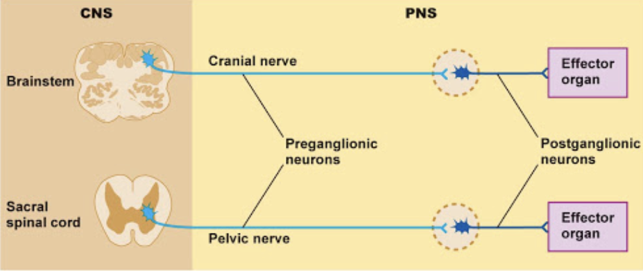
Ganglia
group of neuron cell bodies in the periphery
work as a relay station for nerve signals: one nerve enters (preganglionic) and another nerve exists (postganglionic) from each ganglion
Pre and Post ganglionic Neurons- where are the cell bodies of their neurons?
the cell bodies of the preganglionic neurons are in the brainstem or spinal cord of the CNS
the cell bodies of the postganglionic neurons are in the ganglia
Different Parts of the PNS
Somatic Nervous System
Autonomic Nervous System
Somatic Nervous System (function, what kind of nerves does it include?)
serves the skin, skeletal muscles, and tendons
sensory & motor nerves that innervate the limbs & body
includes nerves that take sensory information from external sensory receptors to the CNS and motor commands away from the CNS to the skeletal muscles
sensory nerve fibers in the peripheral nerves are the peripheral axonal process of neurons in the dorsal root ganglion
Autonomic Nervous System
innervates all effector organs and tissues except for skeletal muscles
it is autonomic bc it functions subconsciously and involuntarily
Autonomic Nervous System Regulates:
activities of glands
smooth muscle function
function of heart & circulatory system
function of digestive system
Visceral organs innervated by the autonomic nervous system
unlike somatic motor neurons (always simulatory), autonomic motor neurons can stimulate OR inhibit
Autonomic Nervous System Responses
Sympathetic: fight or flight
Parasympathetic: rest & digest
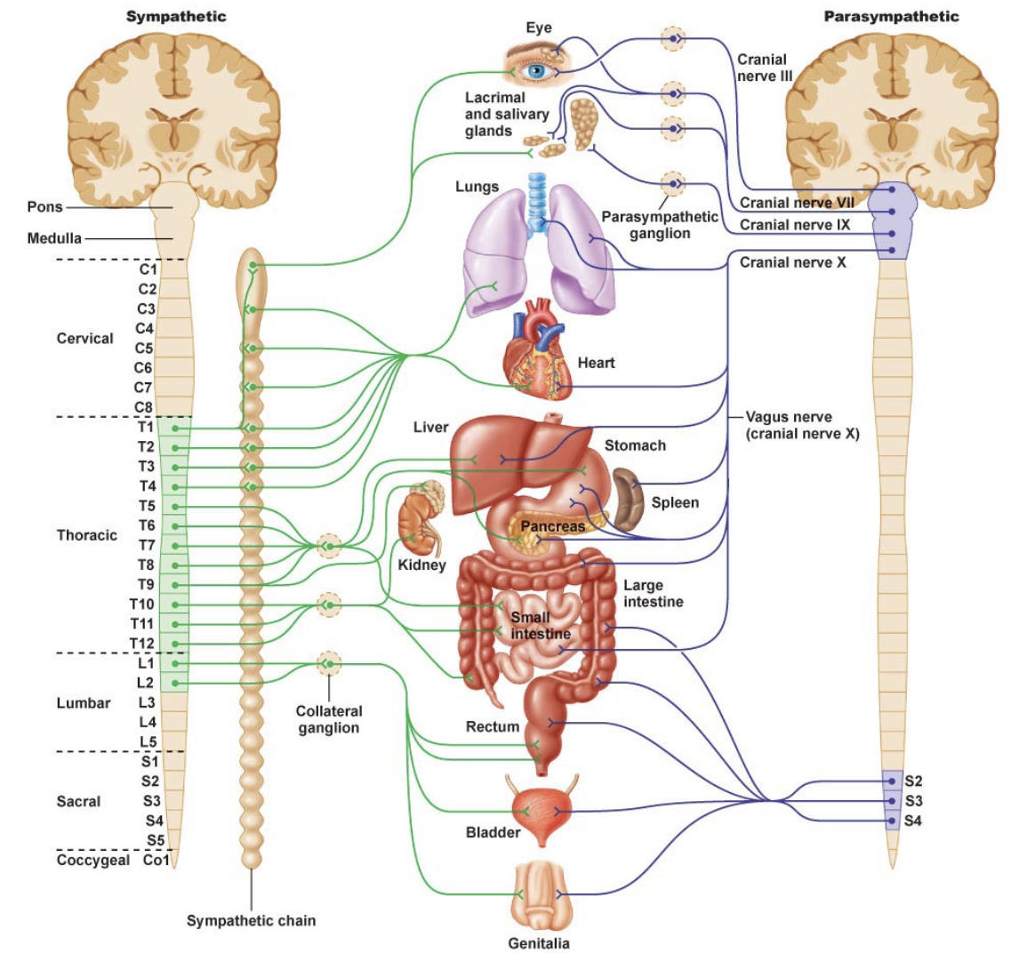
Sympathetic
fight or flight
involved in responses that would be associated with flighting or fleeing, such as increasing heart rate and blood pressure as well as constricting blood vessels in the skin & dilating them in muscles
the sympathetic division is most active during times of excitement and physcial activity
Parasympathetic
rest and digest
involved in energy conservation functions and increases gastrointestinal motility and secretion
increases bladder contractility
parasympathetic division is most active during rest and stimulates digestive activities
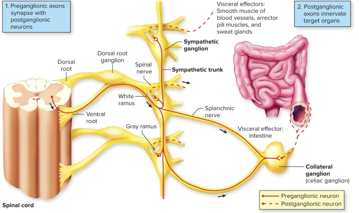
Sympathetic Divison: 3 steps
the axons of the preganglionic neurons come from the thoracic and lumbar regions of the spinal cord
Preganglionic neurons synapse in sympathetic ganglia that run parallel to the spinal cord (not all tho)
called the paravertebral ganglia
paravertebral ganglia are connected forming a sympathetic chain of ganglia
Many of the sympathetic neurons that exit the spinal cord below the diaphragm do NOT synapse in the sympathetic chain of ganglia
Collateral Ganglia: includes celiac, superior mesenteric, and inferior mesenteric ganglia
Sympathetic Division: chain of ganglia- function
it allows nerve fibers to travel to spinal nerve that are superior & inferior to the one in which they originated
Sympathetic Division: Convergence & Divergence
bc preganglionic neurons can branch & synpase in ganglia at any level there is:
Divergence: one preganglionic neuron synapses on several postganglionic neurons at different levels
Convergence: Several preganglionic neurons at different levels synpase on one postganglionic neuron
allows the sympathetic division to act as a SINGLE unit through mass activation & to be tonically active
Parasympathetic Division
preganlionic neurons come from the brain (brainstem) and sacral region of the spinal cord
also called the craniosacral division, some preganglionic neurons synapse on small terminal ganglia or intramural ganglia (interamural- live near or within the organs they innervate)
exception are the 4 parasympathetic ganglia of the head & neck
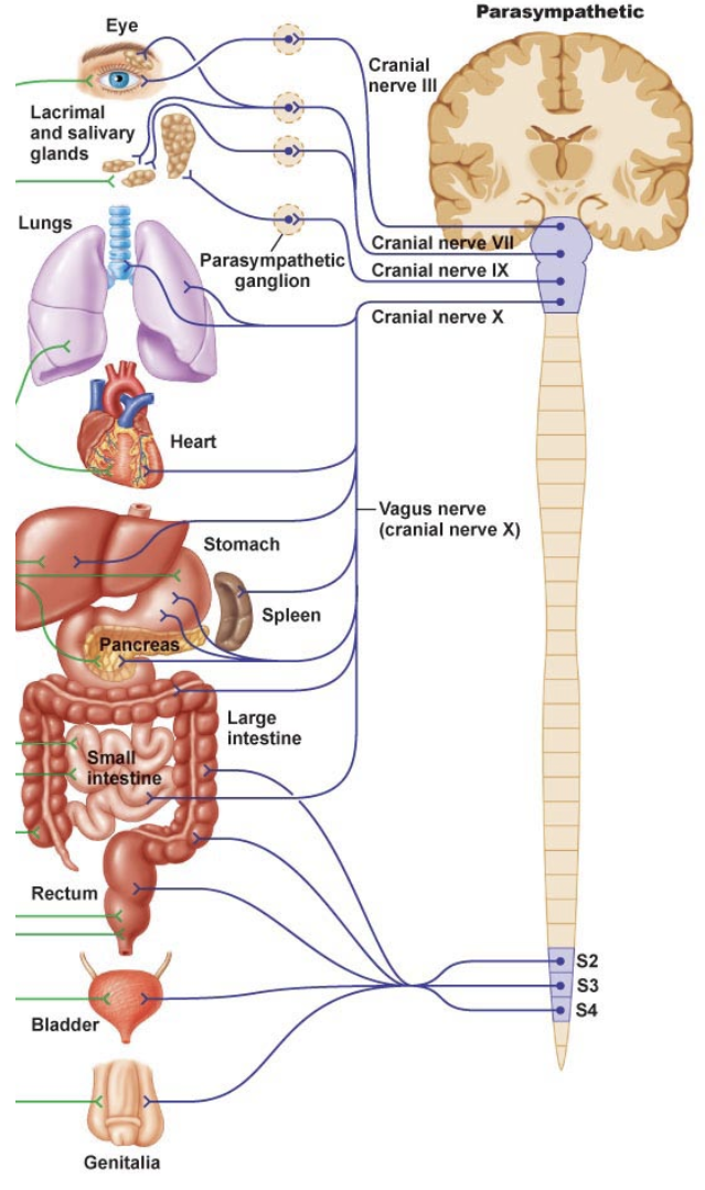
Parasympathetic Divison: Vagus Nerve
vagus nerve (X) represents the main component of the parasympathetic nervous system: oversees a vast array of crucial bodily functions including:
control of mood
immune response
digestion
heart rate
establishes one of the connections between the brain & the gastrointestinal tract & sends the information about the state of the inner organs to the brain via afferent sensory fibers
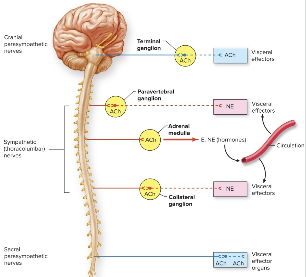
ANS: Cholinergic Synaptic Transmission
a) acetylcholine (ACh) is the neurotransmitter used by all preganglionic neurons (sympathetic & parasympathetic
b) always excitatory- nicotinic receptors
c) it is the neurotransmitter released from most parasympathetic post ganglionic neurons
d) can be excitatory or inhibitory- muscarinic receptors
Adrenergic Synaptic Transmission
a) norepinephrine is the neurotransmitter released by most sympathetic postganglionic neurons
b) can be excitatory or inhibitory- nonadrenergic alpha or beta receptors
Other Autonomic Nuerotransmitters
Some postganglionic autonomic neurons do not release ACh or norepinephrine:
called: “nonadrenergic, noncholinergic fibers”
proposed neurotransmitters include ATP, vasoactive intestinal peptide (VIP), and nitric oxide (NO)
Organs with Dual Innervation
Most visceral organs are innervated by both sympathetic & parasympathetic neurons
The activity of the two divisions of the autonomic system can be:
antagonistic
complementary
cooperative
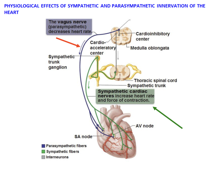
Antagonistic action of sympathetic & parasympathetic autonomic system
occurs when both divisions produce OPPOSITE effects on the same target
Ex:
Heart rate: sym increases, para decreases
Digestive functions: sym decreases, para increases
Pupil diameter: sym dilates, para contricts
Complementary action of sympathetic & parasympathetic autonomic system
occur when both divisions produce similar effects on the same target
Ex:
Salivary Gland Secretion: parasympathetic division stimulates secretion of saliva; sympathetic constricts blood vessels so the secretion is thicker
Cooperative Action action of sympathetic & parasympathetic autonomic system
occur when both divisions produce different effects that work together to promote a single action
Ex:
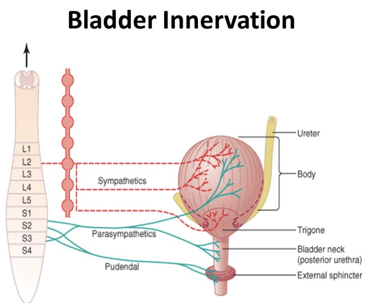
Urination: parasympathetic division aids in urinary bladder contraction; sympathetic helps bladder muscle tone to control urination reflex (triggered when the bladder fills with urine)
Organs without Dual Innervation
Organs that are innervated by sympathetic division ONLY:
Adrenal medulla
Arrector pili muscles in skin
Sweat glands in skin
Most blood vessels
regulated by increases & decrease in sympathetic nerve activity
important for body temperature regulation through blood vessels & sweat glands
Sensory Physiology (sensory system, modality)
a sensory system is a part of the nervous system responsible for processing sensory information, it consists of:
sensory receptors
neural pathways
parts of the brain involved in sensory perception
In Each sensory modality:
a specific type of stimulus energy is transformed into electrical signals by specialized receptors
the sensory information is transmitted to the central nervous system by trains of action potentials (along the neural pathway) that represents aspects of the stimulus