Cardiac Conduction Study Set
1/11
There's no tags or description
Looks like no tags are added yet.
Name | Mastery | Learn | Test | Matching | Spaced | Call with Kai |
|---|

12 Terms
Sinoatrial (SA) node
A specialized area of cardiac tissue, located in the right atrium of the heart, which starts the electrical impulses that determines the heart rate & rhythm.
Atrioventricular (AV) node
A small clump of specialized cardiac muscle fibers found between the wall of the right atrium and right ventricle that receives heartbeat impulses from the sinoatrial node and sends them on to the HIS bundle.
Left & Right Bundle Branches
Fibers that conduct the cardiac impulse down through the septum towards the apex of the heart.
Purkinje Fibers
Conductive fibers that wrap around the outer walls of the ventricles and carry the cardiac impulse around the myocardium of the heart.
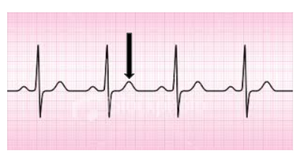
P wave
Part of an EKG that shows the depolarization, or change in cardiac cell charge, and contraction of the atria.
QRS Complex
Part of an EKG wave that shows the depolarization, or change in cardiac cell charge, and contraction of the ventricles.
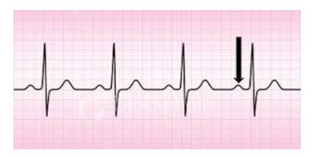
T wave
Part of an EKG wave that shows repolarization, or relaxation, of the ventricles.
Depolarization
The change in electrical charge of cardiac cells that causes a contraction of cardiac muscle tissue.
Repolarization
The return of cardiac cells to their original resting charge and state, causing a relaxation of cardiac muscle tissue.
T Wave
P Wave
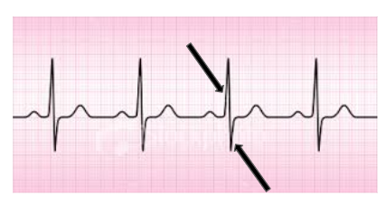
QRS Complex