The Periodontium: Periodontal Ligament
1/37
There's no tags or description
Looks like no tags are added yet.
Name | Mastery | Learn | Test | Matching | Spaced |
|---|
No study sessions yet.
38 Terms
Review: What structures compose the periodontium?
Gingiva, PDL, Cementum, Bone
What structure occupies the space between the root surface and alveolar bone?
Periodontal ligament
What is the radiological presentation of the PDL?
Thin, dark (radiolucent) line between alveolus and root
What is the thickness of the PDL?
0.1 - 0.25 mm
What comprises the PDL?
CT fibers
Cells
Vasculature
Nerves
Ground substance
What are the functions of the PDL?
Formative
Supportive
Nutritive
Sensory
Protective
What specific cells compose the PDL?
Undifferentiated mesenchymal cells
Fibroblasts
Osteoblasts
Cementoblasts
Odontoclasts
Epithelial cells
Immune cells
What are remnants of Hertwig’s epithelial root sheath (islands of squamous epithelium near root surface)?
Epithelial Rests of Malassez

What is labeled CB?
Cementoblasts
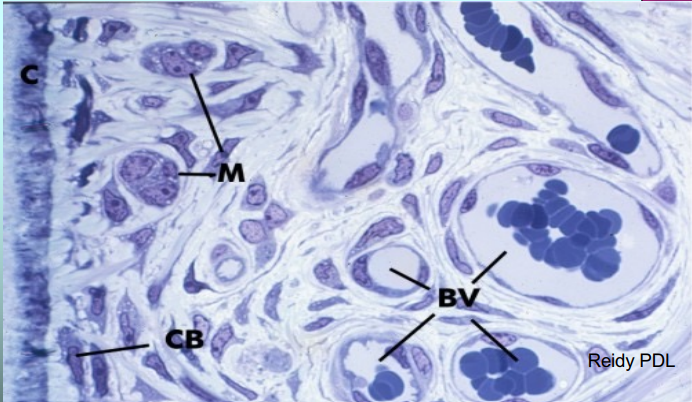
What is labeled M?
Rests of Malassez

What is labeled BV?
Blood vessels
What is the supportive function of the PDL?
Anchor tooth to bone
What is the nutritive function of the PDL?
Contains blood supply and lymphatics

What region is labeled 1?
Periodontal ligament branch

What region is labeled 2?
Interradicular
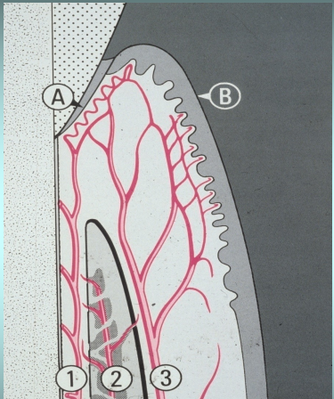
What region is labeled 3?
Periosteal
What are the three types of vessels of the PDL?
Alveolar artery branches
Interradicular arteries
Gingival vessels
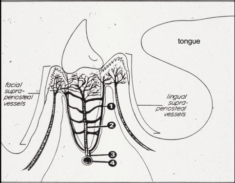
What is the structure labeled 1?
Periodontal membrane branch

What is the structure labeled 2?
Interdental septal arteriole

What is the structure labeled 3?
Dental arteriole

What is the structure labeled 4?
Inferior alveolar artery
What is the sensory function of the PDL?
Transmitting tactile pressure and pain sensations
What is the protective function of the PDL?
Cushions occlusal forces
(T/F) The PDL is static tissue, restricting all movement.
False. It is dynamic, allowing the tooth to move and adapt to functional states.
How are PDL fiber groups identified?
Location and orientation (direction)
What are the different PDL fiber groups?
Alveolar crest group
Horizontal group
Oblique group
Apical group
Interradicular group

What are the fibers labeled 1?
Alveolar crest

What are the fibers labeled 2?
Horizontal

What are the fibers labeled 3?
Oblique

What are the fibers labeled 4?
Apical
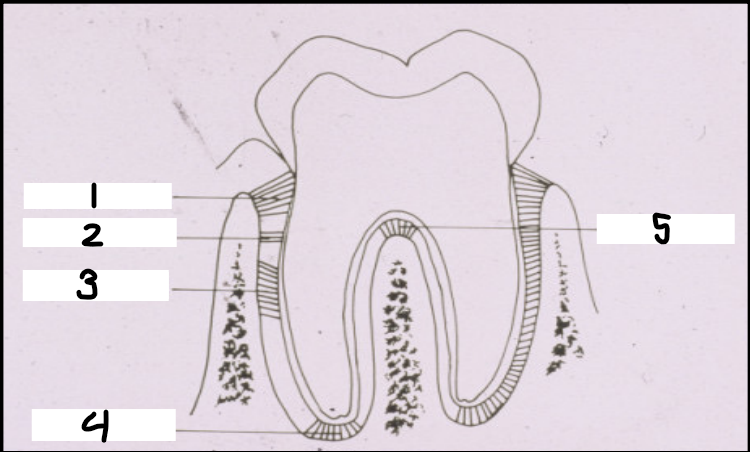
What are the fibers labeled 5?
Interradicular
Which PDL fiber group is only found on teeth with furcated roots?
Interradicular
What are the mineralized ends of principal collagen fibers embedded in cementum and bone?
Sharpey’s Fibers
Where would you find the largest amount of Sharpey’s Fibers?
Cemental surface
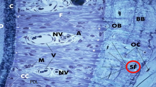
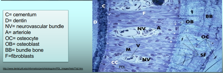
Just a diagram that might be helpful to know. She really likes it.
[ FLIP CARD ]
What is the mineralized CT that covers the roots?
Cementum
What is the main function of the cementum?
Anchor PDL fibers to the tooth