2.3 - ADAPTATIONS FOR TRANSOPRT IN ANIMALS
1/88
There's no tags or description
Looks like no tags are added yet.
Name | Mastery | Learn | Test | Matching | Spaced |
|---|
No study sessions yet.
89 Terms
What components does an effective transport system have?
.Transport medium (blood)
.Haemoglobin —> has high affinity for 02
.Blood vessels —> capillaries , arteries , veins , arterials , venues
.Muscular pump (heart)
.Valves —> ensures uni-directional flow in heart + veins
What organism has an OPEN CIRCULATORY SYSTEM ?
INSECTS
What is an OPEN CIRCULATORY SYSTEM?
Blood is not transported in blood vessels but BATHES THE TISSUE in a body cavity called the HAEMOCOEL (eg insects)
Describe the direction of blood flow in an open circulatory system.
. DORSAL AORTA with thick muscular walls→ runs the length of the body
.Blood pumped at low pressure into HAEMOCOEL → change of materials occur
.Blood returns slowly to tube shaped heart portion → areas with THICKENED MUSCULAR WALLS
.No RBC —> O2 delivered directly to RESPIRING TISSUES by TRACHEA (fluid called HAEMOLYMPH)
Explain how an insect can have a circulatory system which functions at low pressures despite having a high demand for oxygen?
.The trachea and tracheoles transport 02 directly to the respiring tissues.
. Circulatory system not involves in 02 transport —> no RBC or Haemoglobin
What organisms use a CLOSED CIRCULATORY SYSTEM ?
. FISH
. EARTHWORM
What is a CLOSED CIRCULATORY SYSTEM ?
The blood is always transported in blood vessels . There are two types.
Single circulatory system
Double circulatory system
What are the advantages of a closed circulatory system?
. Blood is transported at higher pressures
. Can be transported further ( because os higher pressures)
.Maintains conc grad
What are the disadvantages of a closed circulatory system?
. Higher energy requirement
. Increased complexity
What is a SINGLE CIRCULATORY SYSTEM?
.The blood mores through the heart once in once in one complete circuit of the body
.eg FISH + EARTHWORM
How does the SINGLE CIRCULATORY SYSTEM work in a EARTHWORM?
.Blood moves forward in the DORSAL VESSEL + back through the VENTRAL VESSEL
.Thick muscular walled blood vessels (PSEUDOHEARTS) pump the blood
What is the advantages of a single circulatory system in an earthworm?
ADVANTAGE
→ has blood containing HAEMOGLOBIN
→ high affinity for 02 —> improves efficiency of 02 transportation
→ thin skin is gas exchange surface - short diffusion path
What is the disadvantages of a single circulatory system in an earthworm?
→Reduced blood pressure + flow rate
→ Slower circulation to body tissues
→ Less efficient delivery of 02
→ Limited metabolic capacity
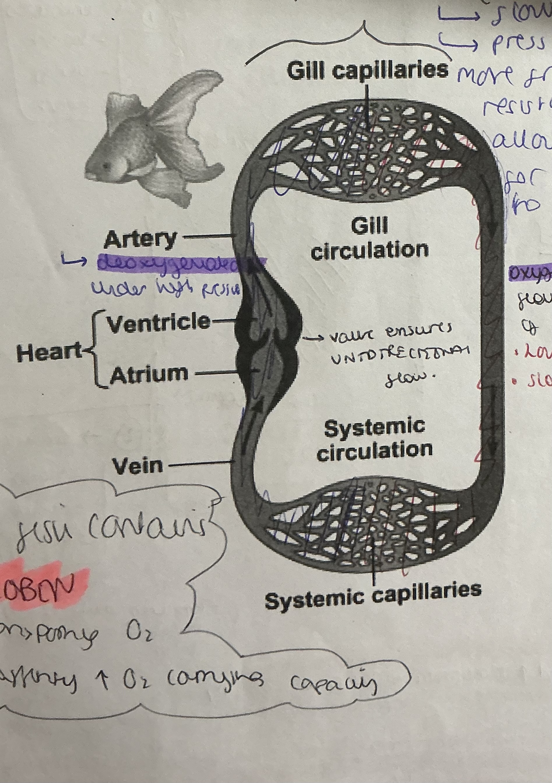
ow does the SINGLE CIRCULATORY SYSTEM work in a FISH?
.Have a TRUE HEART (only 2 chambers)
.Simplicity of structure → less likes of damage - operated under LOWER PRESSURES
.DEOXYGENATED blood pumped under HIGH PRESSURE from ventricle → gill capillaries via AFFERANT ARTERIES
.OXYGENATED BLOOD pumped under LOW PRESSURES + LOW FLPW RATE → GILL CAPILLARIES - increasing FRICTION + resistance to flow - DECREASES pressure
What organisms use a DOUBLE CIRCULATORY SYSTEM?
Mammals
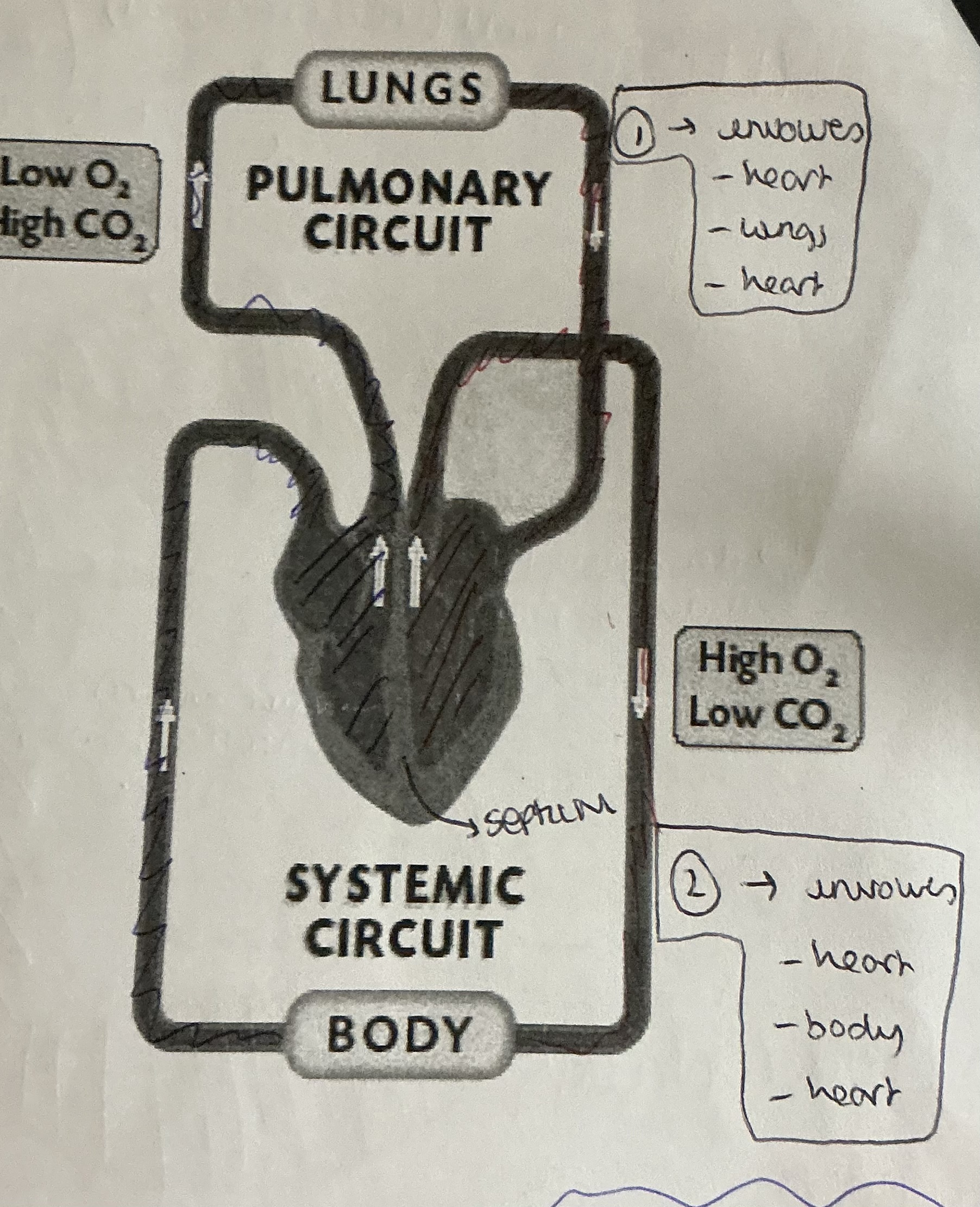
What is a double circulatory system ?
.Blood passes through the heart twice in one complete circuit of the body
.Muscular heart pumps OXYGENATED blood at high pressures with a HIGH FLOW RATE → body
.TISSUE FLUIDS → formed to enable exchange of materials with body tissue
What are the advantages of a double circulatory system?
. OXYGENTATED blood and deoxygenated blood are kept separate
.OXYGENATED BLOOD is pumped at a much HIGHER PRESSURE to our respiring tissues so flow rate is higher. Therefore it reaches tissues faster as OXYGENATED blood is pumped back to the heart before getting to the body so pressure + flow rate INCREASES
.Blood is at different pressures in both circuits . High in SYSTEMATIC CIRCUIT to ensure tissue fluid formation . Low in PULMONARY CIRCUIT to avoid tissue fluid formation
SUMMARY TABLE
What are the 2 separate systems that the mammalian circulatory system can be divided into?
The PULMONARY CIRCULATION (heart-lungs-heart)
The SYSTEMATIC CIRCULATION (heart-body-heart)
Why is mammalian having 2 systems more efficient
Is extremely efficient as it enables oxygenated blood to be pumped around the body at high pressures
What is PULMONARY CIRCULATION VS SYSTEMATIC CIRCULATION
PULMONARY CIRCULATION - transports deoxygenated blood from the heart to the lungs for gas exchange
SYSTEMATIC CIRCULATION - delivers oxygenated blood from the heart to the rest of the body and returns deoxygenated blood back to the heart
What are the 3 main type of blood vessels?
Artery —> Transport blood from the heart to the body tissues
Vein —> Transport blood from the body tissues back to the heart
Capillary —> Facilitates the exchange of substances between the blood and body tissue
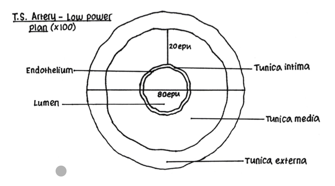
Draw and explain what ARTERIES do
.Transport OXYGENATED BLOOD from heart → body under high pressure + high flow rate
. endothelium/TUNICA INTIMA - ONE CELL THICK inner layer - provides a smooth lining to reduce friction + and ensures minimum resistance to blood flow.
. middle layer/ TUNICA MEDIA is made up of ELASTIC FIBRES + SMOOTH MUSCLE (elastic fibres to enable Bessel to stretch under high pressures + cause ELASTIC RECOIL back to original diameter) (this layer is thicker in ARTERIES - accommodate changes in blood flow and pressure.)
.The outer layer/TUNICA EXTERNA / TUNICA ADVENTIA - made up of COLLAGEN RICH CONNECTIVE TISSUE FIBRES - which are resistant to over-stretching under high hydrostatic pressure of blood.
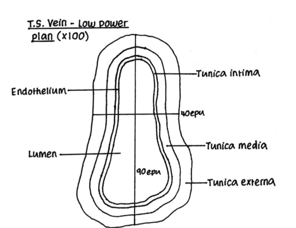
Draw and explain what VEINS do
.The outer layer/TUNICA /TUNICA ADVENTIA - made up of COLLAGEN RICH CONNECTIVE TISSUE FIBRES - which are resistant to over-stretching under high hydrostatic pressure of blood
. endothelium/TUNICA INTIMA - ONE CELL THICK inner layer - provides a smooth lining to reduce friction + and ensures minimum resistance to blood flow.
.TUNICA MEDIA - thinner than in artery - doesn’t need to withstand the high pressure in artery - pressure in veins are lower ( further away from heart)
.Has a LARGER LUMEN- Increased VOL , decreased PRESSURE in vein
.Has VALVES - moving against gravity so blood moves backwards - SEMI LUNAR VLAVE prevents back flow of blood due to decreased PRESSURE in vein + slower FLOW RATE
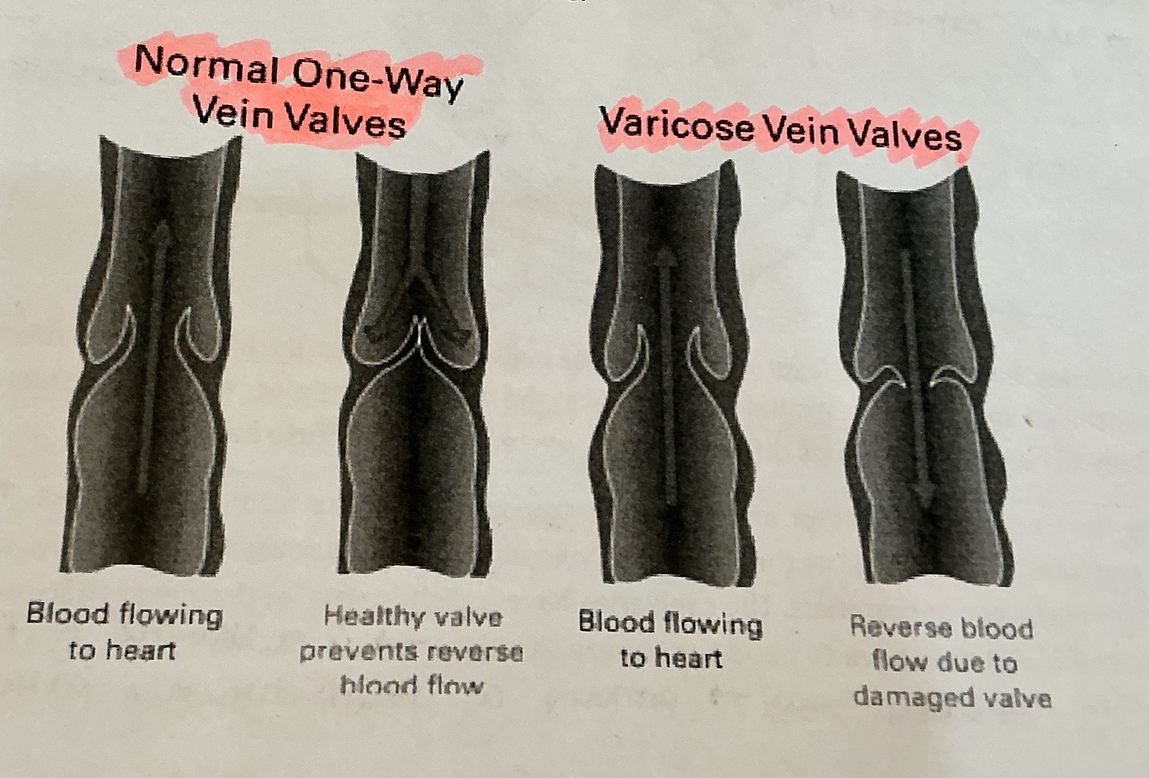
What is the difference between normal one-way valves and VARICOSE valves?
NORMAL ONE WAY VALVES
.blood flowing to heart
.healthy valve prevents reverse blood flow
VARICOSE VEIN VALVES
.Blood flowing to heart
. Reverse blood flow due to damages valve
What do CALF MUSCLES do?
. Helps upward blood flow
.When muscles contract it pinches the vein - decreasing DIAMETER , increasing PRESSURE , pushing blood back up to the heart
Draw and explain what CAPILLARIES do
.Transport both OXYGENATED + DEOXYGENATED blood —> link between arteries + veins
. Single layer of ENDOTHELIAL cells surrounded by BASEMENT MEMBRANE —> provides short diffusion path
.Endothelial layer is FENESTRATED —> makes walks PERMEABLE - enable exchange of materials (Water+solutes diffuse out + waste products diffused in)
.Narrow diameter + numerous —> Increases total SA - increases RESISTANCE TO BLOOD FLOW - velocity of flow decreases lots - allows more TIME for exchange of materials
.Smaller diameter that RBC —> RBC has to bend to squeeze through (Membrane of RBC pushes against capillary wall providing short diffusion path)
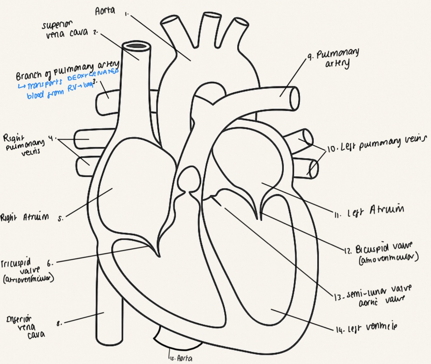
What is the structure of the HEART>
.made of specialised muscular tissues - CARDIAC MUSCLE - never tires
.MYOGENIC - heartbeat is initiated within muscle cells + contract without external stimulation (can be altered by nervous + hormonal stimulation )
.Right side pumps -DEOXYGENTATED blood to LUNGS
.Left side pumps - OXYGENATED blood to body
.Each side kept separate - prevent OXYGENATED + DEOXYGENTATED mixing
Draw a full diagram of the heart
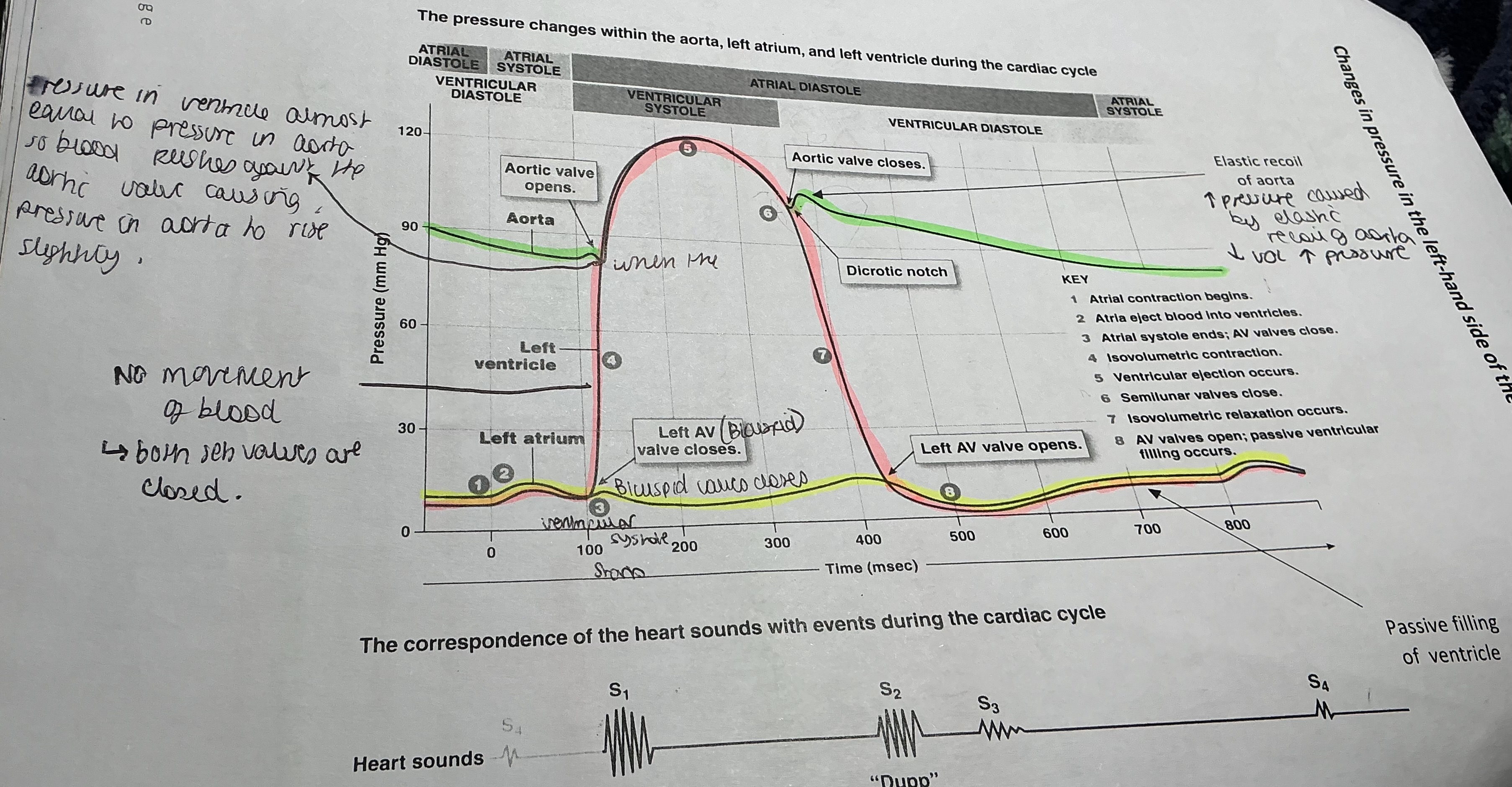
How many main stages are there of the CARDIAC CYCLE?
. 3 Main stages
/ Approximately 0.8 seconds
What are the 3 stages of the cardiac cycle?
Atrial systole (atrial contractions)
Ventricular systole (ventricular contraction)
Diastole (relaxation of cardiac muscle)
What happens during the first stage of the cardiac cycle? - ATRIAL SYSTOLE
. Thin - walled ATRIA CONTRACT - increasing the BLOOD PRESSURE
. ATEIO-VENTRICULAR VALVES (bicuspid+tricuspid) open - due to pressure in ATRIA exceeding pressure in VENTRICLES
. SEMI-LUNAR valves are closed
What happens during the second stage of the cardiac cycle? - VENTRICULAR SYSTOLE (ventricular contraction)
. Thick muscular walls of the VENTRICLES CONTRACT - decreasing volume , increasing pressure
.pressure in VENTRICLES quickly exceeds atrial pressure - blood BACK FLOWS , hitting the ATRIO-VENTRICULAR VALVE forcing them closed - causes sound ‘LUB’
.Pressure i’m the AORTA + PULMONARY ARTERY - still HIGHER than the ventricles - SEMI-LUNAR VALVES remain closed - at this point no movement of blood in ventricles - both set of valves closed
. VENTRICLES continue to contract - increasing pressure - pressure in VENTRICLES exceed that of the AORTA + PULMONARY ARTETY - pressure of blood against the SEMI-LUNAR VALVES forces them open
.PULMONARY ARTERY carries DEOXYGENATED blood to lungs
.AORTA carries OXYGENATED blood to different parts of body
What happens during the third stage of the cardiac cycle? - DIASTOLE (relaxation of cardiac muscle )
. VENTRICLES relax
. As volume increases , pressure inside VENTRICLE drops below in that in ARTERIES
. CAUSES BACKFLOW OF BLOOD - CLOSES THE SEMI-LUNAR valves - preventing blood from going back into ventricles . This produces second heart sound ‘DUB’
.Blood from VENA CAVA + pulmonary vein passively enters ATRIA - relaxed
. Whole cycle starts again
When do BI + TRI VALVES open and close?
BI + TRI VALVES OPEN when —> pressure in ATRIUM > pressure in VENTRICLE (arteriole systole)
BI + TRI VALVES CLOSE when —> pressure in VENTRICLE > pressure in ATRIUM (early ventricular systole)
When do SMEIL LUMNAR VALVES open and close?
SEMI LUNAR OPEN when —> pressure in VENTRICLES > PRESSURE IN AORTA + PUL ARTETY (late ventricular systole)
SEMI LUNAR CLOSE when —> pressure in VENTRICLES < pressure in AORTA + PUL ARTERY (diastole)
Draw a graph to show the changes of pressure in the LEFT-HAND side of the heart?
How is heartbeat controlled ?
. Cardiac muscle is MYOGENIC —> can contract without nervous stimulation
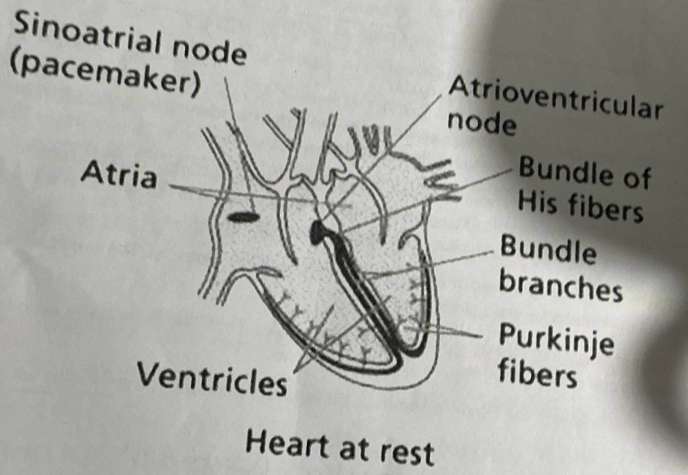
. Wall of RIGHT ATRIUM has cluster of cardiac cells (SINO-ATRIAL NODE) —> acts as a PACEMAKER
What does the SAN (sing-atrial node) do?
. SAN generated an ELECTRICAL IMPULSE / WAVE OF EXCITATION —> spreads across muscular walls of atria - DEPOLARISING them - stimulating ATRIAL SYSTOLE
What does the AVN (ATRIO-VENTRICULAR NODE) do?
. A layer of connective tissue insulates the ventricles apart from another cluster of cells —> AVN
.This introduces a DELAY in the transmitting of electrical stimulation
Why is the delay produces by the AVN necessary ?
So that the ATRIA complete their contractions + empty their blood before VENTRICULAR contact + to allow ventricles to completely fill with blood
What happens after the delay produces by the AVN?
. The AVN passes the next wave of excitation down the SEPTUM via nerves of the BUNDLE OF HIS to the APEX of heart
.Then transfers up to PURKINJIE FIBRES —> in outer muscular walls of VENTRICLES
. This stimulates VENTRICULAR SYSTOLE from APEX upwards —> pushing blood up + out of heart into AORTA + PULMONARY ARTERT
Draw a diagram and label the BUNDLE OF HIS , PURKINJIE FIBRES , SAN , AVN
What is an ECG?
Electrocardiogram / Electrocardiograph
What does an ECG do?
An ECG is a trace of the voltage changes that occurs in the heart detected by electrodes on the skin
What are the 4 stages involved during an ECG?
P wave
PR interval
QRS complex
T wave
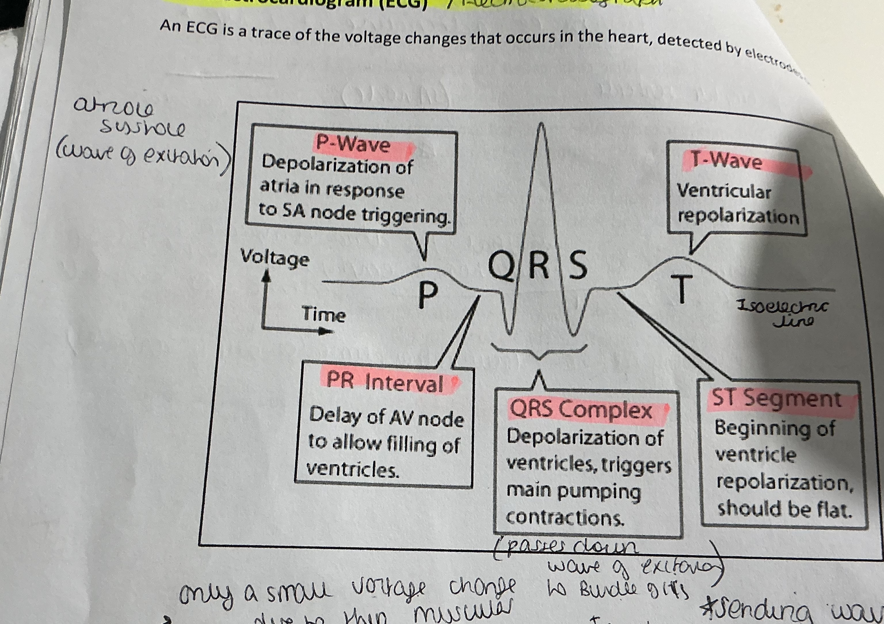
What does the P WAVE show ?
. The VOLTAGE CHANGE generated by the SAN in association with CONTRACTION OF ATRIA —> waves are small due to thin muscle walls
What does the PR INTERVAL show?
. The TIME BETWEEN THE START OF = WAVE AND START OF QURS COMPLEX = time taken for electrical stimulation to spread from ATRIA to VENTRICLES through AVN
.Represents the DELAT introduced by the connective tissue
What does the QRS complex show ?
. The DEPOLARISATION + CONTRACTION of the VENTRICLES
. AMPLITUDE is larger —> thick muscular walls of VENTRICLE
What does the T WAVE show?
. REPOLARIZATION of VENTRICLE muscles
. ST SEGMENT —> period of time between the END OF S WAVE + START OF T WAVE
What is the HORIZONTAL LINE after the T wave known as ?
. ISOELECTRIC line
. Is the baseline voltage
What is ATRIOLE FEBRILATION ?
Irregular heartbeat
Suggest how the cardiac cycle would be affected in a patient with heart defect ?
. Atria will not empty with blood - not enough blood in ventricles
. Many p waves - atria contracting more frequently
. SA nodes firing too rapidly
. Have an irregular heartbeat
What are the advantages of training at HIGH ALTITUDES?
Number of RBC would increase
Number of HB molecules would increase
More 02 delivered to respiratory tissues
Providing more 02 for respiration
Produce more ATP
Allowing athlete to improve performance
What are ERYTHROCYTES ?
Red blood cells - Red due to the presence of the pigment HAEMOGLOBIN
What id the function of red blood cells?
Transport of oxygen from the lungs to the respiring tissues
What are 2 ways in which ERYTHROCYTES RBC are adapted to their functions?
. RBC can change its shape to fit through capillaries
. Biconcave —> provides LARGE SA for diffusion of 02
. Doesn’t contain a NUCLEUS —> more room for heamoglobin
. THIN CENTRE —> short diffusion path
What are LEUCOCYTES?
White blood cells
What are the 2 main groups of LEUCOCYTES (WBC) ?
. AGRANULOCYTES -
—> clear CYTOPLASM + SPHERICAL NUCLEUS
—> produce ANTIBODIES + ANTITOXINS
—> responsible for IMMUNITY
. GRANULOCYTES
—> have a GRANULAR CYTOPLASM
—> have LOBED NUCLEUS
—> Involves in FIRST LINE OF DEFENCE
—> Mainly phagocytosis
What is BLOOD PLASMA ?
.A pale yellow liquid = 90% water
. Has many solutes = WATER , BLOOD PROTEINS , NUTRIENTS , HORMONES , ELECTROLYTES
. Distributes heat release during RESPIRATION
How is OXYGEN transported around the body ?
O2 is Transported from the LUNGS —> RESPIRING TISSUE bound to the Haemoglobin molecule
How is O2 transported effcientely?
. Haemoglobin must bind to the molecule readily when there is a HIGH PARTIAL PRESSURE (conc) of 02 ( ALVEOLI)
.Must also readily DISSOCIATE the 02 when LOW PARTIAL PRESSURE OF 02 (in respiring tissues - muscles)
. The degree to which Haemoglobin is attracted to and binds to 02 molecule = AFFINITY
What does the affinity of Haemoglobin depend on?
The CONCENTRATION of 02
What type of binding is it when Haemoglobin binds to 02 ?
Cooperative binding
What is cooporative bonding
.When the first molecule of O₂ binds to the haemoglobin, it changes the shape of the haemoglobin molecule.This makes it easier for the second oxygen molecule to bind.
.The second binding causes a further shape change, making it easier for the third O₂ molecule to attach.
.The binding of the 3rd molecule has no further affect on shape so it takes a large increase in the partial pressure of 02 to enable the 4th 02 molecule to bind
What is the equation for when Haemoglobin binds to oxygen via cooperative binding?
402 + H6 —> H608 (oxyhemoglobin )
What type of curve is shown in an OXYGEN DISSOCIATION CURVW FOR ADULT HAEMOGLOBIN?
Sigmoidal curve (never reaches 100% saturation)
Why is a sigmoidal curve shown in a oxygen dissociation curve for adult Haemoglobin?
.Due to cooperative binding - Haemoglobin exposed to increases partial pressures of 02 shows a SIGMOIDAL CURVE
. At LOW PARTIAL PRESSURES OF 02 —> difficult for Haemoglobin to load 02
What is the acronym to use when taking about 02 dissociation curve?
S aturation
A ffinity of Haemoglobin
D issociation / unloading
Draw and explain the graph of an oxygen dissociation curve for adult Haemoglobin . A
. At HIGH partial pressures , Haemoglobin has a HIGH AFFINITY for 02 —> binds readily + REACHES SATURATION , does not RELEASE the 02 readily
.As partial pressures of 02 DECREASES , Haemoglobin AFFINITY DECREASES . Oxygen is DISSOCIATED READILY to respiring tissues to maintain aerobic respiration
.Steep part of graph - SMALL change in partial pressure of 02 results in a LARGE CHANGE OF Haemoglobin % SATURATION (cooperative binding) —> tissues can respire aerobically
What are the advantages of a sigmoid curve for adult Haemoglobin?
. Change in partial pressure of 02 doesn’t result in a large change in %saturation of Haemoglobin
. A higher % SATURATION is achieved at all partial pressures of oxygen
. Can reach 95-98% saturation at much lower partial pressure of 02
What is the OXYGEN DISSOCIATION curve like in ADULT HAEMOGLOBIN?
. Rapid saturation of 02 in lungs
. Rapid dissociation of 02 as the conc decreases
. Oxygen release to respiring tissues for aerobic respiration
What is the oxygen dissociation curve like in FOETAL HAEMOGLOBIN
.Curve to LEFT of adult Haemoglobin
.Higher affinity for 02 than adult Haemoglobin + higher % saturation at all partial pressures
.Oxygen can be absorbed from maternal Haemoglobin to foetal Haemoglobin in placenta
What is the oxygen dissociation curve like in HAEMOGLOBIN HAEMOGLOBIN
. To the LEFT of foetal haemoglobin
. Extremely high affinity for oxygen and only dissociates during INTENSE PHYSICAL ACTIVITY when partial pressures are very low
. Had a higher % saturation at all partial pressures oxygen at all partial pressures of 02 compared to adult Haemoglobin
. Only SINGLE haemoglobin unit therefore only binds to one molecule of oxygen
. Acts as an OXYGEN STORE IN MUSCLE CELLS
. A small decrease in partial pressures of 02 results in an extremely large decrease in saturation of myoglobin
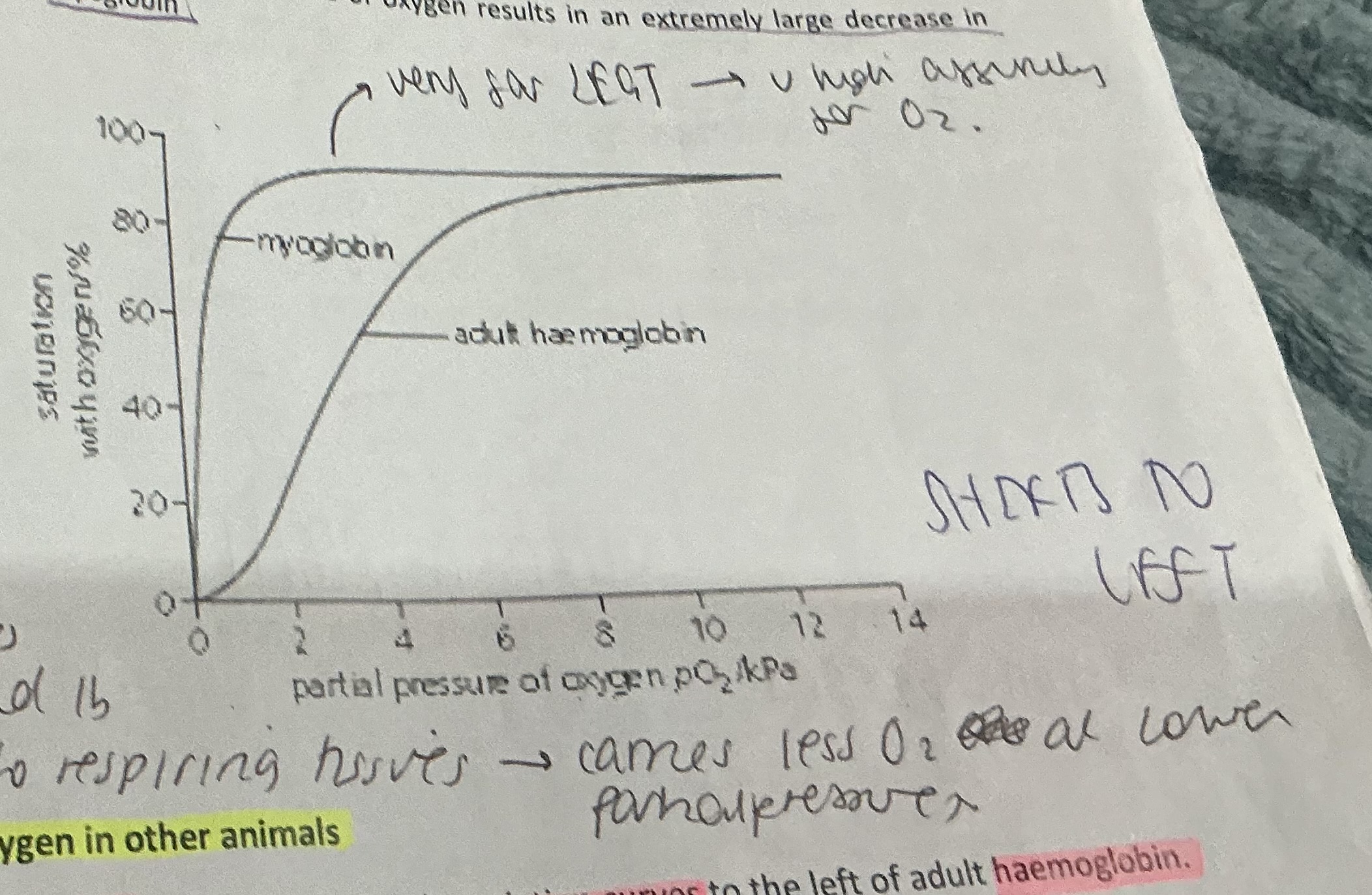
What are the DISADVANTAGES of the MYOGLOBIN oxygen dissociation curve?
When partial pressure decreases it doesn’t dissociate its 02 as readily to respiring tissues —> carries less 02 at lower partial pressures
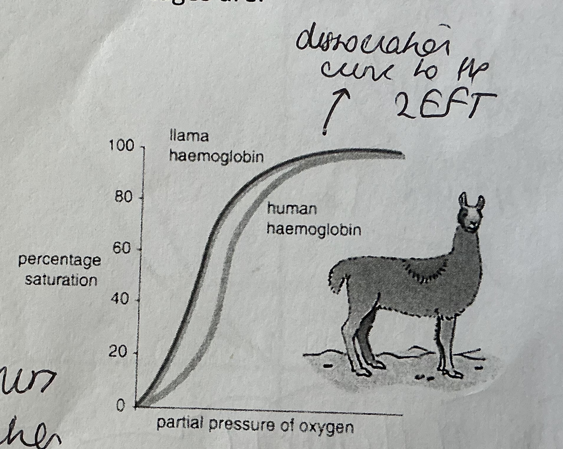
What is the TRANSPORT OF OXYGEN like in OTHER ANIMALS ?
Llama + Lugwoem —> 02 dissociation curves to the LEFT of adult Haemoglobin
Why is the 02 dissociation curve for llamas to the left?
. Live in HIGH ALTITUDES —> undies
. Live in habitats with LOWER PARTIAL PRESSURE OF 02
. Has 02 dissociation curve to the LEFT of ours (humans) —> has HIGHER AFFINITY FOR 02
. ADVANTAGE —> HB reaches 95-98% saturation at lower partial pressure of 02 available in its habitat
. DISADVANTAGE —> low partial pressures does not dissociate 02 as readily
Why is the 02 dissociation cure of a LUGWORM to the LEFT?
. LOWER partial pressure of 02
. Lives near sea —> when tide comes in burrow fills with water , when tide out burrow empties with water - DRIES OUT
.02 dissociation curve to LEFT of HB —> higher affinity for )2
. ADVANTAGE —> Can reach 95-98% saturation at LOWER partial pressure of 02 available in its habitat
. DISADVANTAGE —> low partial pressures does not dissociate 02 as readily
What are some ADAPTATIONS of LUGWORM?
. Has EXTERNAL GILLS —> Increases SA for gas exchange
. Water flows through burrow in ONE DIRECTION —> maintains conc grad
. COUNTER CURRENT FLOW —> maintains a conc grad over entire surface
. Has HAEMOGLOBIN —> has high affinity for 02
What is the effect of INCREASING C02 CONCENTRATION on the 02 dissociation curve called?
The BOHR effect
Why does a shift to the RIGHT of the 02 dissociation curve occur during the BOHR effect?
. 02 dissociation curve moves to the RIGHHT
. More 02 released to RESPIRING TISSUES
. Active respiration increases C02 —> partial pressure of C02 INCREASES
. Increases ACIDITY LEVELS (lowered PH due to formation of hydrogen ions)
. Hydrogen ions bind to OXYHEMOGLOBIN —> change in shape which LOWERS HB affinity for 02 (shown as shift to right of curve)
. 02 then released from OXYHEMOGLOBIN to respiring tissues so AEROBIC RESPIRATION MAINTAINED
List 3 ways that C02 is transported in blood?
Dissolves in plasma (5%)
Bound to HB as CARBAMINOHAEMOGLOBIN (10%)
As HYDROGEN CARBONATE ions (85%)
Describe fully hoe carbon dioxide is transported in the blood? ( BOHR EFECT)
C02 in the blood DIFFUSES into the RBC
CARBONIC ANHYDRASE enzyme catalyses the addition of C02 to water - CARBONIC ACID
Carbonic acid dissociated to form HYDROGEN IONS + HYDROGEN CARBONATE IONS
Hydrogen carbonate ions diffuse out of RBC into plasma .
To negate the outward flow of negative hydrogen carbonate ions , CHLORIDE IONS DIFFUSE into the RBC from the plasma - CHLORIDE SHIFT - occurs to MAINTAIN-ELECTROCHEMICAL NEUTRALITY
Hydrogen ions BINDS RO OXYHEMOGLOBIN at an allosteric site. Changing shape of HB molecule - LOWERING its affinity for 02 which is then DISSOCIATED
Hydrogen ions that are bound to HB form HAEMOGLOBONIC ACID (HHb) - removes hydrogen ions + acts as a BUFFER to maintain pH within RBC
OXYGEN DIFFUSES out of cell and into the RESPIRING TISSUES
What is TISSUE FLUID?
Blood plasma MINUS the plasma proteins
What are the 2 major functions of tissue fluid?
SUPPLY body tissues with oxygen + other solutes (glucose , fatty acids , salts , amino acids) to RESPIRING TISSUES
To TRANSPORT waste products such as C02 + urea (in liver) from the CELLS —> BLOOD
What are 3 ways that the CAPILLARIES are adapted for its function of GAS EXCHANGE?
. Have FENESTRATIONS —> allow substances to move out
. Only a SINGLE ENDOTHELIAL thick → short diffusion path
. Close association with BODY CELLS / BODY TISSUES
What is KWASHIORKOR ?
. Condition occurring in children
. Diets CONTAINING LOW QUANTITIES OF PROTEIN
. Results in SWOLLEN LIMBS + FACE + ABDOMEN
What would a diet lacking in protein result in? (LEARN)
.FEWER PLASMA PROTEINS
.Which raises the WATER POTENTIAL of BLOOD PLASMA
. Reduces WATER POTENTIAL GRADIENT
. Less in REABSORBED BY OSMOSIS
. Resulting in severe OEDEMA