AQA A Level Psychology - Biopsychology
1/168
There's no tags or description
Looks like no tags are added yet.
Name | Mastery | Learn | Test | Matching | Spaced | Call with Kai |
|---|
No analytics yet
Send a link to your students to track their progress
169 Terms
What components make up the central nervous system?
the brain and spinal cord
What is the function of the brain?
The brain is the central organ of awareness, enabling consciousness and thought.
It controls perception and motor control, processing sensory information and coordinating movement.
It regulates bodily processes and maintains homeostasis by responding to information from the peripheral nervous system (e.g. regulating body temperature and hormone levels).
Describe the divisions of the nervous system.
CNS (Central Nervous System): Brain + spinal cord; processes information.
PNS (Peripheral Nervous System): Nerves outside CNS; sends messages to/from CNS.
Somatic NS: Voluntary movement.
Autonomic NS: Involuntary functions.
Sympathetic: Fight or flight.
Parasympathetic: Rest and digest.
What is the function of the spinal cord?
Relays information between the brain and body
Coordinates reflexes via reflex arc (automatic responses)
Transmits sensory signals from body to brain
Sends motor commands from brain to muscles/glands
What is the structure of the peripheral nervous system?
The PNS consists of all nerves outside the CNS.
It is divided into two main branches:
Somatic Nervous System:
- Controls voluntary movements via skeletal muscles.
- Carries sensory information to the CNS and motor commands from the CNS.Autonomic Nervous System:
- Controls involuntary bodily functions (e.g. heart rate, digestion).
- Subdivided into:
- Sympathetic Nervous System: Prepares the body for fight or flight.
- Parasympathetic Nervous System: Supports rest and digest functions.
What is the function of the peripheral nervous system?
Connects CNS to the body (limbs, organs, etc.)
Transmits sensory information from body to CNS
Carries motor commands from CNS to muscles/glands
Controls voluntary movements (somatic nervous system)
Regulates involuntary functions (autonomic nervous system: heart rate, digestion)
The peripheral nervous system has two divisions, what are they?
autonomic nervous system and somatic nervous system
What is the role of the autonomic nervous system?
- Made of sensory and motor neurones
- Regulates involuntary functions (e.g., heart rate, breathing, digestion)
- Maintains homeostasis (internal balance)
- Divided into two branches:
Sympathetic Nervous System: activates "fight or flight" response (increases heart rate, dilates pupils)
Parasympathetic Nervous System: triggers "rest and digest" response (slows heart rate, promotes digestion)
What is the role of the somatic nervous systems?
- Made of sensory and motor neurones
- Voluntary body movement and sensing external stimuli
- It inputs from sense organs (5 senses) and outputs to muscles, skin and joints by receiving a signal to respond to the changes detected.
Example: catching a ball-your eyes detect the ball moving towards you, brain assesses info, then instructs the muscles of your arm to reach out and catch the ball.
Which division of the Peripheral Nervous System contains the sympathetic and parasympathetic divisions?
Autonomic
What is the role of the sympathetic nervous systems?
Made of motor neurones
Activates "fight or flight" response during stress or danger
Increases arousal: raises heart rate, blood pressure, and breathing rate
Dilates pupils for better vision
Inhibits digestion to conserve energy
Releases adrenaline from adrenal glands
Redirects blood flow to muscles
What is the role of the parasympathetic nervous systems?
Made of motor neurones
Activates "rest and digest" response after stress
Reduces arousal: lowers heart rate, blood pressure, and breathing rate
Promotes digestion: stimulates saliva, increases digestive enzyme production
Conserves energy: promotes relaxation and recovery
Constricts pupils
Encourages waste elimination (e.g., bladder function)
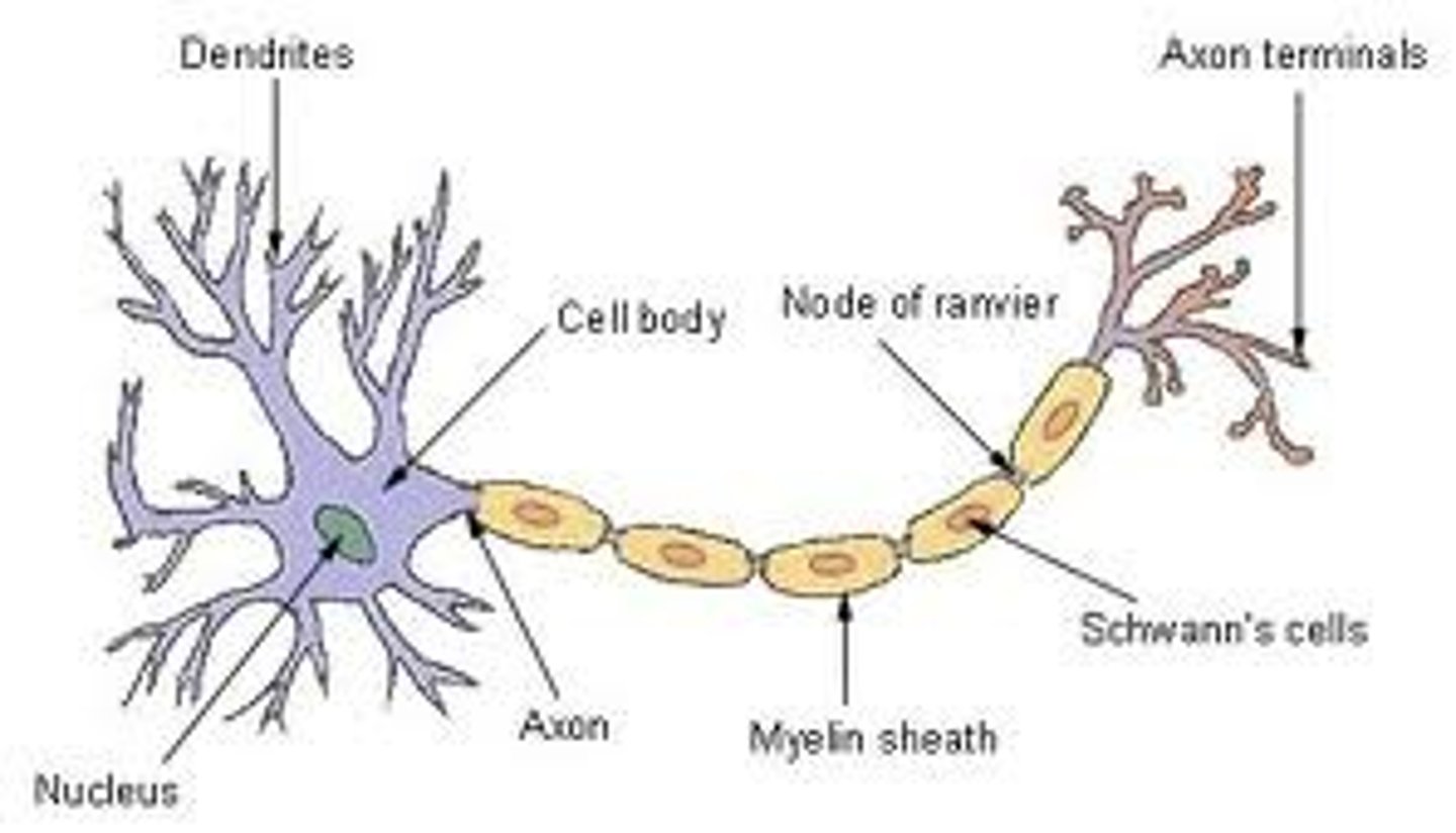
Describe the structure of a neuron.
Cell Body (Soma)
Dendrites
Axon
Myelin Sheath
Node of Ranvier
Axon Terminals (Synaptic Boutons)
What are the functions of the cell body (soma) in a neuron?
Contains the nucleus and organelles
Integrates incoming signals
What are dendrites, and what do they do?
Branch-like structures
Receive signals from other neurons or sensory receptors
What is the role of the axon in a neuron?
Long, slender projection
Transmits electrical impulses away from the cell body
What is the myelin sheath, and what is its function?
Fatty layer surrounding the axon
Increases speed of impulse transmission (saltatory conduction)
What are the Nodes of Ranvier, and why are they important?
Gaps in the myelin sheath
Facilitate rapid signal conduction
What are the functions of the axon terminals (synaptic boutons)?
End of the axon branches
Release neurotransmitters to communicate with other neurons or muscles
What is the structure of sensory neurones?
Long dendrites and short axons
Receptor endings (e.g., skin, eyes)
What is the function of sensory neurones?
Transmit sensory information (e.g., touch, pain, temperature) from peripheral receptors to the CNS
What is the structure of relay neurones?
Short dendrites and axons
Located within the CNS (brain and spinal cord)
What is the function of relay neurones?
Connect sensory and motor neurons
Process and relay information between neurons
What is the structure of motor neurones?
Short dendrites, Long axons
Cell bodies located in the CNS, extending to muscles
Which part of the central nervous system is in control of reflexes?
Spinal cord
Describe synaptic transmission.
- transmission involves impulses crossing a space or gap between an axon terminus and the adjacent neuron (the synapse/synaptic cleft)
- neurotransmitters are chemicals released from vesicles on the presynaptic neuron
- electrical impulses (action potentials) reach the presynaptic terminal
- electrical impulses (action potentials) trigger release of neurotransmitter
- neurotransmitters cross the synapse from vesicles
- neurotransmitters combine with receptors on the postsynaptic membrane
- stimulation of postsynaptic receptors by neurotransmitters result in either excitation (depolarisation) or inhibition (hyperpolarisation) of the postsynaptic membrane.
What are excitatory neurotransmitters?
neurotransmitters that make the post synaptic neuron is more likely to fire an impulse
Are neurotransmitters excitatory or inhibitory?
They can be both but GABA is purely inhibitory.
What is a neurotransmitter?
Chemical substances that transmit signals across synapses from one neuron to another or to target cells (such as muscles or glands).
What are inhibitory neurotransmitters?
neurotransmitters that make the post synaptic neuron is more likely to fire an impulse
What is the function of motor neurones?
Carry motor commands from the CNS to effectors (muscles and glands)
Control voluntary and involuntary movements
Explain why neurons can only transmit information in one direction at a synapse.
- the synaptic vesicles containing the neurotransmitter are only present on the presynaptic membrane
- the receptors for the neurotransmitters are only present on the postsynaptic
membrane
- it is the binding of the neurotransmitter to the receptor which enables the signal transmitted to the next neuron
- diffusion of the neurotransmitters mean they can only go from high to low
concentration, so can only travel from the presynaptic to the postsynaptic
membrane.
How do drugs affect synaptic transmission?
Psychoactive drugs work by affecting (increasing or inhibiting) the transmission of neurotransmitters across the synapse
Describe what is meant by summation.
During synaptic transmission, the excitatory and inhibitory influences are summed, if the net effect on the post synaptic neuron is inhibitory, the neuron will be less likely to 'fire' and if the net effect is excitatory, the neuron will be more likely to fire.
Example
Stimulus --> receptor --> sensory --> relay --> motor --> effector ---> response.
Outline the structures involved in synaptic transmission.
The synaptic cleft, pre and postsynaptic membranes, postsynaptic receptor sites, neurotransmitters in vesicles in the presynaptic terminal
What glands and hormones make up the endocrine system, and what do they produce?
1. Pituitary gland --> HGH, TSH, ACTH, LH, FSH, ADH,
2. Pineal --> melatonin
3. Pancreas --> insulin, glucagon
4. Adrenal gland -->. adrenaline
5. Thyroid gland --> thyroxine
6. Testicles --> testosterone
7. Ovaries --> oestrogen
What are the differences between the endocrine system and the nervous system?
- Endocrine system gives chemical messengers vs nervous system giving electrical impulses.
- Endocrine system has long-lasting effects whilst the effects of nervous system is short-lived.
- Endocrine system takes much longer to work whilst the nervous system is very quick (e.g. reflexes)
- Endocrine system has more permanent and wide effects whilst the effects of nervous system is more localised and temporary.
When and how do endocrine and nervous system work together?
- The endocrine and ANS often work in parallel with one another, e.g. a stressful event.
- When a stressor is perceived, the hypothalamus triggers the sympathetic nervous system and the ANS changes from its usual resting state (the parasympathetic state).
- Adrenaline is released from the adrenal medulla into the bloodstream, resulting in the physical arousal needed for fight/flight.
- Once the threat has passed, the parasympathetic nervous system kicks in and returns the body to its natural resting state (decreases heart/breathing rate etc.)
What is the fight or flight response?
A sequence of activity within the body that is triggered when the body prepares itself for defending or attacking (fight) or running away to safety (flight). This activity involves changes in the NS and the secretion of hormones that are necessary to sustain arousal.
What changes occur in the body during the fight or flight response?
- increase heart rate
- constricts blood vessels, increasing rate of blood flow and raising blood
pressure
- diverts blood away from the skin, kidneys and digestive system
- increases blood to brain and skeletal muscle
- increases respiration and sweating
Why is adrenaline released?
released from the adrenal medulla in response to activation of
the sympathomedullary pathway
What are the general effects of adrenaline?
- prepare the body for action, fight or flight
- increase blood supply/oxygen to skeletal muscle for physical action
- increase oxygen to brain for rapid response planning
Evaluate the flight or fight response (1): Limited explanation
POINT: One issue with the fight or flight explanation is that human behaviour is not limited to just two response.
EVIDENCE: Gray suggests that the first response to danger is to avoid confrontation altogether, which is demonstrated by a ‘freeze’ response.
EXPLAINATION: During the freeze response, humans are hyper-vigilant while they appraise the situation to decide the best course of action for that particular event.
EVALUATION: This suggests that the fight or flight explanation of behaviour is limited and doesn’t fully explain the complex cognitive and biological factors that underpin human response in stress/danger.
Evaluate the fight or flight response (2): Androcentrism/Beta Bias
POINT: Another issue with the fight or flight explanation is that it doesn’t fully explain the stress response in females.
EVIDENCE: Taylor suggested that females adopt a ‘tend to befriend’ response in stressful situations.
EXPLAINATION: Women are more likely to protect their offspring and form alliances with other women, rather than an adversary or flee.
EVALUATION: This is a limitation because it highlight’s the beta bias within this area of psychology as psychologists assumed that females responded in the same way as males until Taylor provided evidence. This has prompted more recent research which has provided an alternate explanation which is applicable to females
Evaluate the fight or flight response (3): Detrimental health effects
PONT: A final issue is that it can have a detrimental effect on health, especially in modern day life.
EXPLAINATION: While it may have been a useful survival mechanism for our ancestors who faced genuinely life threatening situations, modern day life rarely requires such an intense biological response. This matters because the activation of fight or flight can increase blood pressure and cause damage to blood vessels and contribute to heart disease.
EVALUATION: This suggests that fight or flight is a maladaptive response to modern day life (lacks temporal validity).
What is the cerebrum divided into?
The largest section of the brain which is divided into four sections. The frontal, parietal, occipital and temporal lobe.
What joins each hemisphere of the brain?
The corpus callosum
Where in the brain is the motor cortex?
At the back of the frontal lobe in both hemispheres
What is the function of the motor cortex?
Controls voluntary movements on the opposite side of the body
What happens if the motor cortex is damaged?
Loss of control over skeletal muscles
Where in the brain is the somatosensory cortex?
At the front of the parietal lobe in both hemispheres.
What is the function of the somatosensory cortex?
Receives information from sensory receptors in the skin and skeletal muscle
What happens if the somatosensory cortex is damaged?
Loss of sensation to Skin and Skeletal muscle.
Where in the brain is the visual centres?
They are located on the occipital lobe
What is the function of the visual centres?
Receives impulses conveying visual information. Each eye sends visual information from the right visual field to the left visual cortex and vice versa.
What happens if the visual centres is damaged?
Loss of vision. For example damage to the left visual centre causes blindness to the right visual field
Where in the brain is the Auditory centre?
On the temporal lobe in both hemispheres.
What is the function of the Auditory centre?
Receives and responds to auditory stimulus. Analyses speech based information.
What happens if the Auditory centre is damaged?
Loss of hearing.
Where in the brain is Broca's Area?
Left Frontal lobe.
What is the function of Broca's Area?
Controls muscles responsible for the production of speech.
What happens if Broca's area is damaged?
Affects speech production. Speech is often described as slow and lacking fluency ( Broca's Aphasia.
Broken speech.
Where in the brain is Wernike's area?
On the left of the temporal lobe.
What is the function of Wernike's area?
Understanding and formulating speech.
Responsible for language comprehension.
What happens if Wernikes area is damaged?
Loss of ability to understand and produce coherent speech.
Can produce fluent speech though however it is meaningless
What is hemispheric lateralisation?
The brain is divided into two symmetrical halves called hemispheres.
Who conducted split brain research?
Sperry
Why did Sperry carry out split brain research?
To prevent severe and frequent epileptic seizures.
What was Sperry's procedure?
- engineered a study on split brain patients that had their corps callosum severed in a procedure to manage the symptoms of epilepsy.
- separated the right and the left visual field and displayed stimuli to each side then asked them questions
- the right hemisphere was responsible for the left visual field and the left hemisphere was responsible for the right visual fields
What did Sperry find?
- RVF (LH) viewing allows verbal description of objects.
- LVF (RH) viewing results in "nothing there" response; verbal descriptions not possible due to split-brain.
- RH to LH language center communication is blocked in split-brain individuals.
- Despite lack of verbal labels for LVF images, participants can match objects with their left hand (RH).
- Left hand (RH) selects objects or related items based on LVF presentations (e.g., ashtray for a cigarette).
- Emotional responses (e.g., giggling) occur to LVF (RH) images, like pinups, without conscious image recognition.
What did Sperry conclude?
- usually the information is relayed to the other side of the brain via the corpus callosum however in these patients it was severed
- when the participant was asked to describe an object shown to the right visual field (RVF) the patient was easily able to describe it
- when shown to the left visual field (LVF) The patient said There's nothing there?'
- therefore right hemisphere that language centres
- when the patient was asked to pick a matching object using the left visual field they were able to do so and they could also pick an object associated with the stimuli
e.g cigarette, the participant picked an ashtray
- shows that they were able to comprehend the stimulate they just couldn't say it due to the right hemispheres lack of language centre
- these observations show how certain functions are lateralised in the brain and support the view that the LH is verbal and the RH is 'silent' but emotional.
What would splitting the brain in half achieve?
Removing the main line of communication between the two hemispheres.
Why is split brain research useful?
Allows us to investigate to what extent each hemisphere specialises in certain functions.
How was split brain research standardised?
The item was only displayed for a very short amount of time to prevent giving them enough time to move their heads to both sides.
Hemispheric lateralisation:
-brain plasticity
Point: The recruitment of homologous areas in the brain illustrates the flexibility of brain function and challenges the traditional notion of strict hemispheric lateralization.
Evidence: For instance, if Broca's area is damaged on the left side of the brain, the right-sided equivalent may take over some of its functions. Over time, functionality could potentially shift back to the left side as recovery progresses.
Analysis: This phenomenon demonstrates that the brain is capable of adapting to injury by utilizing homologous areas to compensate for lost functions. Such flexibility suggests that the division of labor between the two hemispheres is not as absolute as previously believed. Instead, the brain exhibits a degree of plasticity, allowing for functional reassignment in response to damage or changes in environmental demands.
Link: This understanding of homologous area recruitment enhances our knowledge of neuroplasticity and recovery, emphasizing the brain's resilience and adaptability in the face of injury.
Hemispheric lateralisation:
+lab experiment
Point: Lab experiments in split-brain research utilize standardized procedures to investigate the functional differences between the two hemispheres of the brain.
Evidence: In one procedure, participants were instructed to stare at a fixation point while one eye was blinded. When an image was projected to the visual field for one-tenth of a second, the participant did not have time to move. This setup ensured that information could not be processed by both sides of the visual field simultaneously, adhering to the principles of split-brain research.
Analysis: By isolating visual information to one hemisphere, researchers can establish clear cause-and-effect relationships regarding hemispheric specialization and the distinct roles of each hemisphere. This methodological rigor allows for a deeper understanding of cognitive processes and lateralization in the brain.
Link: The use of such controlled conditions strengthens the validity of findings in split-brain research, as they provide a reliable framework for exploring the division of labor in the brain.
Counterpoint: However, it is important to note that the patient group in these studies often had epilepsy, and the degree of disconnection of the corpus callosum varied among individuals. This variability may lead to a lack of internal validity, as the cause-and-effect relationships established may not apply universally to all patients, limiting the generalizability of the findings.
Hemispheric lateralisation:
+supporting research
oint: Supporting research by Gereon Fink et al. (1996) provides evidence for hemispheric specialization in visual processing tasks.
Evidence: The researchers utilized PET scans to identify active brain areas during a visual processing task. Participants with intact corpus callosa were instructed to attend to global elements of an image, such as a picture of an entire forest. The results showed significantly increased activity in the right hemisphere (RH) during this global processing. Conversely, when participants were required to focus on finer details, like individual trees, specific areas in the left hemisphere (LH) became dominant.
Analysis: These findings suggest that the RH is more involved in processing holistic or global aspects of visual information, while the LH is more specialized for detailed, analytical tasks. This supports the idea of functional specialization within the hemispheres, enhancing our understanding of how the brain processes visual information differently depending on the task.
Link: Fink et al.'s research contributes to the growing body of evidence supporting the concept of lateralization, reinforcing the notion that each hemisphere plays distinct roles in cognitive functioning.
Counterpoint: However, one limitation of using PET scans is that they provide substantially lower resolution than imaging techniques like CT and MRI. PET scans are generally poor at delineating anatomical detail, which can lead to imprecise localization of brain activity and obscure the borders of lesions, potentially affecting the interpretation of the results.
What is Brain Plasticity?
The brains ability to adapt and change as a result of experience.
What are the 4 processes involved in functional recovery?
Neuronal unmasking
Axonal sprouting
Reformation of blood vessels
Recruitment of homologous areas
What is neuronal unmasking?
The activation of dormant synapses in the brain that are not typically used to continue function.
What is synaptic pruning?
At a young age rarely used connections are deleted and frequently used ones are strengthened.
What is recruitment of homologous areas?
Moving the specific function to the opposite side of the brain to continue working.
What is reformation of blood vessels?
Re routing blood from damaged areas to increase oxygen uptake.
Which types of experiences can chantge neural organisation?
Life experiences.
Playing video games.
Meditation.
What is functional recovery?
The brains ability to recover after damage by moving the function of the damaged area to an undamaged area.
What is brain plasticity?
the brains ability to adapt and change as a result of experience
How do life experiences change neural organisation?
Nerve pathways we use frequently develop stronger. Through the process of developing new connections and pruning weak ones the brain is able to adapt to a changing environment.
How does playing video games change neural organisation?
Playing video games involves many complex cognitive and motor demands.
Kuhn et al found an increase in grey matter in group of people who played video games for 30 minutes a day for 2 months compared to a control group who didn't.
How does meditation effect neural organisation?
Meditation can change the inner workings of the brain.
Brain plasticity:
+supporting evidence
Point: Maguire et al. (2000) provide compelling evidence for brain plasticity through their study of London taxi drivers, highlighting significant structural changes in the hippocampus related to spatial navigation skills.
Evidence: The researchers found that taxi drivers had a larger posterior hippocampus compared to a control group, suggesting that their brains had adapted in response to the demands of their profession, which requires extensive spatial knowledge and navigation abilities.
Analysis: These findings illustrate that brain plasticity enables structural changes as a direct response to environmental demands, underscoring the brain's capacity for adaptation. The increased size of the posterior hippocampus in taxi drivers reflects enhanced spatial memory and navigational skills, supporting the notion that experience and learning can lead to physiological changes in the brain.
Link: This research not only reinforces the concept of brain plasticity but also highlights its practical implications, demonstrating that such adaptive abilities are beneficial in various contexts, including learning new skills and recovering from brain injury.
Brain plasticity:
-overlook age related decline
Point: Research indicates that while neuroplasticity is most pronounced in childhood, there is a notable age-related decline in the brain's capacity for reorganization and adaptation.
Evidence: As individuals age, the brain undergoes a process known as synaptic pruning, where there is a reduction in its ability to form new synaptic connections. This phenomenon can hinder the brain's adaptability and its capacity to respond to new experiences or injuries.
Analysis: The decline in neuroplasticity with age suggests that older adults may face greater challenges in learning new skills or recovering from brain injuries compared to younger individuals. This reduction in plasticity may limit the effectiveness of rehabilitation strategies that rely on the brain's ability to reorganize and adapt to new demands.
Link: While neuroplasticity remains an important concept in understanding brain function, its limitations in older age groups highlight the need for tailored approaches in rehabilitation and recovery, as the principles of neuroplasticity cannot be universally applied across all age demographics.
Brain plasticity:
+animal studies
Point: The animal studies conducted by Hubel and Wiesel (1963) provide important insights into neuroplasticity and the brain's adaptability.
Evidence: In their experiment, the researchers sewed one eye of a kitten shut and analyzed the cortical responses in the brain. Contrary to their predictions, they found that the area of the visual cortex associated with the closed eye was not idle; instead, it continued to process information from the open eye, demonstrating the brain's ability to reorganize itself in response to altered sensory input.
Analysis: This finding illustrates the concept of neuroplasticity, as the visual cortex adapted to the loss of input from one eye by enhancing its responsiveness to stimuli from the other. The study suggests that the brain is capable of adjusting its functions based on experience, highlighting its inherent adaptability during critical developmental periods.
Link: Hubel and Wiesel's research underscores the importance of understanding neuroplasticity in both developmental and rehabilitative contexts, showcasing the brain's remarkable capacity for reorganization.
Counterpoint: However, there are generalization issues when applying findings from animal studies to humans, as the neural processes in animals may differ significantly from those in humans. Additionally, ethical concerns arise regarding the treatment of animals in research, particularly when invasive procedures are involved, which can complicate the interpretation of such studies in the context of human neuroplasticity.
Name the types of neurons labelled A, B and C on the figure below. Write your answers in the boxes provided.
sensory, relay, motor
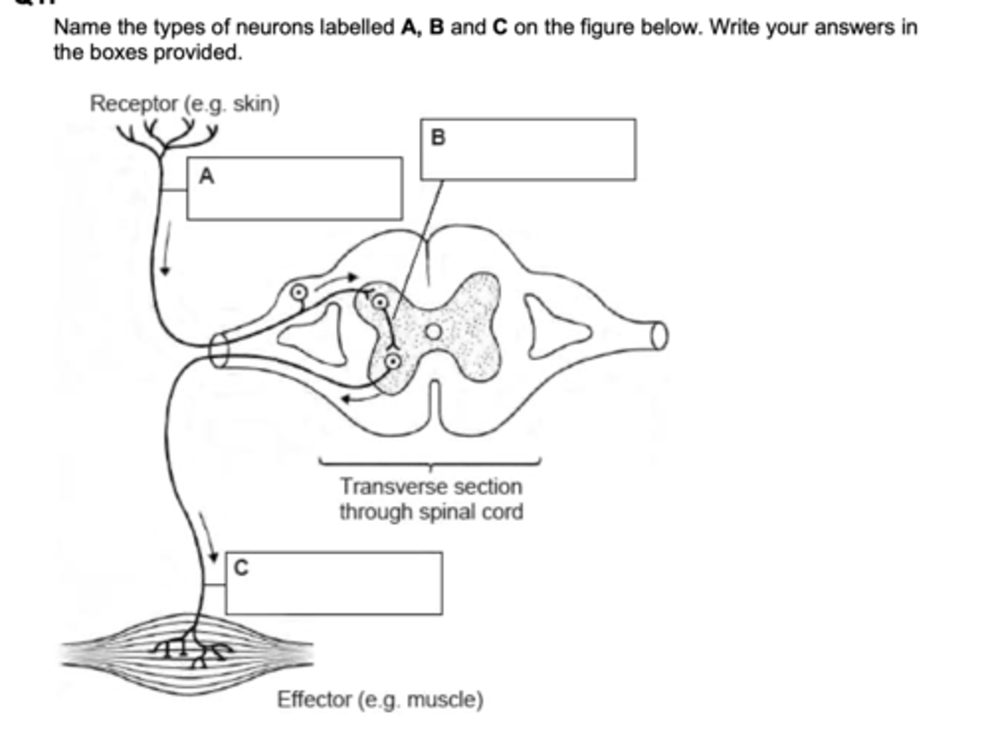
Jeremy is digging in the garden. He feels the spade hit a rock and stops digging
Explain how sensory, relay and motor neurons would function in this situation.
Sensory neurons send information from the senses to the brain - here receptors in Jeremy's hand would sense the jolt of the spade hitting the rock and send that information via the peripheral nervous system to his brain/CNS.
Relay neurons connect with other neurons, mostly found in the brain/CNS
- here they would be involved in analysis of the sensation, what it means, deciding about how to respond to it, thus acting between the sensory and motor neurons.
Motor neurons send messages via long axons from the brain to the muscles or effectors
- here the message from the brain instructs Jeremy's arm muscles to stop working and stop the digging action.
Raoul has recently been prescribed a drug for a mental illness. He looks on the internet to find out more about the drug but he does not understand the phrase 'synaptic transmission.
Write a brief explanation of synaptic transmission in the brain to help Raoul understand how his drug might work.
Synaptic transmission is how neurons pass messages using neurotransmitters across a tiny gap called the synapse.
Neurotransmitters are released from one neuron
They cross the synapse and bind to receptors on the next neuron
This can either excite or inhibit the next neuron
Raoul’s drug may:
Increase or decrease neurotransmitter levels
Block or boost receptor activity
Affect how neurotransmitters are broken down or taken back up
This helps change brain activity and improve symptoms.
Localisation of function in the brain:
+case study evidence
Point: The case study of KF, an amnesic patient, provides valuable evidence for the localization of function in the brain, particularly regarding memory processes.
Evidence: KF exhibited poor short-term memory (STM) for verbally presented digits; however, his recall improved significantly when the digits were presented visually for self-reading.
Analysis: This pattern suggests that different types of stimuli are processed by distinct areas of the brain, supporting the existence of separate auditory and visual memory centers. Specifically, it indicates that auditory stimuli are processed in areas associated with hearing, while visual stimuli are handled in areas related to vision, illustrating the brain's functional specialization.
Link: While the findings from KF's case support the theory of localized brain functions, the implications of this evidence should be considered carefully.
Counterpoint: A notable limitation is that KF's case may lack population validity. As an individual case study, the results may not be generalizable to the broader population, raising questions about the robustness of conclusions drawn from such specific evidence.
Further Link: Nevertheless, KF's case remains significant as it highlights the importance of studying individual differences in brain function, ultimately contributing to a deeper understanding of the complex nature of memory systems and the potential for rehabilitation in memory impairments.
Localisation of function in the brain:
+brain scan evidence
Point: Tulving's (1972) study utilizing PET scans provides crucial evidence for the localization of different memory types within specific brain regions.
Evidence: His findings indicated that episodic memory, which involves the recall of personal experiences, is associated with the right prefrontal cortex, while semantic memory, relating to general knowledge, is linked to the left prefrontal cortex.
Analysis: This differentiation suggests that distinct memory processes are localized in different areas of the brain, supporting the idea of functional specialization. The use of PET scans allows for real-time observation of brain activity, reinforcing the notion that various cognitive functions, such as memory recall, are distributed across specific brain regions.
Link: Tulving's research enhances our understanding of how different types of memory are processed in the brain, supporting the concept of localized functions.
Counterpoint: However, one limitation of PET scans is that they provide substantially lower resolution than imaging techniques like CT and MRI. PET scans are generally poor at delineating anatomical detail, which can lead to inaccurate localization of lesions and unclear demarcation of lesion borders.
Further Link: Despite these limitations, Tulving's work remains pivotal in the field of neuropsychology, as it illustrates the importance of employing various methodologies to investigate brain function, while also highlighting the need for complementary techniques to achieve a more comprehensive understanding of the brain's complex structures and their roles in memory.
Localisation of function:
-contradictory research
Point: Lashley's (1950) research provides contradictory evidence to the theory of localized brain functions, challenging the notion that specific brain areas are essential for particular memory tasks.
Evidence: In his experiments, Lashley removed between 10% to 50% of the cortex in rats while they were learning to navigate a maze. He found that no single area of the cortex was more critical than others for successful maze learning.
Analysis: These findings suggest that memory may not be strictly localized but rather distributed across various areas of the brain, indicating a more holistic approach to brain function. Lashley’s results imply that, to some extent, other parts of the brain can compensate for the loss of function, contradicting the idea that certain regions are solely responsible for specific cognitive tasks.
Link: Lashley's work raises important questions about the validity of strict localization theories and suggests a more interconnected understanding of brain function.
Counterpoint: However, one major limitation of Lashley’s research is the issue of extrapolation. The study involved animal subjects, specifically rats, which may not accurately reflect human brain function and memory processes, limiting the generalizability of the findings to human cognition.
Further Link: Nonetheless, Lashley’s findings remain significant as they encourage further investigation into the complexities of brain function and memory, emphasizing the need for a nuanced understanding of how cognitive processes are distributed and interrelated across different brain regions.
Evaluation for the localisation of function.
(+)- Brain scan evidence: Brain scans show that different areas of the brain are responsible for different functions. Eg Brocas area was activated during a reading task.
(+)- Neurosurgical evidence: Suggests that mental health disorders are localised. For example OCD can be treated using cingulotomy suggesting that the cause for OCD may be located in the cingulate gyrus.
(-)- Plasticity: the idea that after trauma the brain can regain its original function in another area of the brain goes against the idea of localisation as it suggests the brain is equipotential.