Week 12: MANUAL DIFFERENTIAL WBC COUNT
1/40
There's no tags or description
Looks like no tags are added yet.
Name | Mastery | Learn | Test | Matching | Spaced | Call with Kai |
|---|
No analytics yet
Send a link to your students to track their progress
41 Terms
MANUAL DIFFERENTIAL WBC COUNT
PRINCIPAL (TURGEON)
● A stained smear is examined to determine the
percentage of each type of leukocyte present and
assess the erythrocyte and platelet morphology.
Peripheral blood, bone marrow, or body fluid sediments
Manual differential WBC count specimen are obtained from?
Ethylenediaminetetraacetic acid tube
manual differential blood should be collected in what container?
within 1 hour
Smears should be made how long from collection in ETA specimens?
room temperature/25 °C
what temperature should manual diff. count specimen be stored?
true
true or false: Unstained smears can be stored for indefinite periods, but stained smears gradually fade
Back-and-forth serpentine/battlement track pattern
pattern used to read WBCs
Oil immersion objective
best/preferred lens when counting WBCs
100 WBCs
Number of WBCs that are counted and classified through the use of pushdown button counters or newer computer interfaced keypads
Neutrophils
Majority of WBCs in adults are _
Lymphocytes
Majority of WBCs in children are _
SCHILLING’S HEMOGRAM
In this method, all the leukocytes (granulocytes and agranulocytes) are classified and grouped into: (according to maturity).
Polymorphonuclear neutrophils
What are these?:
○ Myelocytes (if seen, possible of cancer)
○ Metamyelocytes (if seen, possible of cancer)
○ Bands or stabs (normal in peripheral blood)
○ Segmenters
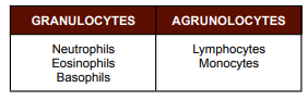
Neutrophils, Eosinophils, Basophils
Enumerate granulocytes from most to least
Lymphocytes, monocytes
Enumerate agranulocytes from most to least
Myelocytes, Metamyelocytes
Polymorphonuclear neutrophil that if seen, possible indication of cancer
Bands/stabs
Polymorphonuclear neutrophil that is normally seen in peripheral blood
Myelocytes
Metamyelocytes
Bands/stabs
Segmenters
Enumerate Polymorphonuclear neutrophil according to maturity of cells
ARNETH’S CLASSIFICATION
● In this method, Polymorphonuclear neutrophils (PMNs) are classified according to the number of lobes which their nuclei possess.
● The more lobes, the older the cells.
From Discussion:
● >5 lobes are hypersegmented neutrophils (olds cells)
● 2–5 lobes are normal lobes
HADEN’S CLASSIFICATION
This method classifies the neutrophils according to the presence of filaments. And classifies under FILAMENTED more mature vs NON-FILAMENTED cells or blast/immature cells
Filamented
more mature classification under Haden’s classification
Non-filamented
immature cells using Haden’s classification
Monocytes
have irregular cytoplasms and are sometimes vacuolated (because they phagocytes)
True
True of false: To increase accuracy, it is advisable to count at least 200 cells when the WBC count is higher than 40 x 109 /L.
● If the WBC count is 100 x 109 /L or greater, it would be more precise and accurate to count 300 or 400 cells.
Formula: (Total count / total cells counted) x 100
Poor staining
MOST COMMON ERROR IN DIFFERENTIAL WBC COUNT
Sampling error, Inadequate mixing, Poor staining, Errors in cell identification
enumerate: ERRORS IN DIFFERENTIAL WBC COUNT
(SeIm PEid)
Flow cytometry
Electronic counting WBC most common principle employed in machines
Speed of performance, Elimination of visual fatigue, Improved precision
Enumerate electronic counting advantages
nucleated RBC, unlysed RBC, cryoglobin, platelet clumping, heparin, Monoclonal CHON
ERRONEOUS RESULTS IN AUTOMATED CELL COUNTS (WBC COUNT): Causes of Suprious INCREASE
clotting of specimen, smudge cells, uremia
ERRONEOUS RESULTS IN AUTOMATED CELL COUNTS (WBC COUNT): Cause of Spurious DECREASE
(CS, SC, U)
Cryoglobin
atypical proteins in blood
Platelet clumping
occur as EDTA induced: EDTA causes platelet aggregation
● Recollection using heparin tubes
50-70%
Reference intervals for Neutrophils %
18-42%
Reference intervals for Lymphocytes %
2-11%
Reference intervals for Monocytes %
1-3%
Reference intervals for Eosinophils
0-2%
Reference intervals for Basophils
Shift to the left
Increase in young forms of cells and has 2 types: Degenerative shift to the left & Regenerative shift to the right
Degenerative shift to the left
● Increase in the number of young forms
● Cause: Normal or decreased WBC count
Regenerative shift to the left
● Increase in the number of young forms
● Cause: Increased WBC count
● Occurs mostly in cancer patients
Shift to the right
Increase in the number of old forms of cells