Muscle Anatomy and Strengthening Exercises: Head, Neck, Chest, Back, Limbs
1/63
There's no tags or description
Looks like no tags are added yet.
Name | Mastery | Learn | Test | Matching | Spaced | Call with Kai |
|---|
No analytics yet
Send a link to your students to track their progress
64 Terms
Frontalis
Location: On the front of the head, In the forehead. Bones Associated: Frontal Bone on the Cranium. Origin: Epicranal Aponeurosis. Insertion: Skin of the eyebrows, blends in with Orbicularis Oculi.
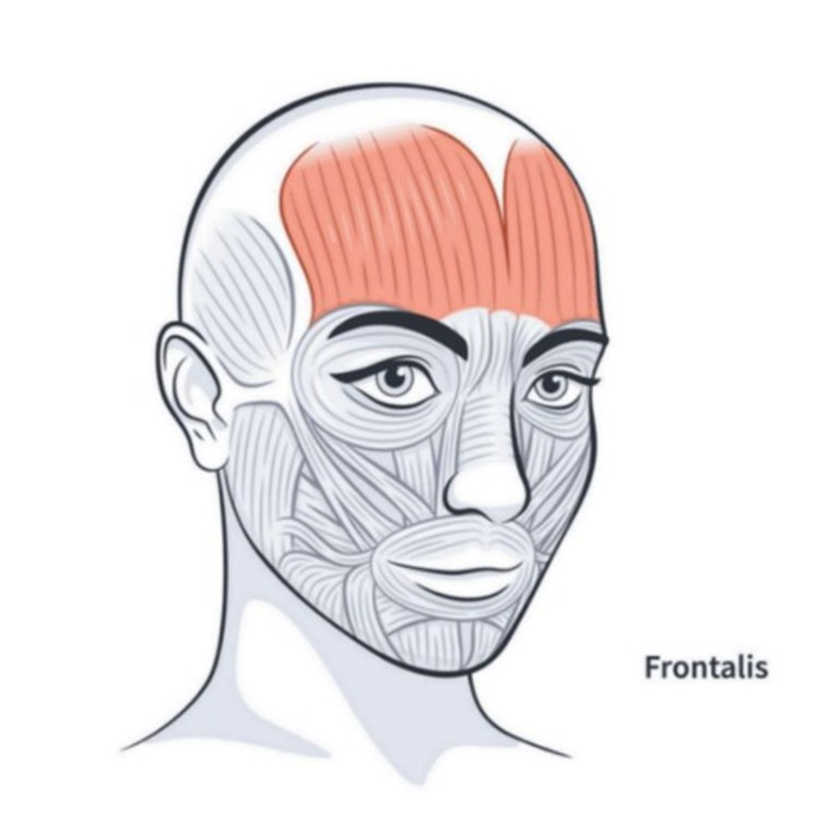
Resistance Brow Lifts
Uses forehead muscles to lift eyebrows up against the force of your hands/fingers.
Orbicularis Oculi
Location: Around the eyes. Bones Associated: Frontal & maxilla. Origin: Medial orbit. Insertion: Eyelid skin.
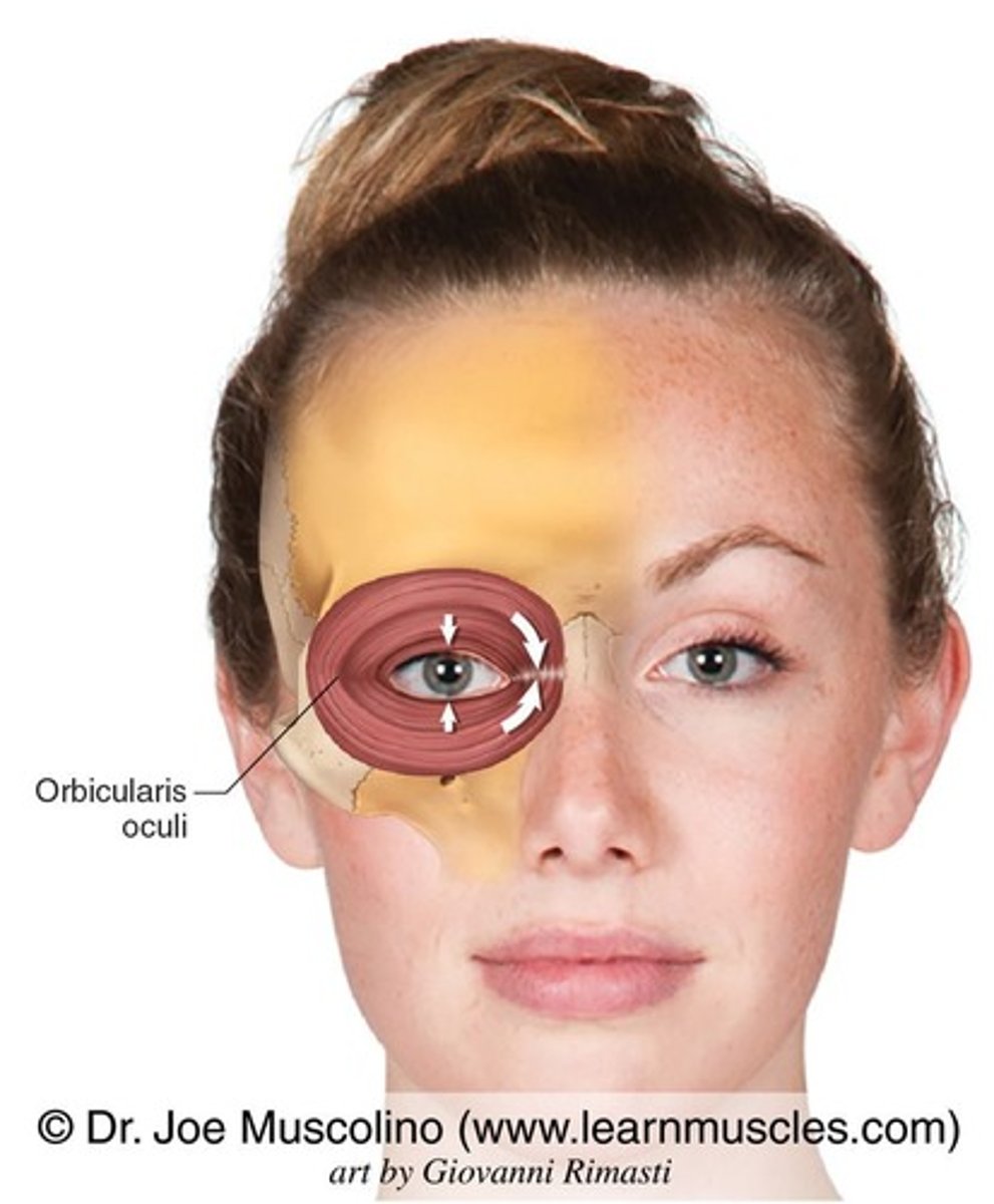
Tight Eye Squeezes
Forcefully closing the eyelids causes concentric contraction of the circular muscle fibers, strengthening them as they generate tension to fully compress the eyelids.
Orbicularis Oris
Location: Around the mouth. Bones Associated: Maxilla, mandible. Origin: Maxilla & mandible. Insertion: Lips.
Lip Puckers
Puckering or pressing lips tightly together activates the circular fibers around the mouth. The resistance created by air pressure or hand pressure makes the muscle contract harder, building strength.
Sternocleidomastoid
Location: Side of neck. Bones Associated: Sternum, clavicle, temporal bone. Origin: Sternum & clavicle. Insertion: Mastoid process.
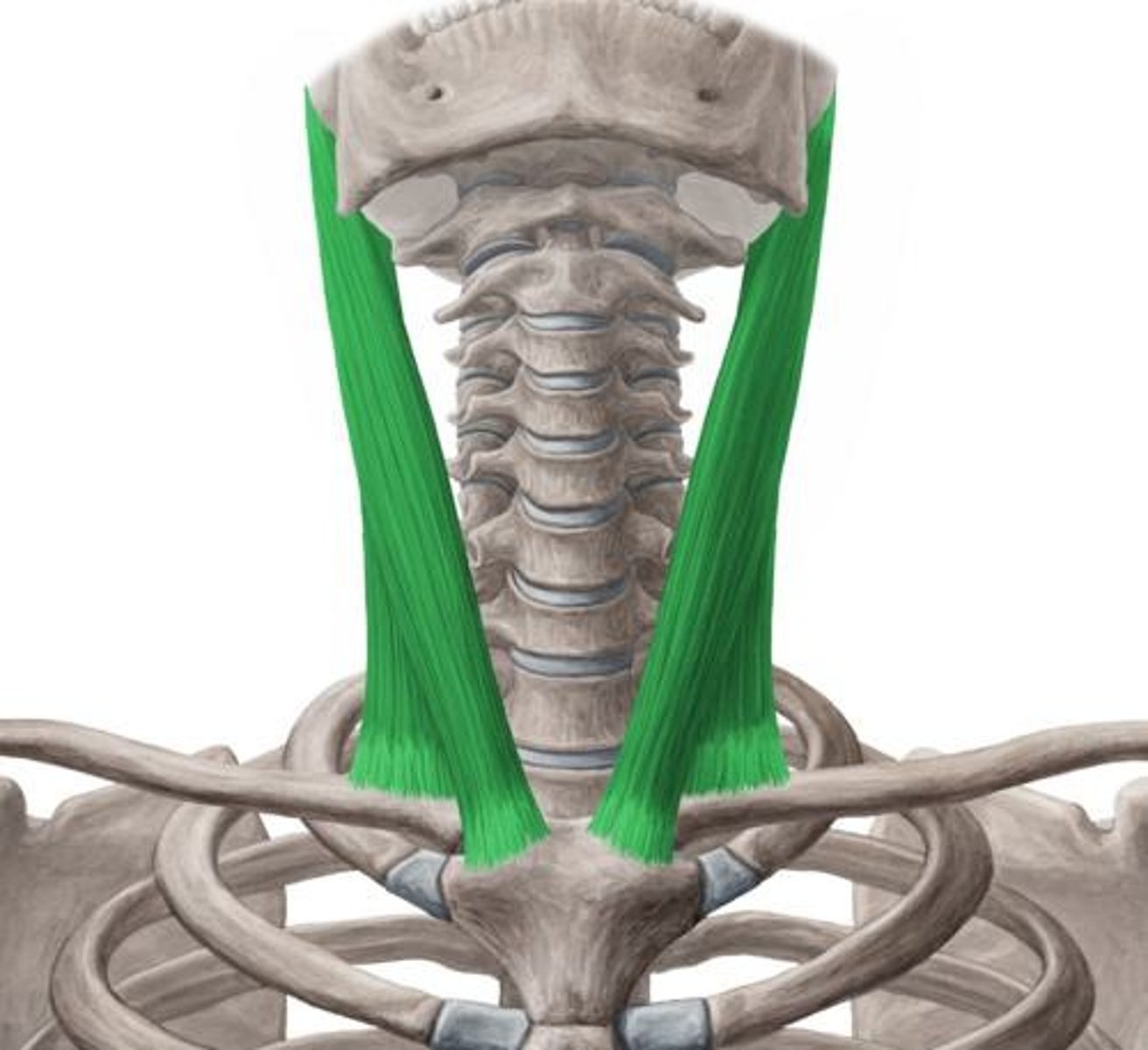
Neck Resistance Rotation
Pushing your head against your hand while rotating recruits the SCM to turn the head against resistance. This eccentric and concentric loading strengthens the muscle.
Platysma
Location: Front of neck. Bones Associated: Mandible, clavicle. Origin: Chest fascia. Insertion: Mandible & lower lip skin.
Platysma Tighten-Lifts
Lowering the jaw and tightening the neck skin forces the platysma to contract strongly, strengthening the thin muscle fibers through repeated tension.
Deltoid (3 headed)
Location: Shoulder cap. Bones Associated: Clavicle, scapula, humerus. Origin: Clavicle & scapula. Insertion: Deltoid tuberosity of humerus.
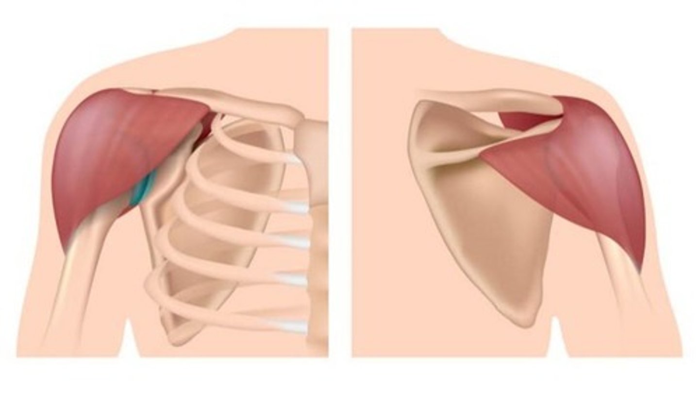
Lateral raises
Lifting the arm sideways loads the deltoid fibers concentrically to raise the arm and eccentrically to lower it, strengthening the shoulder.
Pectoralis Major
Location: Chest. Bones Associated: Sternum, clavicle, humerus. Origin: Clavicle & sternum. Insertion: Humerus (greater tubercle).
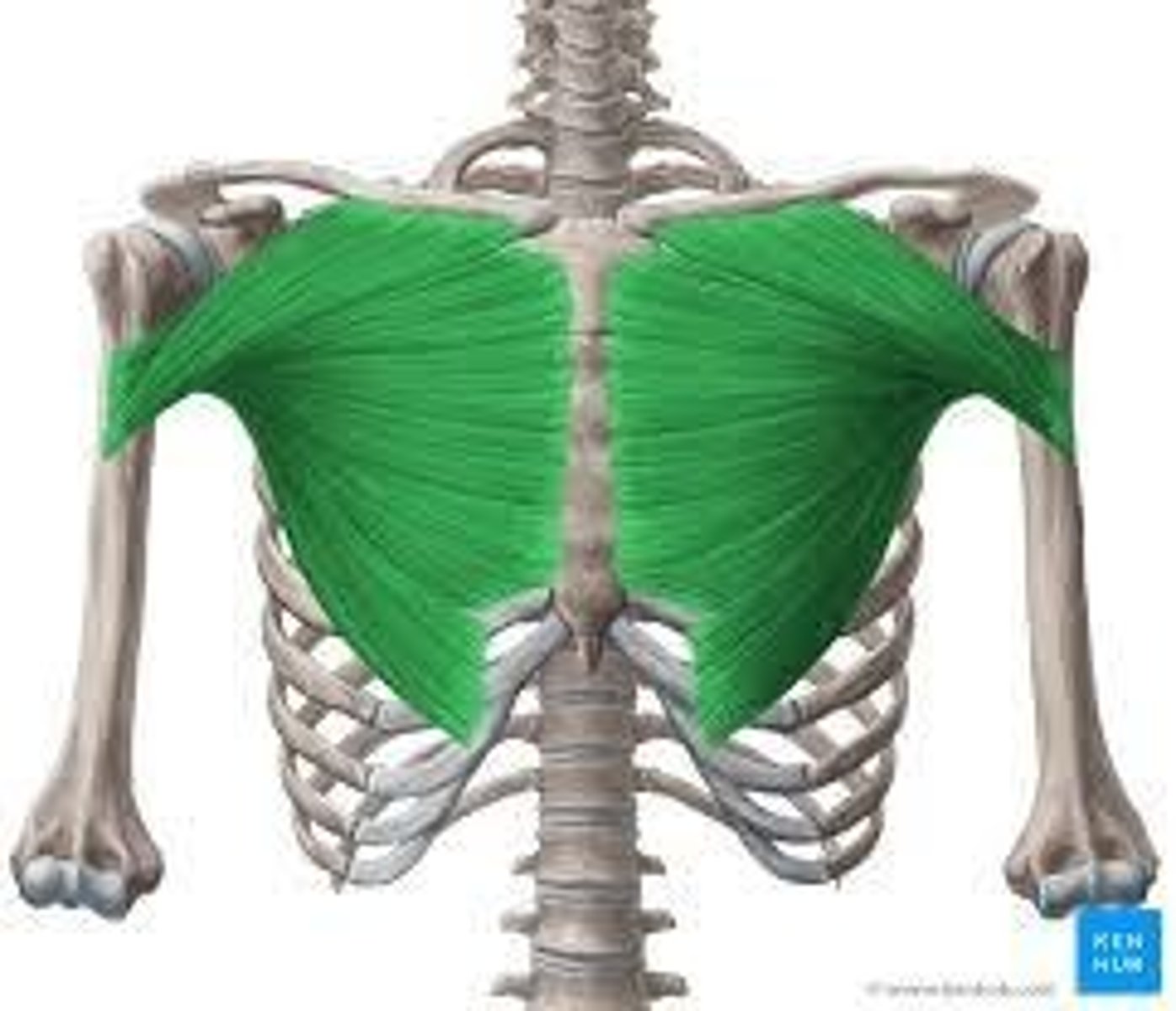
Push-ups
Pushing the body upward requires strong horizontal adduction of the arms, activating pectoral fibers under resistance.
Internal Intracostals
Location: Deep rib spaces. Bones Associated: Ribs. Origin: Rib above. Insertion: Rib below.
Forced exhalation drills
Blowing air out forcefully contracts internal intercostals to compress rib cage, strengthening expiratory muscle action.
External Intracostals
Location: Outer rib spaces. Bones Associated: Ribs. Origin: Rib above. Insertion: Rib below.
Deep inhalation breathing
Expanding the chest during deep breaths forces these muscles to lift ribs, strengthening inspiratory movement.
Rectus Abdominus
Location: Front abdomen. Bones Associated: Pubis, ribs. Origin: Pubic crest. Insertion: Ribs 5-7 & xiphoid process.
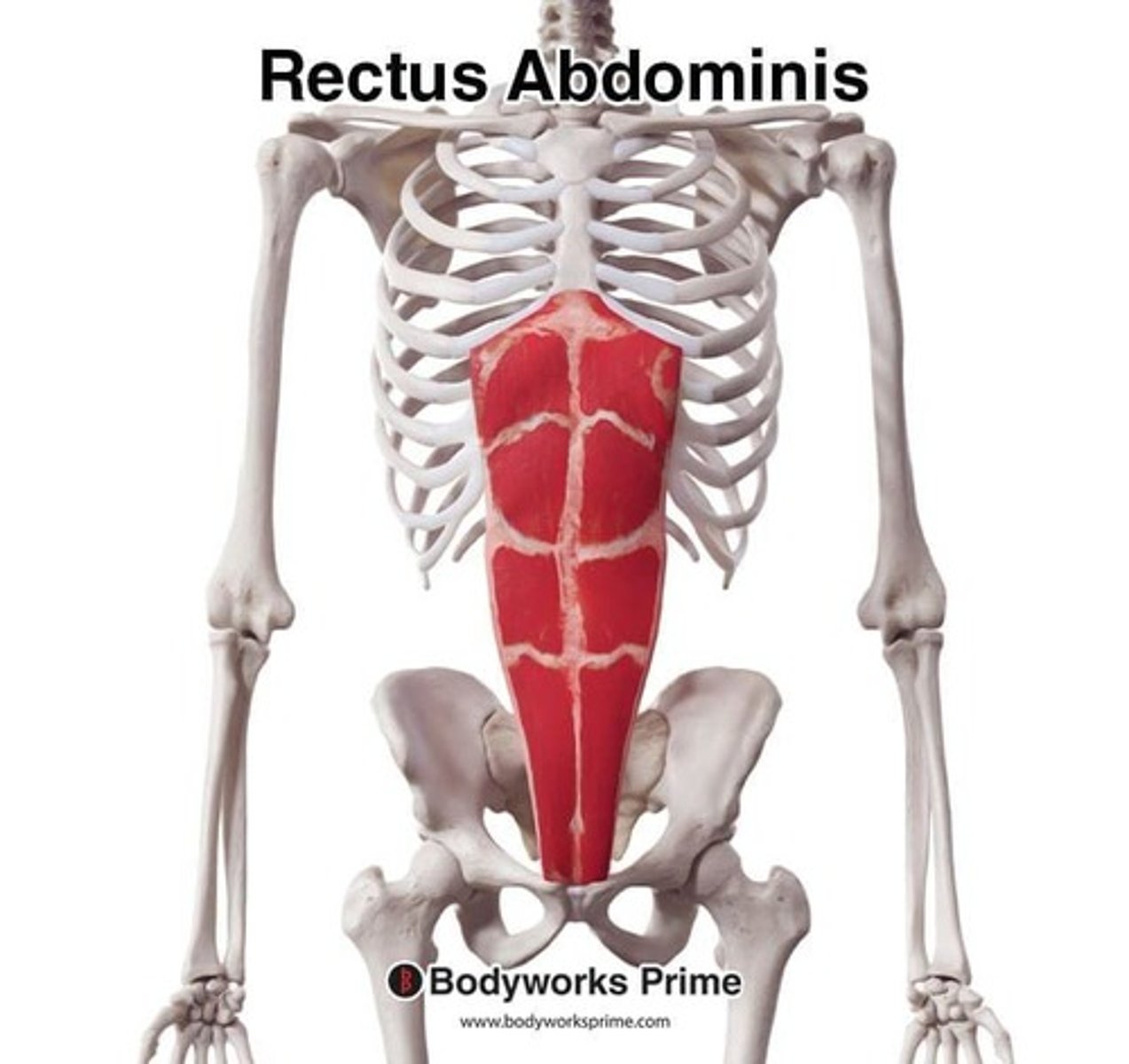
Crunches
Curling the trunk forward forces the muscle to shorten, strengthening spinal flexion through repeated contraction.
Internal Obliques
Location: Deep side abdomen. Bones Associated: Ilium, ribs. Origin: Iliac crest. Insertion: Ribs 10-12 & linea alba.
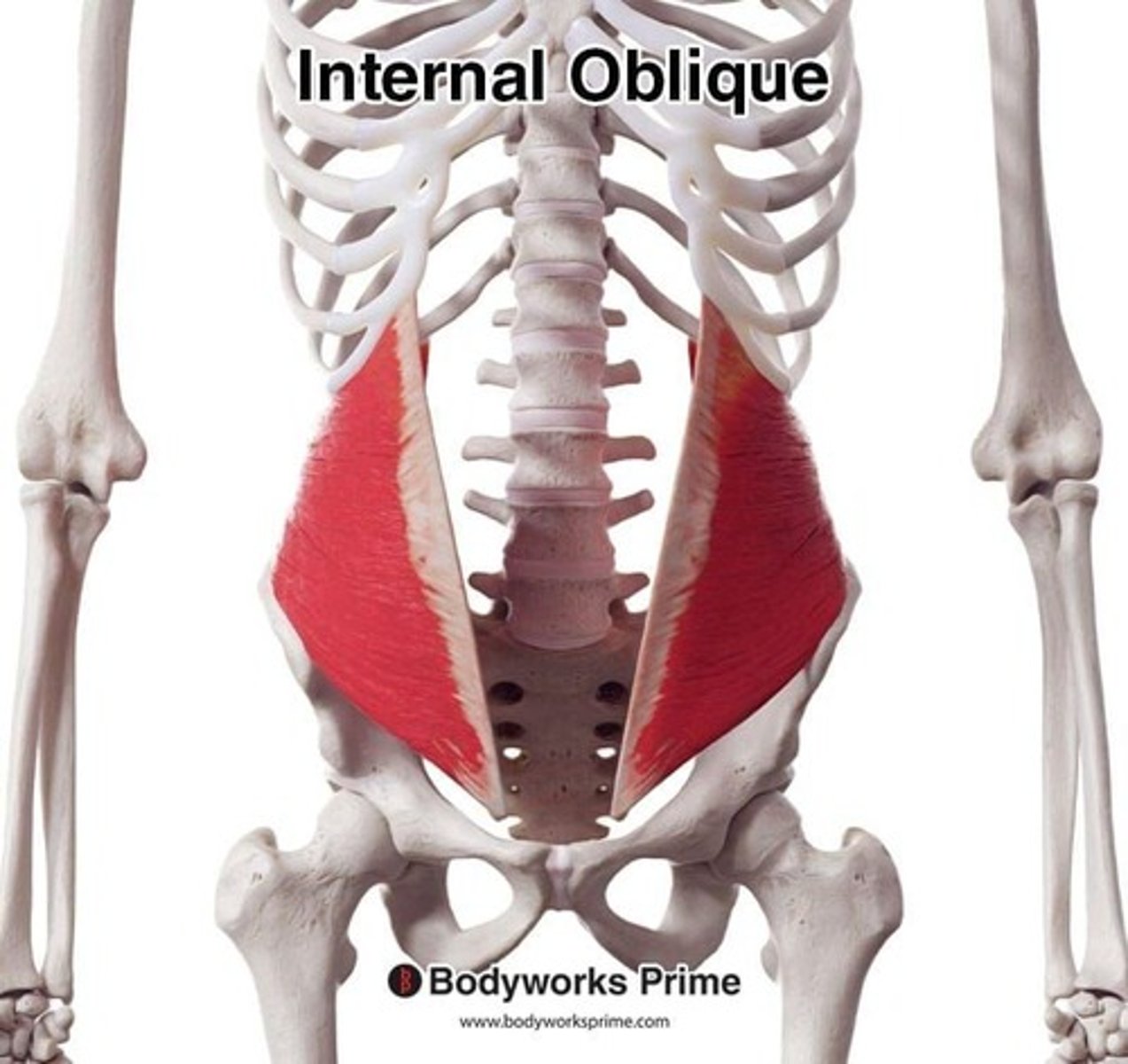
Bicycle crunches
Opposite elbow-to-knee motion activates internal obliques in trunk rotation and flexion under load.
External Obliques
Location: Side abdomen. Bones Associated: Ribs, ilium. Origin: Ribs 5-12. Insertion: Iliac crest & linea alba.
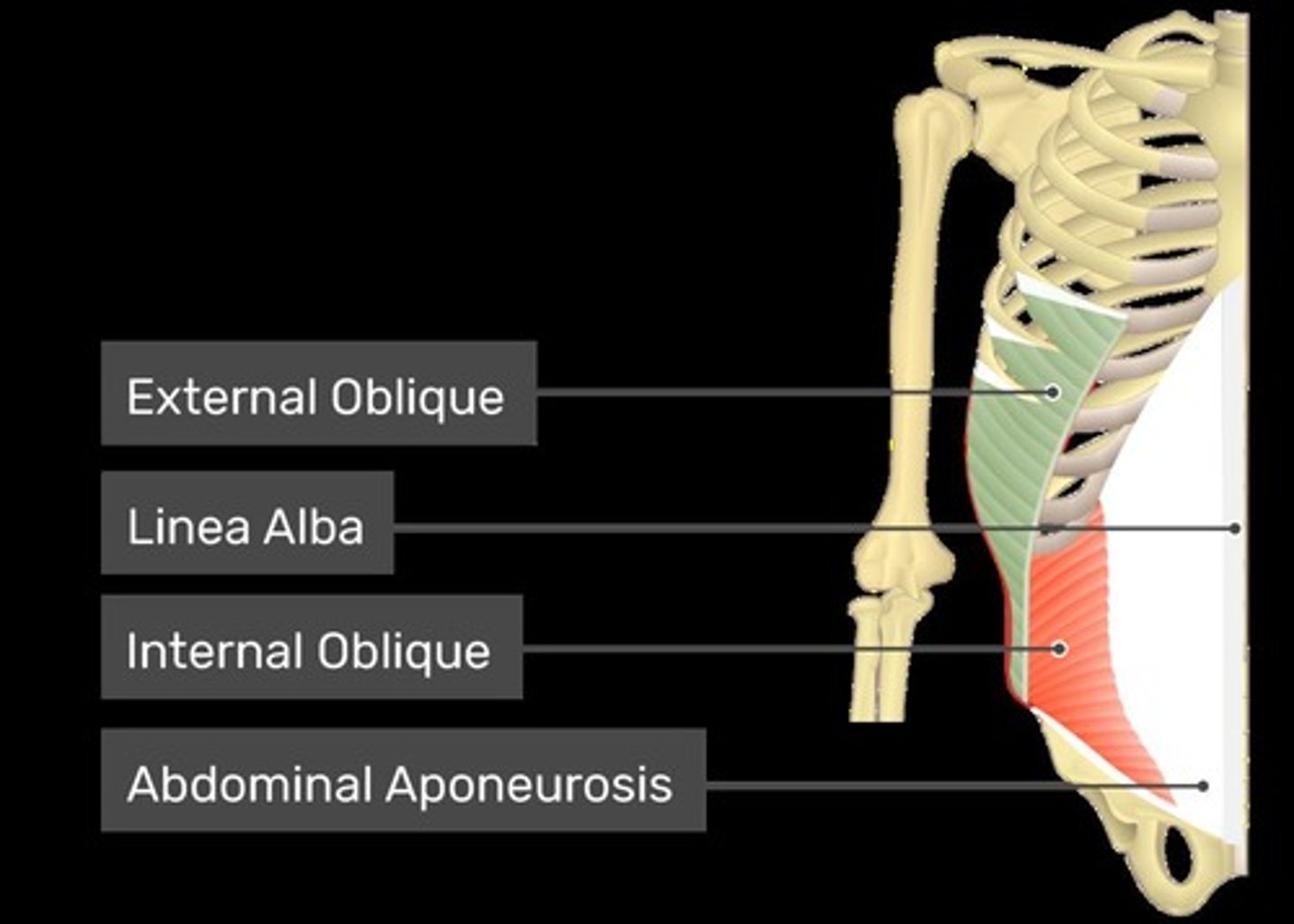
Russian twists
Rotating the torso engages obliques concentrically and stabilizes core eccentrically as the weight moves side to side.
Triceps Brachii
Location: Front upper arm. Bones Associated: Scapula, radius. Origin: Scapula. Insertion: Radius.
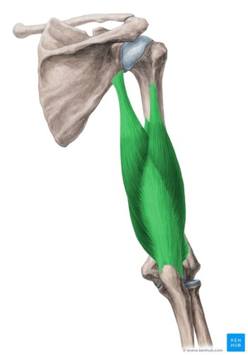
Bicep curls
Flexing the elbow against weight loads the biceps concentrically while lowering the weight strengthens through eccentric control.
Biceps Brachii
Location: Back upper arm; Bones Associated: Scapula, humerus, ulna; Origin: Scapula & humerus; Insertion: Olecranon of ulna.
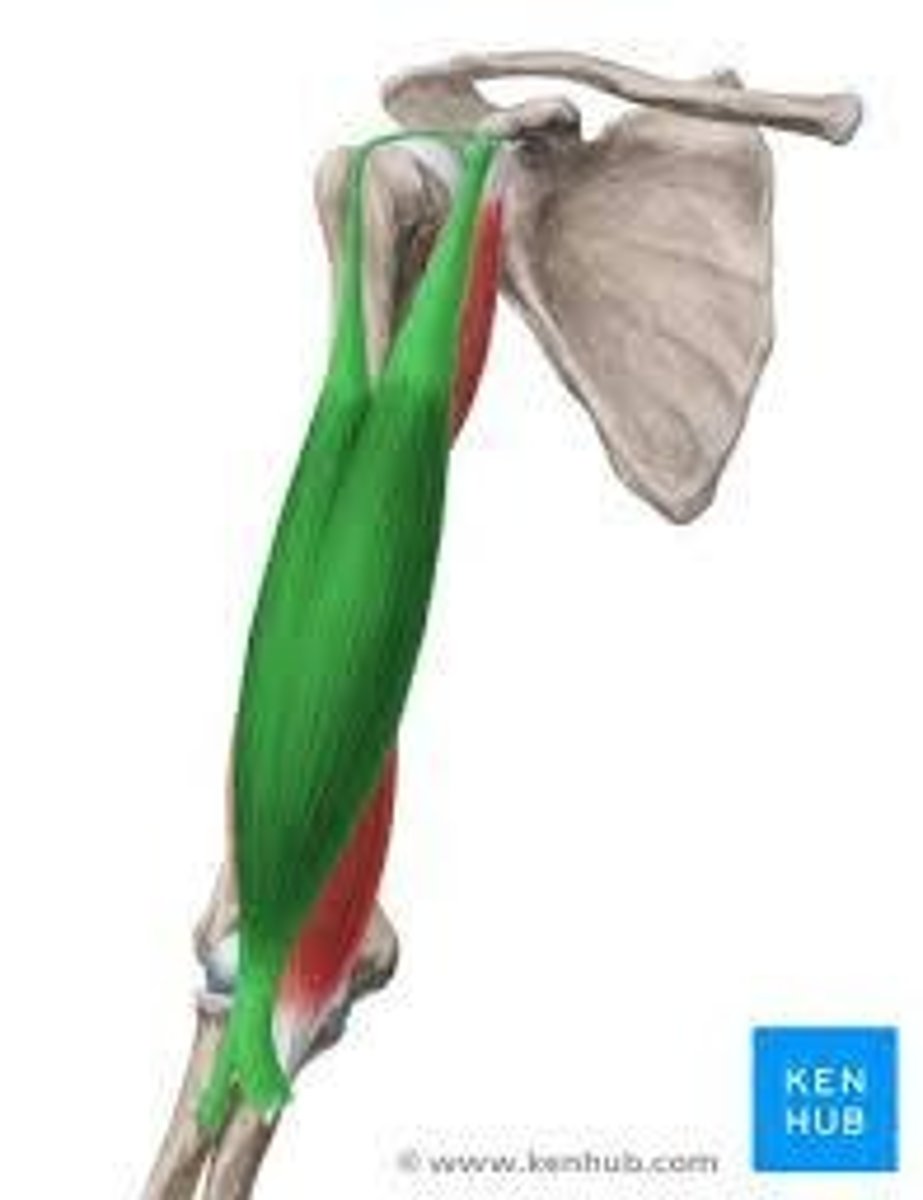
Tricep dips
Exercise: Extending the elbow to push the body upward contracts triceps against bodyweight resistance.
Pronator Teres
Location: Anterior forearm; Bones Associated: Humerus, radius; Origin: Humerus & ulna; Insertion: Radius.
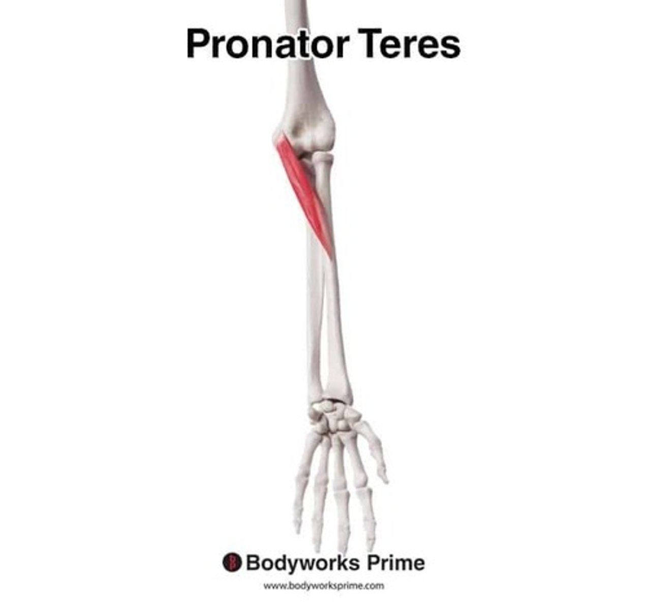
Resistance forearm pronation
Exercise: Turning the palm downward against resistance makes pronator teres work harder to rotate the radius over the ulna.
Flexor Carpi Radialis
Location: Anterior forearm (thumb side); Bones Associated: Humerus, metacarpals; Origin: Medial epicondyle; Insertion: 2nd-3rd metacarpals.
Wrist flexion
Exercise: Curling the wrist upward against weight loads the flexor tendons, strengthening wrist flexion.
Extensor Carpi Ulnaris
Location: Posterior forearm (pinky side); Bones Associated: Humerus, ulna, metacarpal; Origin: Lateral epicondyle; Insertion: 5th metacarpal.
Wrist extension
Exercise: Lifting the wrist upward against resistance strengthens the extensor group by loading extension.
Iliopsoas
Location: Deep hip flexor; Bones Associated: Lumbar vertebrae, ilium, femur; Origin: Iliacus & psoas origins; Insertion: Lesser trochanter.
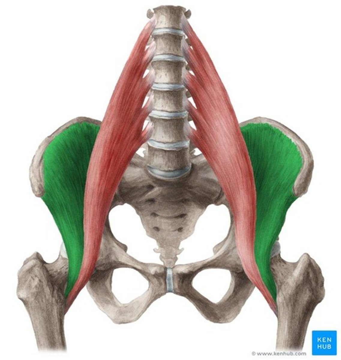
Leg raises
Exercise: Lifting the leg upward requires hip flexion; the iliopsoas contracts intensely to raise and control the leg.
Sartorius
Location: Runs diagonally across thigh; Bones Associated: Ilium, tibia; Origin: Anterior iliac spine; Insertion: Tibia.
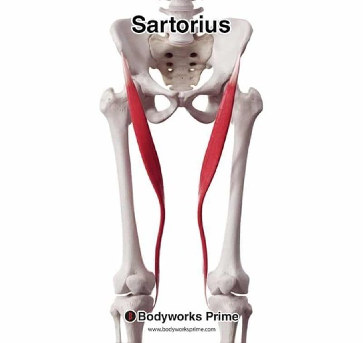
High knee marches
Exercise: Lifting knee engages sartorius in hip flexion and knee movement under repeated contraction.
Adductor Longus
Location: Inner thigh; Bones Associated: Pubis, femur; Origin: Pubis; Insertion: Femur.
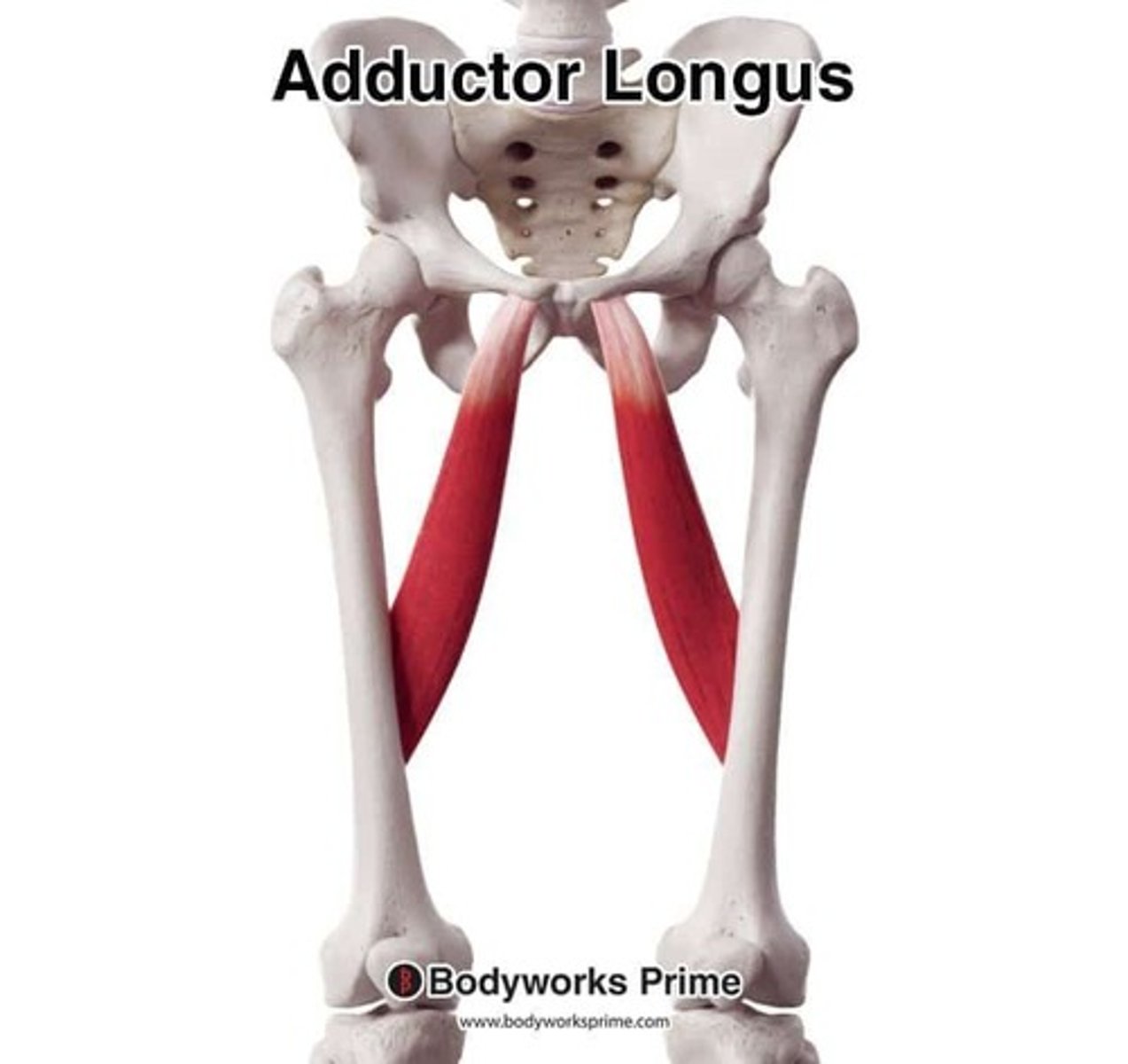
Adductor squeezes
Exercise: Squeezing legs together against resistance activates hip adduction, strengthening inner thigh fibers.
Adductor Magnus
Location: Deep inner thigh; Bones Associated: Pubis, ischium, femur; Origin: Pubis & ischium; Insertion: Femur.
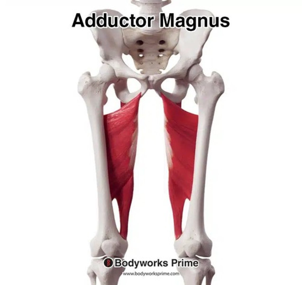
Side lunges
Exercise: Returning from lateral lunge requires strong hip adduction, engaging adductor magnus under load.
Gracilis
Location: Inner thigh surface; Bones Associated: Pubis, tibia; Origin: Pubis; Insertion: Tibia.
Band adduction
Exercise: Pulling leg inward against band tension loads gracilis in hip adduction.
Quadriceps
Location: Front thigh; Bones Associated: Femur, tibia; Origin: Femur & ilium; Insertion: Tibial tuberosity.
Squats
Exercise: Straightening the knee under bodyweight strengthens the quads through resisted knee extension.
Hamstrings
Location: Back of the thigh; Bones Associated: Pelvis (ischial tuberosity), femur, tibia, and fibula; Origin: The ischial tuberosity; Insertion: The proximal ends of the tibia.
Romanian Deadlifts (RDLs)
Exercise: Bending the knee (knee flexion) and extending the hip (hip extension). They also act eccentrically to control and decelerate the leg as it straightens during movements like running and walking.
Tibialis Anterior
Location: Front shin; Bones Associated: Tibia, foot; Origin: Lateral tibia; Insertion: 1st metatarsal & medial cuneiform.
Toe raises
Exercise: Pulling toes upward loads dorsiflexion, strengthening tibialis anterior as it lifts the foot.
Gastrocnemius
Location: Calf; Bones Associated: Femur, calcaneus; Origin: Femur; Insertion: Calcaneus.
Calf raises
Exercise: Pushing heels upward contracts calf muscles powerfully to plantarflex the ankle under bodyweight.
Peroneus Brevis
Location: Deep lateral leg; Bones Associated: Fibula, foot; Origin: Fibula; Insertion: 5th metatarsal.
Lateral ankle raises
Exercise: Lifting outside of foot upward engages brevis to stabilize and evert the foot under tension.
Peroneus Longus
Location: Lateral lower leg; Bones Associated: Fibula, foot; Origin: Fibula; Insertion: 1st metatarsal.
Ankle eversion
Exercise: Turning foot outward against resistance strengthens lateral stabilizers by loading eversion.
Trapezius
Location: Upper back. Bones Associated: Scapula, clavicle, spine. Origin: Occipital bone & vertebrae. Insertion: Scapula & clavicle. Exercise: Shoulder shrugs. Mechanism: Lifting shoulders upward loads the upper traps; holding at the top increases isometric tension for strength.
Erector Spinae
Location: Along spinal column. Bones Associated: Vertebrae, ribs, pelvis. Origin: Iliac crest & vertebrae. Insertion: Vertebrae & ribs. Exercise: Back extensions. Mechanism: Extending the back upward forces erector spinae to straighten and support spine, strengthening postural control.
Infraspinatus
Location: Back of scapula. Bones Associated: Scapula, humerus. Origin: Infraspinous fossa. Insertion: Greater tubercle of humerus. Exercise: External band rotations. Mechanism: Rotating arm outward against resistance strengthens shoulder stabilizers by loading external rotation.
Teres Major
Location: Underside of scapula. Bones Associated: Scapula, humerus. Origin: Inferior angle of scapula. Insertion: Humerus. Exercise: Lat pulldowns. Mechanism: Pulling weight downward activates teres major as it assists internal rotation and adduction under resistance.
Teres Minor
Location: Posterior shoulder. Bones Associated: Scapula, humerus. Origin: Lateral scapula. Insertion: Greater tubercle. Exercise: Band external rotations. Mechanism: Rotating arm outward engages teres minor in stabilizing humeral head during resisted movement.
Latissimus Dorsi
Location: Mid-lower back. Bones Associated: Spine, pelvis, humerus. Origin: Thoracic/lumbar spine & iliac crest. Insertion: Humerus. Exercise: Pull-ups. Mechanism: Pulling the body upward requires strong shoulder adduction and extension, heavily loading the lats.
Gluteus Medius
Location: Outer upper buttock. Bones Associated: Ilium, femur. Origin: Ilium. Insertion: Greater trochanter. Exercise: Side leg raises. Mechanism: Moving leg sideways against gravity strengthens hip abduction and stabilizing fibers.
Gluteus Maximus
Location: Buttocks. Bones Associated: Ilium, sacrum, femur. Origin: Ilium & sacrum. Insertion: Femur & IT band. Exercise: Squats. Mechanism: Standing up from squat position forces glutes to extend the hip powerfully under load.