Transport in animals
1/29
There's no tags or description
Looks like no tags are added yet.
Name | Mastery | Learn | Test | Matching | Spaced | Call with Kai |
|---|
No analytics yet
Send a link to your students to track their progress
30 Terms
Describe circulatory systems
Transport gases and nutrient around in a transport liquid
This liquid is transported around in vessels and there is a pump to move the liquid (heart)
describe open circulatory systems
Invertebrates have this (insects)
Transport medium (haemolymph) is pumped directly to open body cavity (haemocoel), few transports vessels
Transport medium is pumped at low pressure and transport food and nitrogenous waste - not gases, they are transported via teh tracheal system
Exchange takes place at cells and tissues, transport medium return to heart through through open ended vessel
Close circulatory system
Vertebrates and some invertebrates
Transport medium (blood) remains inside of vessels
Gases and small molecules leave blood by diffusion or high hydrostatic pressure
Transport oxygen and carbon dioxide , oxygen transported by a pigmented protein (hameoglobin)
Describe single closed circulatory systems
Blood only passes through heart once per cycle
Fish
Blood passes through two sets of capillaries
Immediately after pumped out of heart, blood flows through capillaries in gills to become oxygenated
Blood flows through capillaries delivery ig blood to body, then return to heart
Not enable efficient gas exchange for mammals, but work for fish due to counter current flow mechanism in gas exchange
Double closed circulatory system
Blood passes through heart twice per cycle
Birds and mammals
One circuit of blood vessels carry blood from heart to lungs for gas exchange
Second circuit carries blood from heart to rest of body to deliver oxygen and nutrients and collect waste
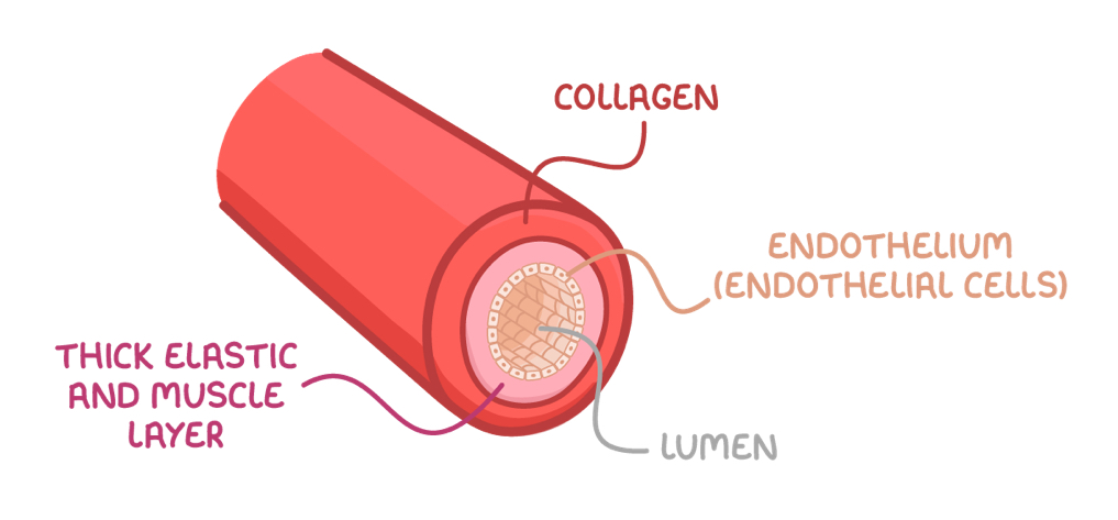
Describe the arteries
Collagen - provide strength to prevent vessel from bursting and to maintain vessel shape
Elastic fibres = elastin lets them stretch and recoil to minimise change in pressure
Thick smooth muscle layer = contracts/ relax to dilate lumen and control blood flow
Describe arterioles
Smaller than arteries, with larger lumen
Their walls have more smooth muscle and less elastin as they withstand less pressure
What is vasoconstriction and vasodilation?
Vasoconstriction = smooth muscle contracts, constricts blood vessel and decreases blood flow
Vasodilation = smooth muscle relaxed, dilate blood vessel and increasing blood flows
Describe capillaries
Form extensive networks between arterioles and venules, area between blood and tissue where exchange of substances occur
Narrow lumen = red blood cells close to body cells
Thin walls =- substances can be exchanged across a short distance by diffusion
Highly branched = large SA for diffusion
Describe veins
Carry blood towards heart at low pressure
Collagen = provide strength to prevent vessel from bursting and maintain vessel shape
Little smooth muscle and elastic fibre = low blood pressure, and thin walls allow veins to be compressed
Valves = pocket valves shut to prevent back flow of blood when wins are squashed by surrounding skeletal muscles
Pocket valves are the only type of value controlled by skeletal muscle
Describe venules:
Venules are smaller than veins
Thin walls, very little smooth muscle
They have valves
What does blood consists of?
Plasma = mostly water, transports substances
red blood cells = carry oxygen
White blood cells = immune cells
Platelets = blood clotting
Function of blood
Transport oxygen and carbon dioxide
Transports nutrients from digestion
Transports waste for excretion
Transports hormones
Transports food for storage
Transports clotting factors
What is tissue fluid?
Fills space between space
Site of diffusion between blood and body cells, provide cells with nutrients and oxygen while also removing waste products
Helps fight infection
Tissue fluid vs plasma
Tissue fluid has no red blood cells
Few proteins
Few white blood cells
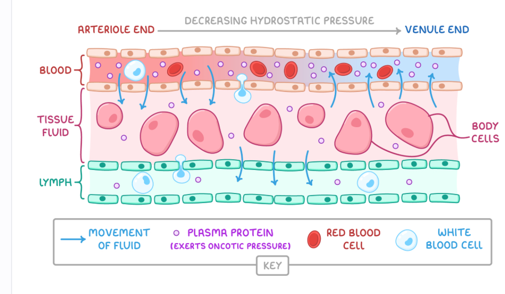
How is tissue fluid formed from arterioles end?
High hydrostatic pressure exerted by force of heart pumping forces fluid out of capillaries
Forms fluid tissue surrounding body cells
How is tissue fluid formed at the venule end of capillaries?
Hydrostatic pressure is lower
Proteins in blood exert a high oncotic pressure, type of osmotic pressure in capillaries
The water potential is lower in capillaries than in tissue fluid due to fluid loss
Some tissue fluid move back into capillaries by osmosis

What is lymph and its composition comapred to tissue fluid?
Fluid that flows around the lymphatic system via lymph vessels
Has less oxygen and nutrients
More fatty acids
More white blood cells
How is lymph formed and transported?
Formed from tissue fluid
Some tissue fluid doesn’t re enter capillaries from tissue fluid
This fluid drains lymph vessels forming lymph
Lymph is transported through lymph vessels by muscles contractions
It passes through lymph nodes to filter pathogens
Lymph returned to blood
Describe the heart chambers:
Human heart has 4 chambers
Left and right atria, left and right ventricle
Atria = top chambers in heart that collect blood from blood vessels (veins)
Ventricles = bottom chambers in heart that pump blood into blood vessels (arteries)
Describe the pumping mechanisms in the heart:
Left side has oxygenated blood + Right side has deoxygenate blood = two seperate pumping machines beacuse:
Blood pressure drops in lungs as it flows through capillaries
Single pump would slow the blood flow to body cells
Two pumps increase the pressure before blood circulates
What does septum do?
Seperate two sides of heart, prevent oxygenated and deoxygenated blood from mixing
Describe the atrioventricular valve in heart:
Tricuspid valve - located between right atrium and right ventricle
Bicuspid = left atrium and left ventricle
Both valves prevent back flow of blood into atria when ventricles contract
Describe the semi lunar valves
Located between the ventricles and pulmonary artery and the aorta
Prevent back flow of blood into ventricle when they relax
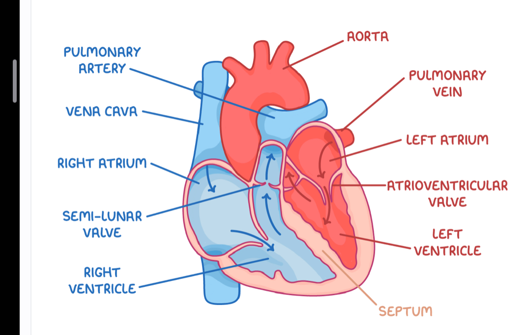
Diagram of heart
Pulmonary vein = moves oxygenated blood into left atrium from lungs
Aorta = moves oxygenated blood from left ventricle to body
Vena cava = moves deoxygenated blood into right atrium from body
Pulmonary artery = moves dpexyganetd blood from right ventricle to lungs
Why are the ventricle walls thicker than the atria walls
They have more muscle beacuse:
Atria only need enough pressure to pump blood a short distance into ventricle
Ventricle need more pressure to pump blood a long distance out of heart to other organs
Why is the left ventricle wall thicker than the right ventricle wall?
More muscle
Right ventricle needs enough pressure to pump deoxygenated blood a short distance to the lungs
Left ventricle needs a lot of pressure to pump oxygenated blood to other more distant organs of body
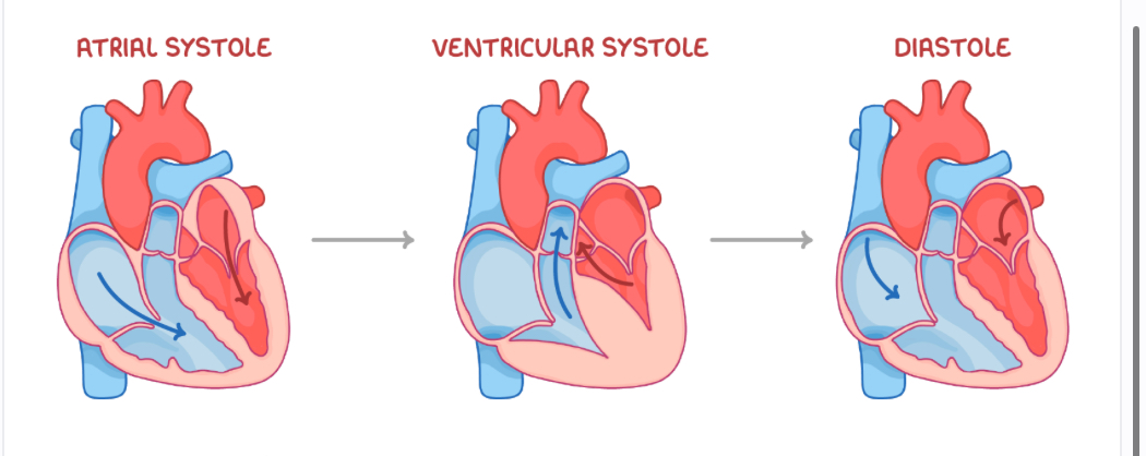
cardiac cycle - stage 1: atrial systole
cardiac cycle is the sequence of contraction and relaxation of cardiac muscle in the walls of the heart -
Ventricle relax, atria contract = increases atrial pressure
Atrioventricular valanced open = blood flows into ventricle
Cardiac cycle - stage 2 = ventricular systole
Ventricles contract, atria relax
Ventricular pressure increases
Semi lunar valves open and atrioventricular valves close
Blood flows into arteries
Cardiac cycle - stage 3 - diastole
Ventricles and atria relax
Semi lunar valves close
Blood flows passively into atria