WSU BIO 315: Lab 1 Exam; WEEK 3 TERMS
1/130
There's no tags or description
Looks like no tags are added yet.
Name | Mastery | Learn | Test | Matching | Spaced | Call with Kai |
|---|
No study sessions yet.
131 Terms
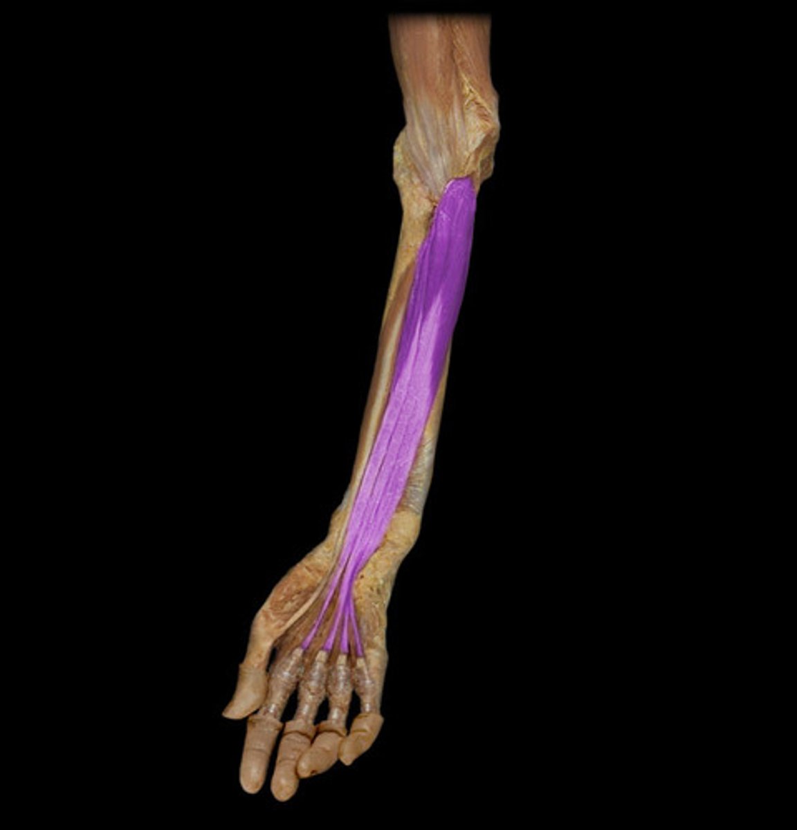
Flexor digitorum profundus
Deep inside arm, white strands in the cadaver, inserts on the distal phalanges
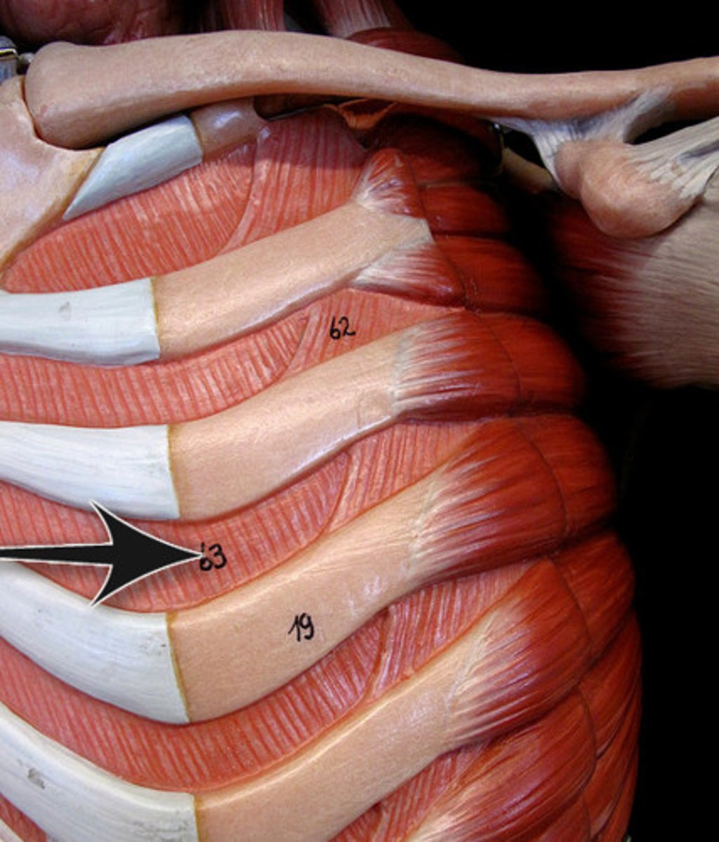
External Intercostal Muscles
Slant downward and forward between ribs- like putting your hands in your front pocket. Elevate ribs when you breathe in.
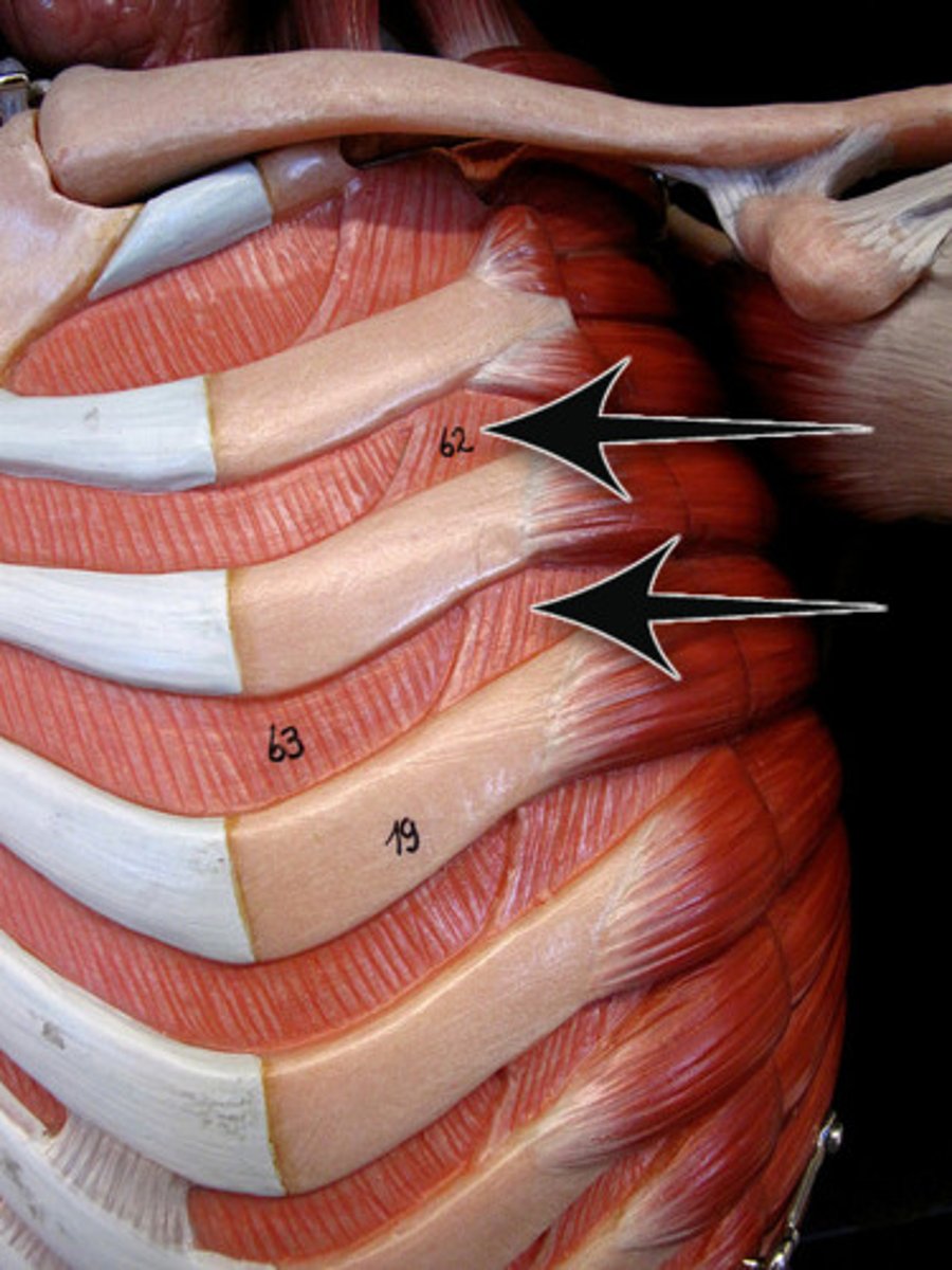
Internal Intercostal Muscles
Slant downward and outward. Depress ribs when you forcefully breathe out.
External Oblique Muscle
Slants same direction as external intercostal muscles.
Inguinal Ligament
BQ: What is the thick inferior edge of the oblique aponeurosis.
Hint: "V line"
Internal Oblique Muscle
Slants the same direction as internal intercostals muscles.
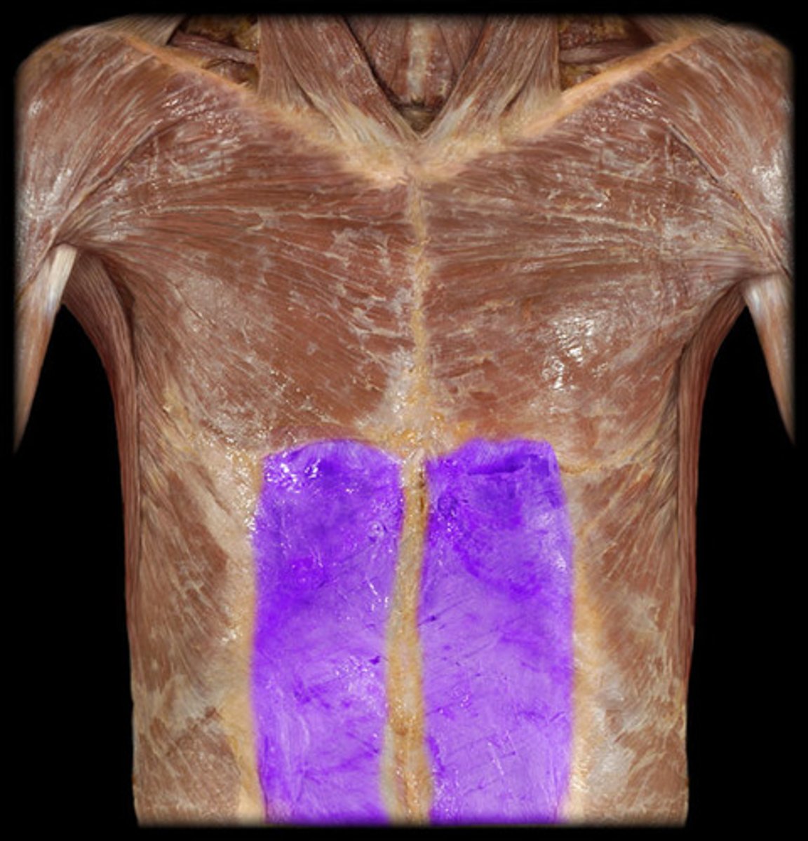
Transversus Abdominis
Fascicles run horizontally
Aponeuroses
BQ: Strong flat tendons are called ____________.
Linea Alba
-Division
"White line" where the abdominal muscles fuse
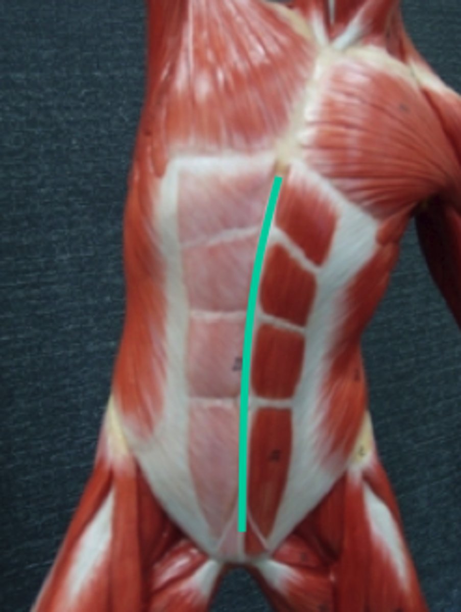
Rectus Abdominis
Fascicles run vertically, 6 pack of abs.
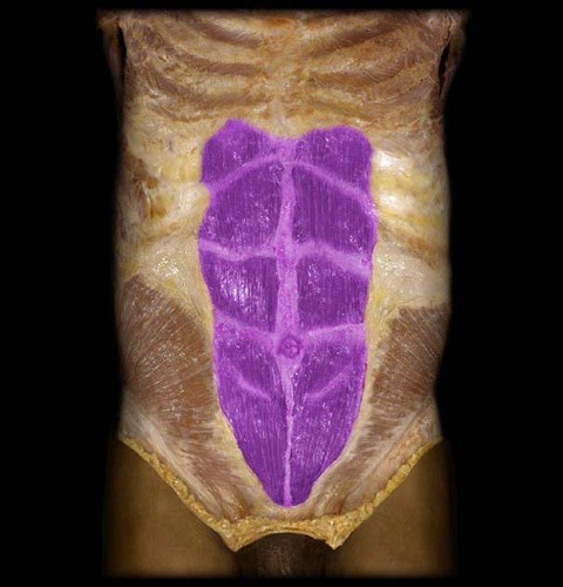
Tendinous Intersections
They break the "six pack" muscle into segments.

Rectus Sheath
Underneath the rectus abdominis muscles.
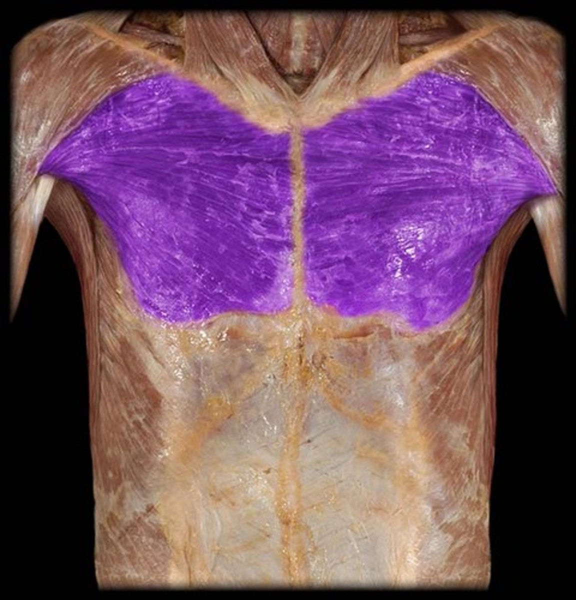
Pectoralis Major
Attaches the humerus to the clavicle and sternum.
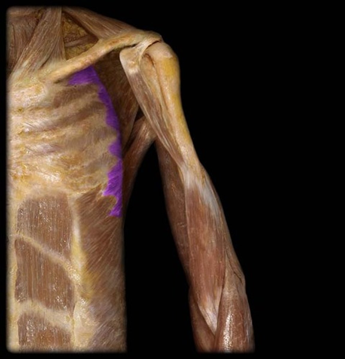
Pectoralis Minor
Attaches scapula to the ribs.
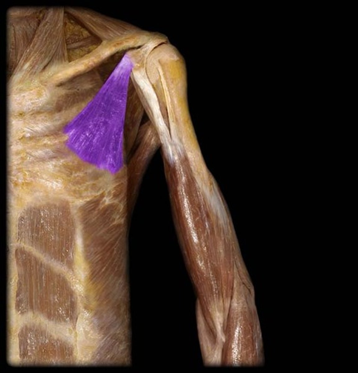
Serratus Anterior
Think saw, like teeth. Attaches the medial border of the scapula to the ribs.
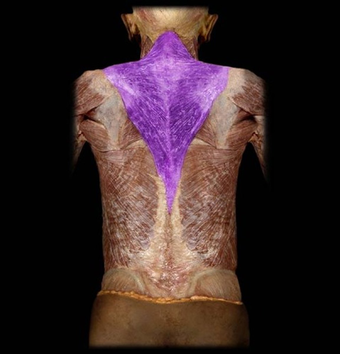
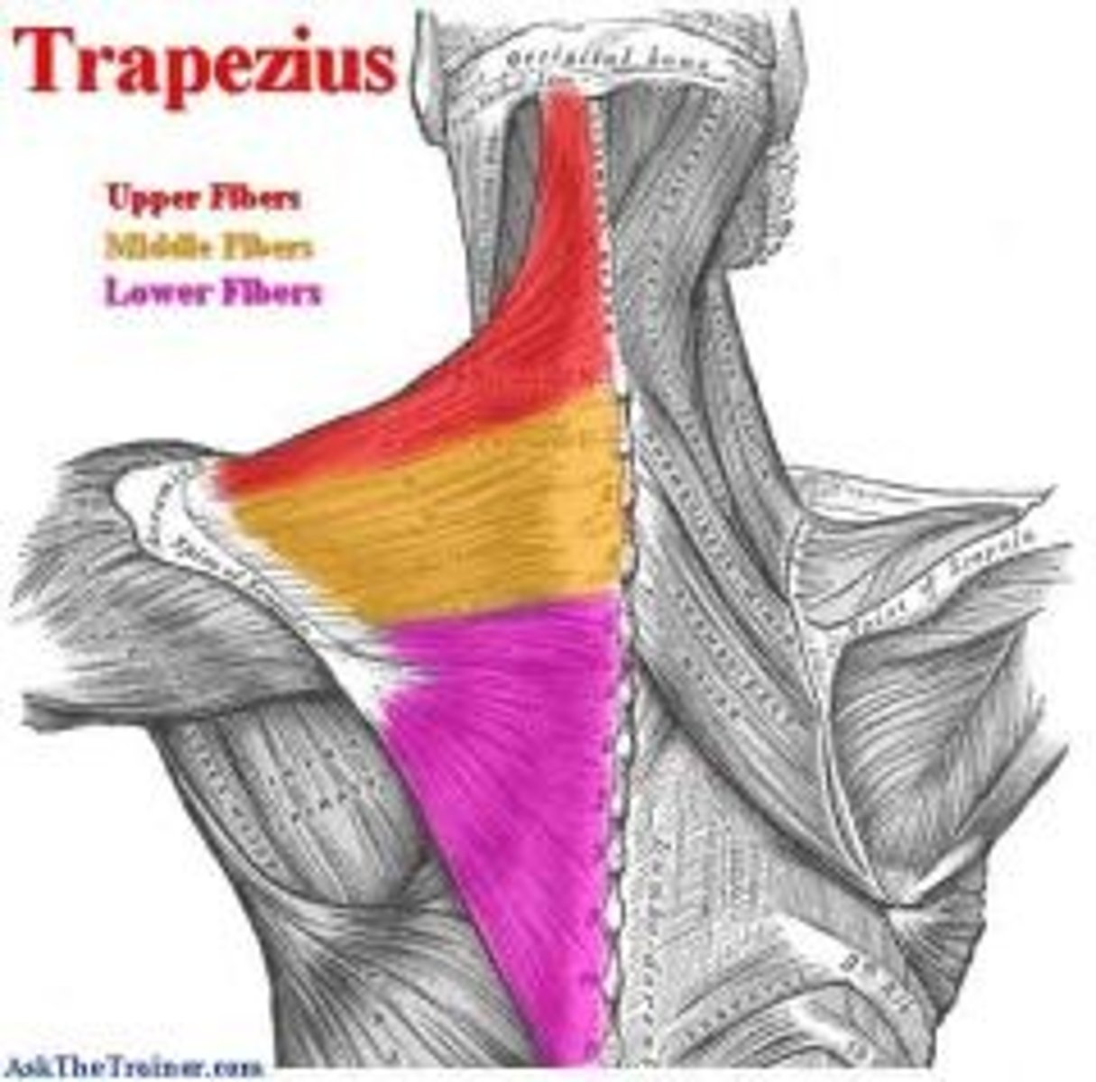
Trapezius Muscle
Attaches pectoral girdle to the vertebral column.
Descending Part of the Trapezius Muscle
Red in the picture, closest to neck

Transverse Part of the Trapezius Muscle
Yellow in the picture, middle piece

Ascending Part of the Trapezius Muscle
Pink in the picture, very bottom of trapezius
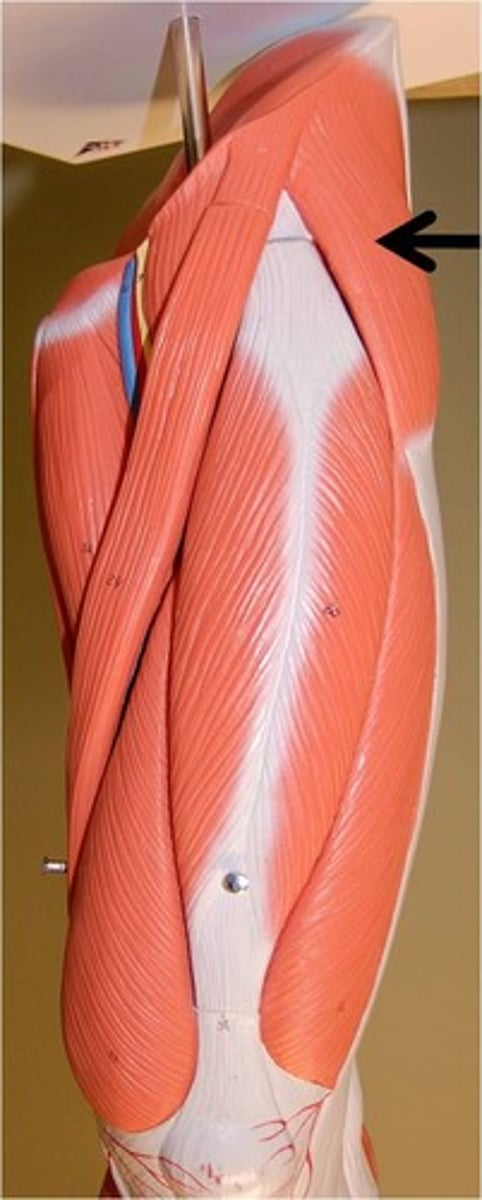
Latissimus Dorsi Muscle
Lats, "wings"
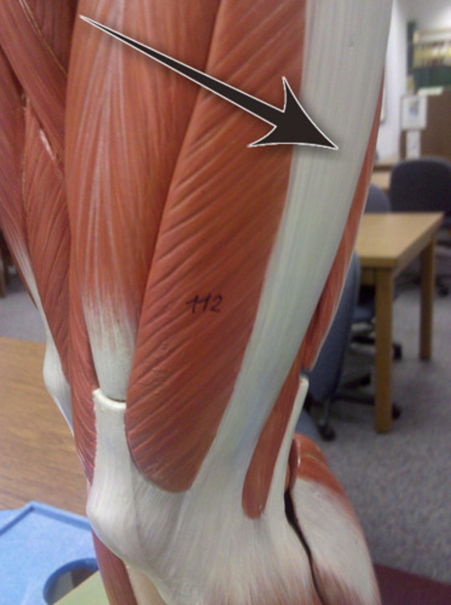
Rhomboid Major Muscle
Triangle shaped on the back, underneath trapezius
Rhomboid Minor Muscle
Y shaped on the back, underneath trapezius
Levator Scapulae Muscle
Branch off of the rhomboid minor, attaches the scapula to cervical vertebrae. Think shoulder shrugs.
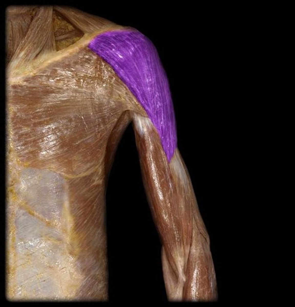
Deltoid Muscle
Shoulder muscle, upside down delta (triangle)
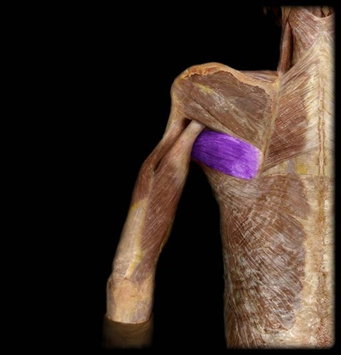
Teres Major Muscle
In your armpit area
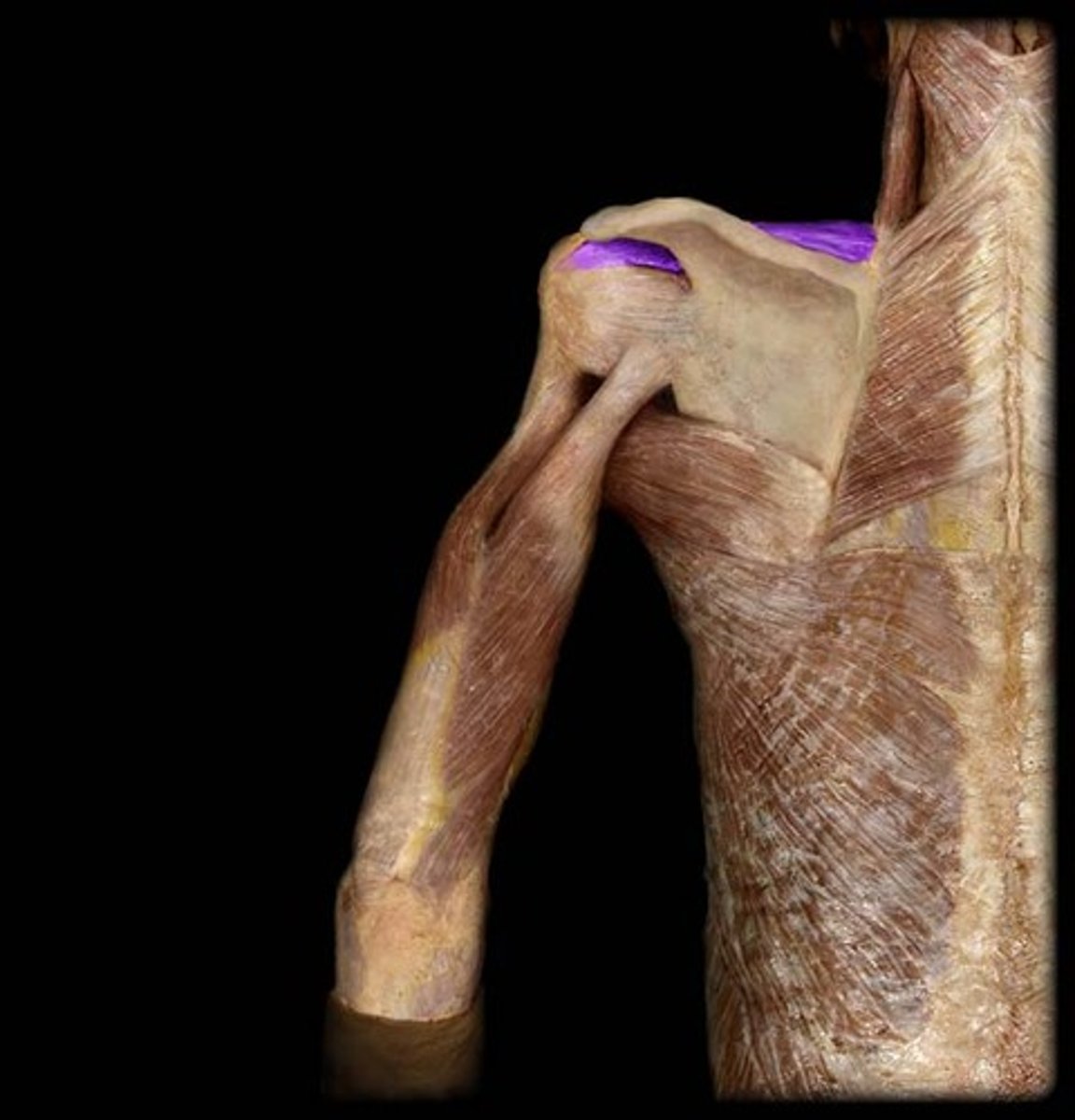
Supraspinatus Muscle
Fills the supraspinous fossa
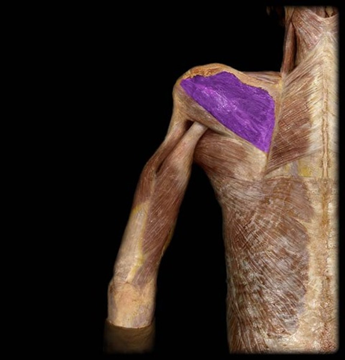
Infraspinatus Muscle
Fills the infraspinous fossa
Teres Minor Muscle
On the lateral border of the scapula
Subscapularis Muscle
Fills the subscapular fossa
Supraspinatus, infraspinatus, teres minor, subscapularis
BQ: Know 2 of the 4 Rotator Cuff Muscles
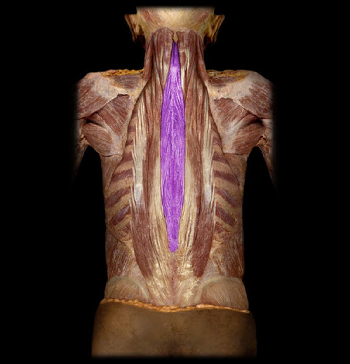
Erector Spinae Muscles
Make the spine erect after it has been flexed
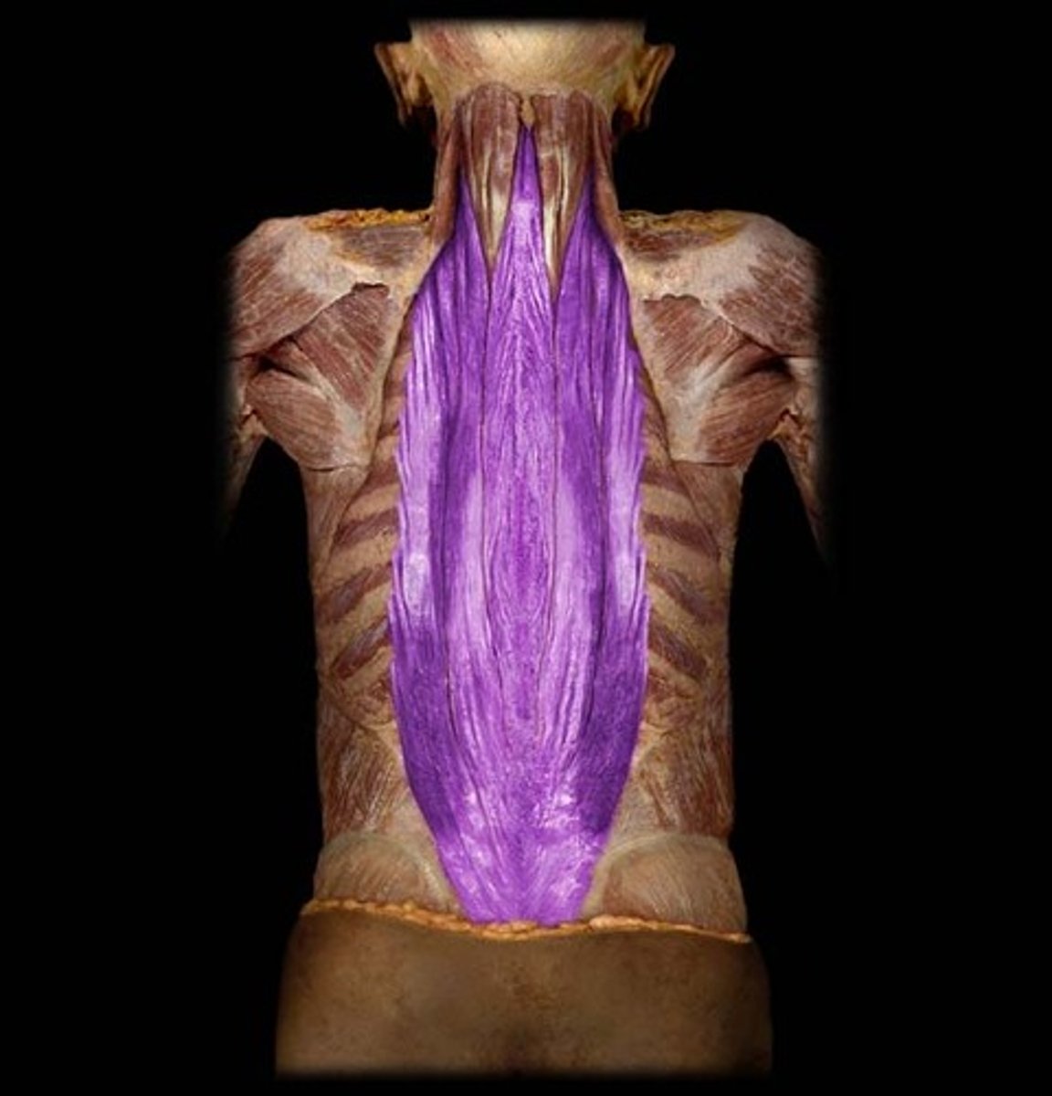
Spinalis Muscle
Medial part of erector spinae, attaches to spinous processes
Longissimus Muscle
Intermediate part of erector spinae, attaches to transverse process of vertebrae
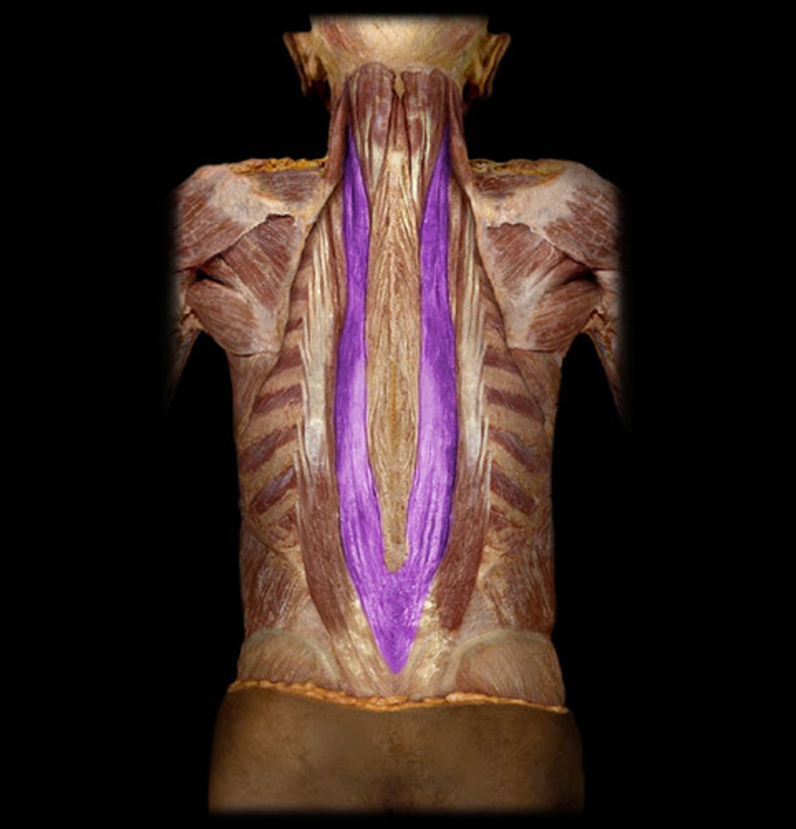
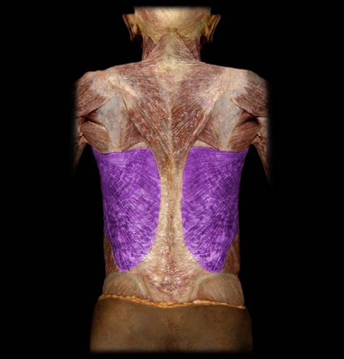
Iliocostalis Muscle
Lateral part of erector spinae, attaches to ribs
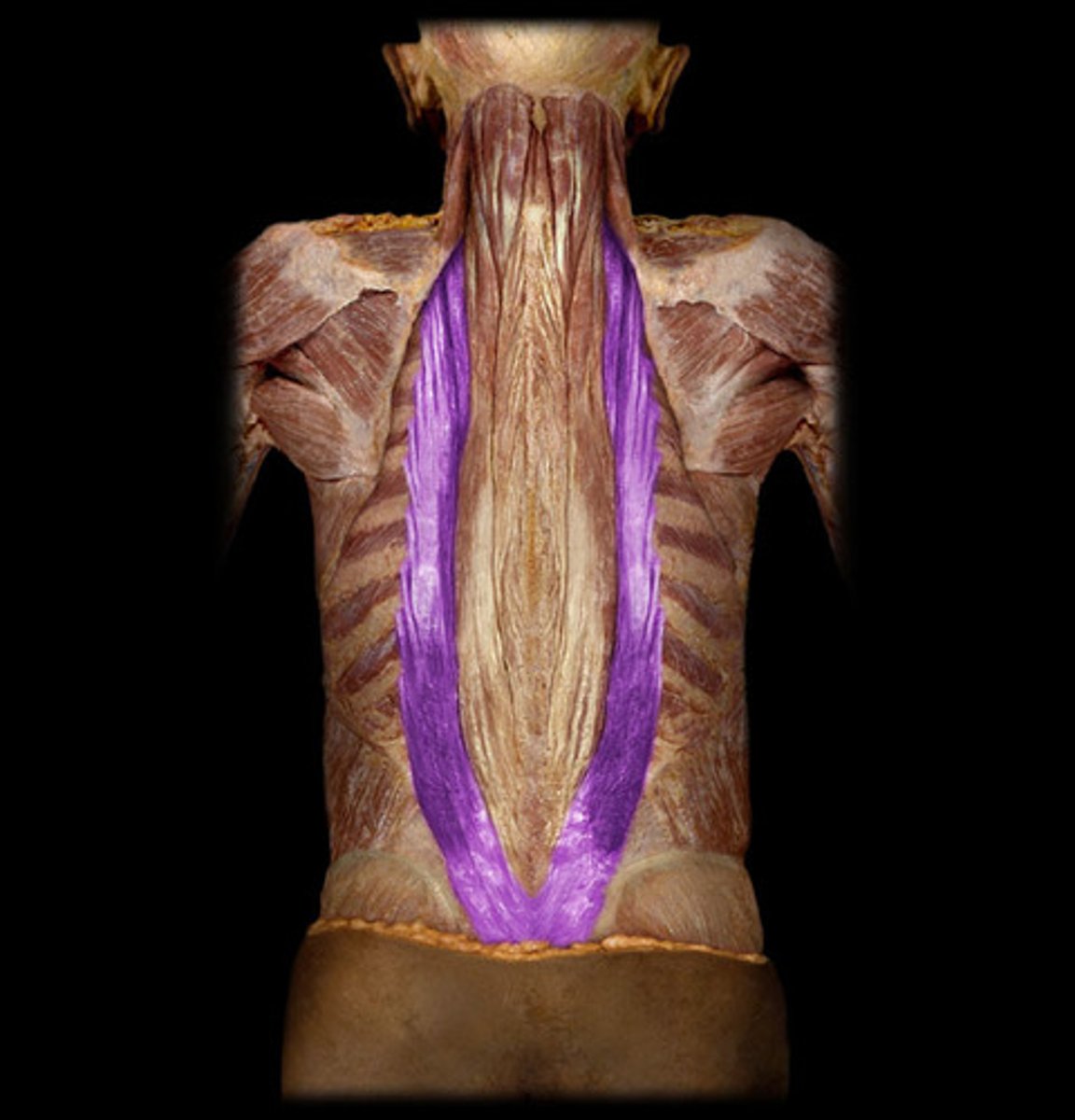
Spinalis, Longissimus, Iliocostalis
BQ: Know 2 of the 3 parts of the erector spinae muscle
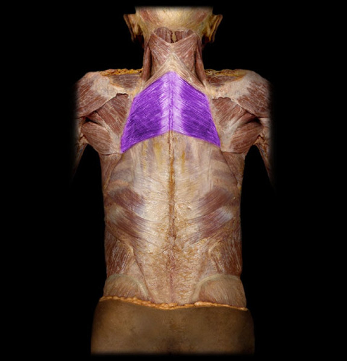
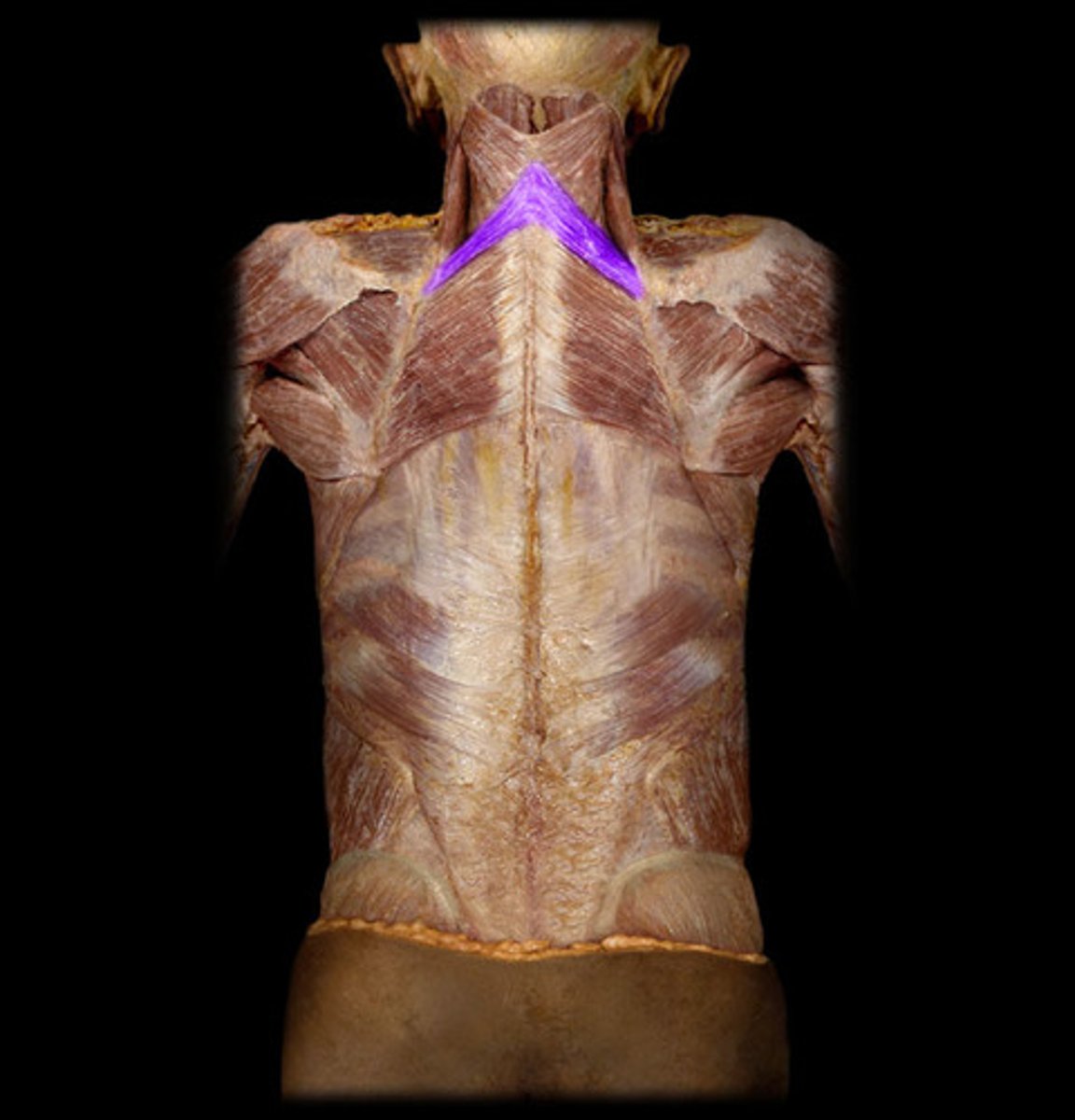
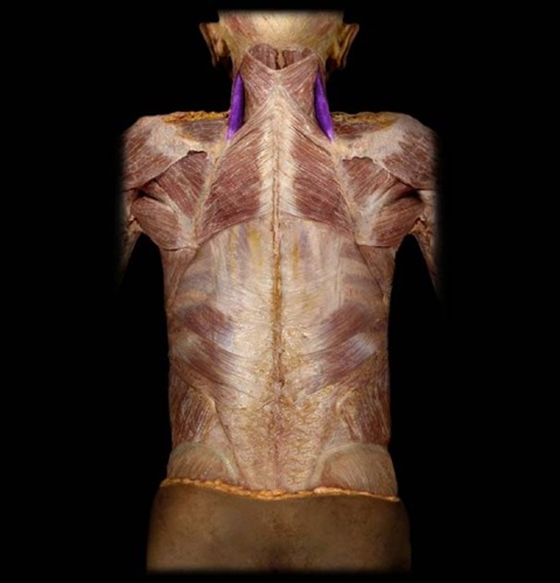
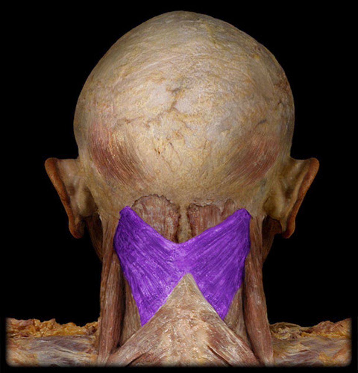
Splenius Capitis Muscle
Attaches the posterior of the skull to the vertebral column, bow tie shaped
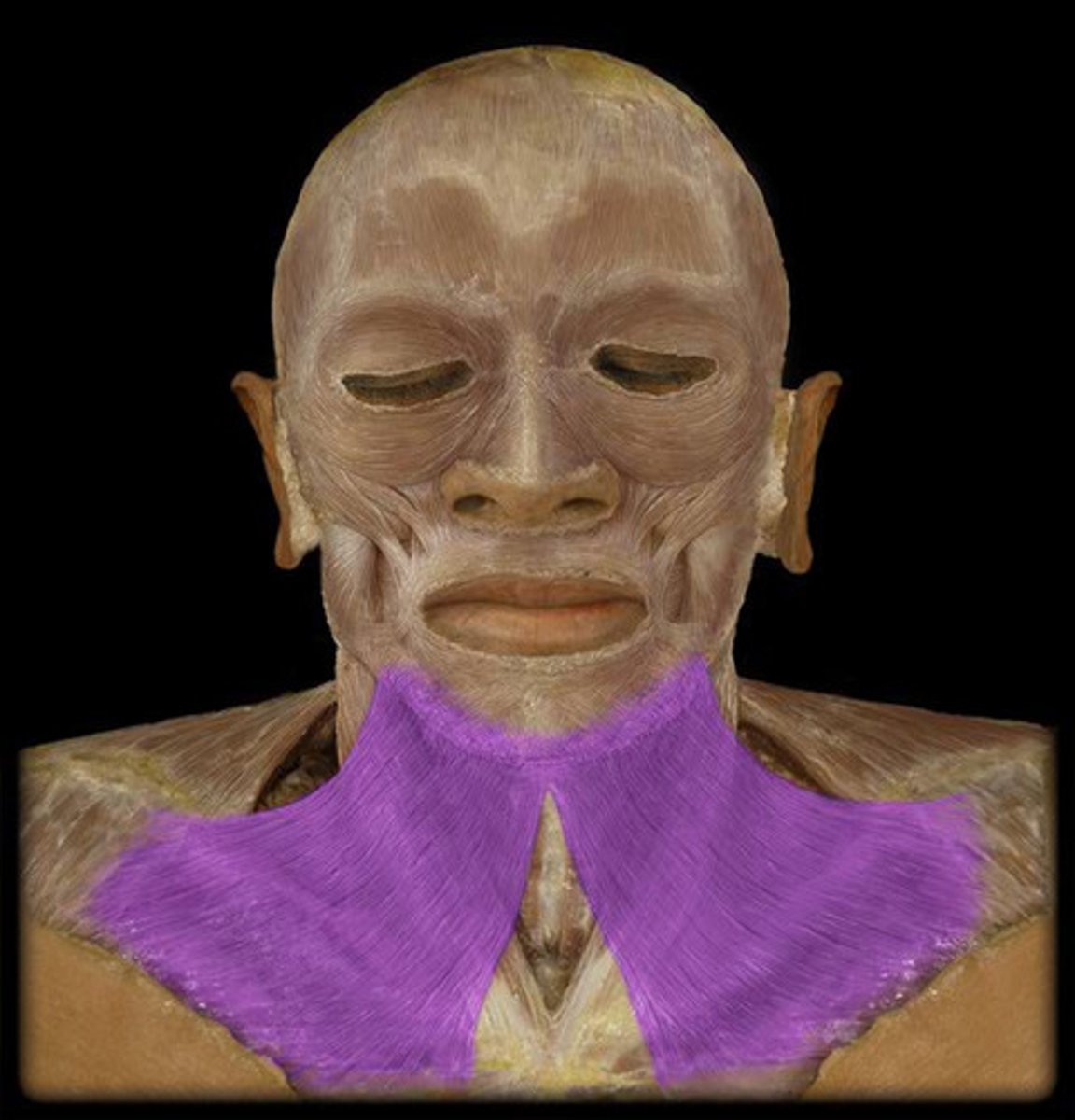
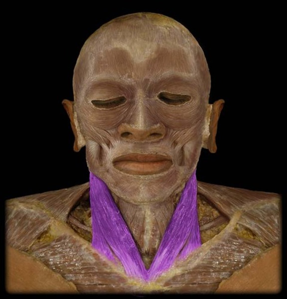
Platysma Muscle
Tenses the skin of the neck
Sternocleidomastoid Muscle
Helps bend the neck and rotate the head
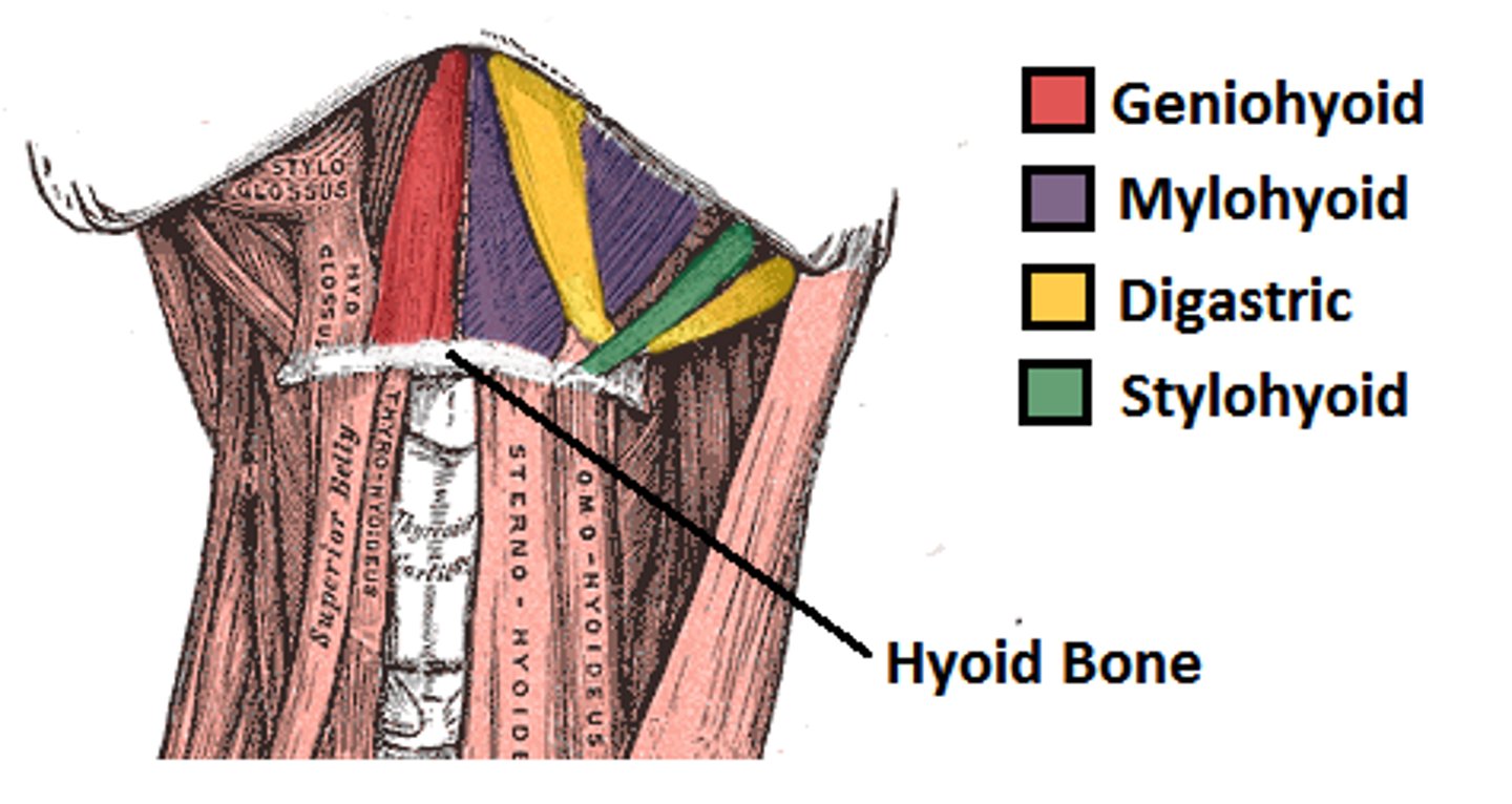
Digastric Muscle
Sides, underneath the chin, yellow in the pic

Mylohyoid Muscle
Sits in between the digastric muscles, purple in the pic
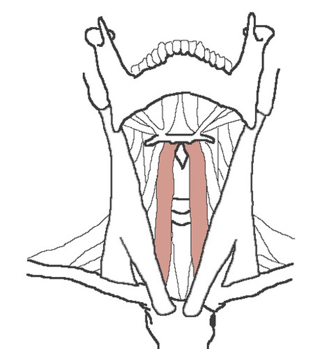
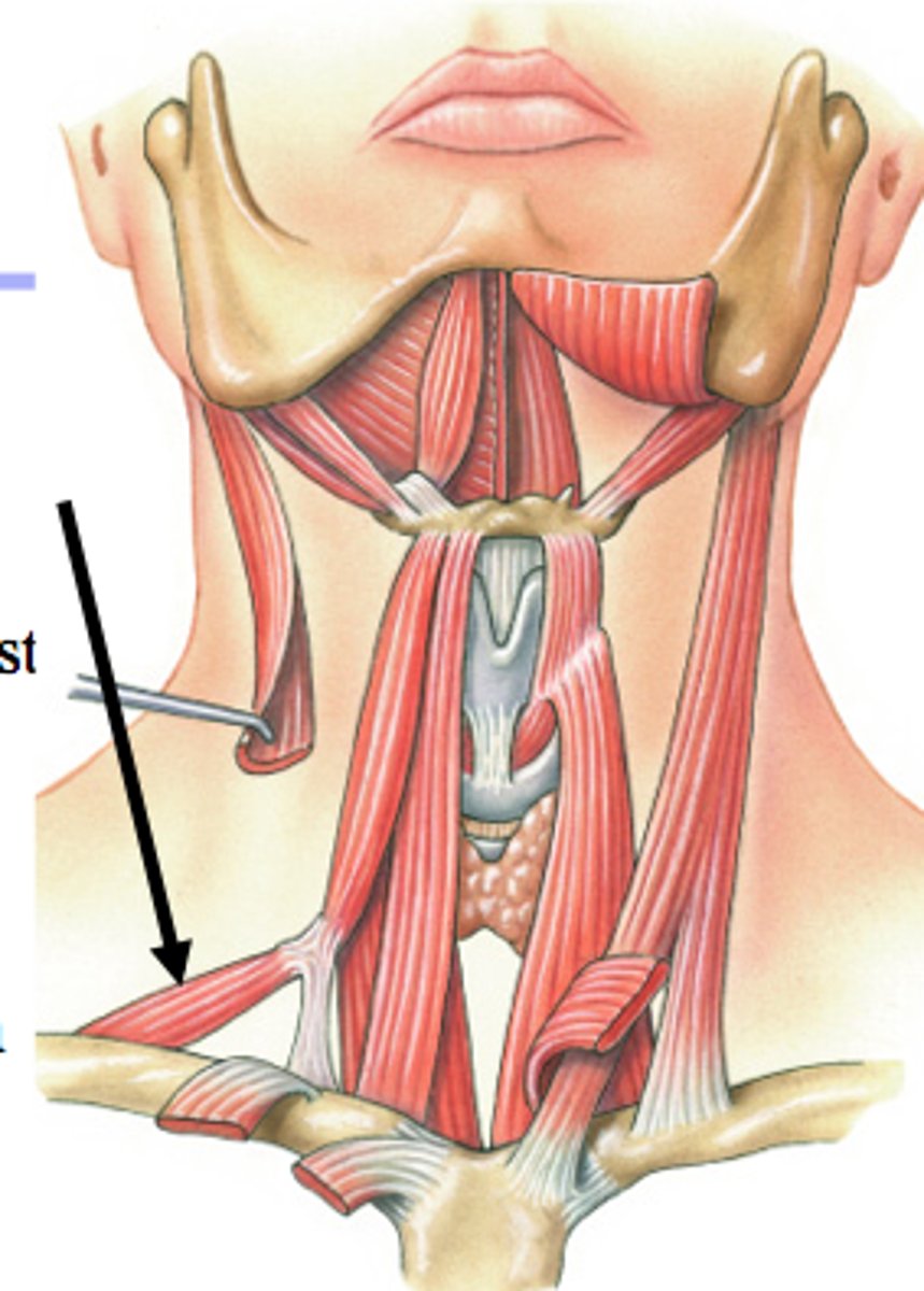
Sternohyoid Muscle
On both sides of the throat
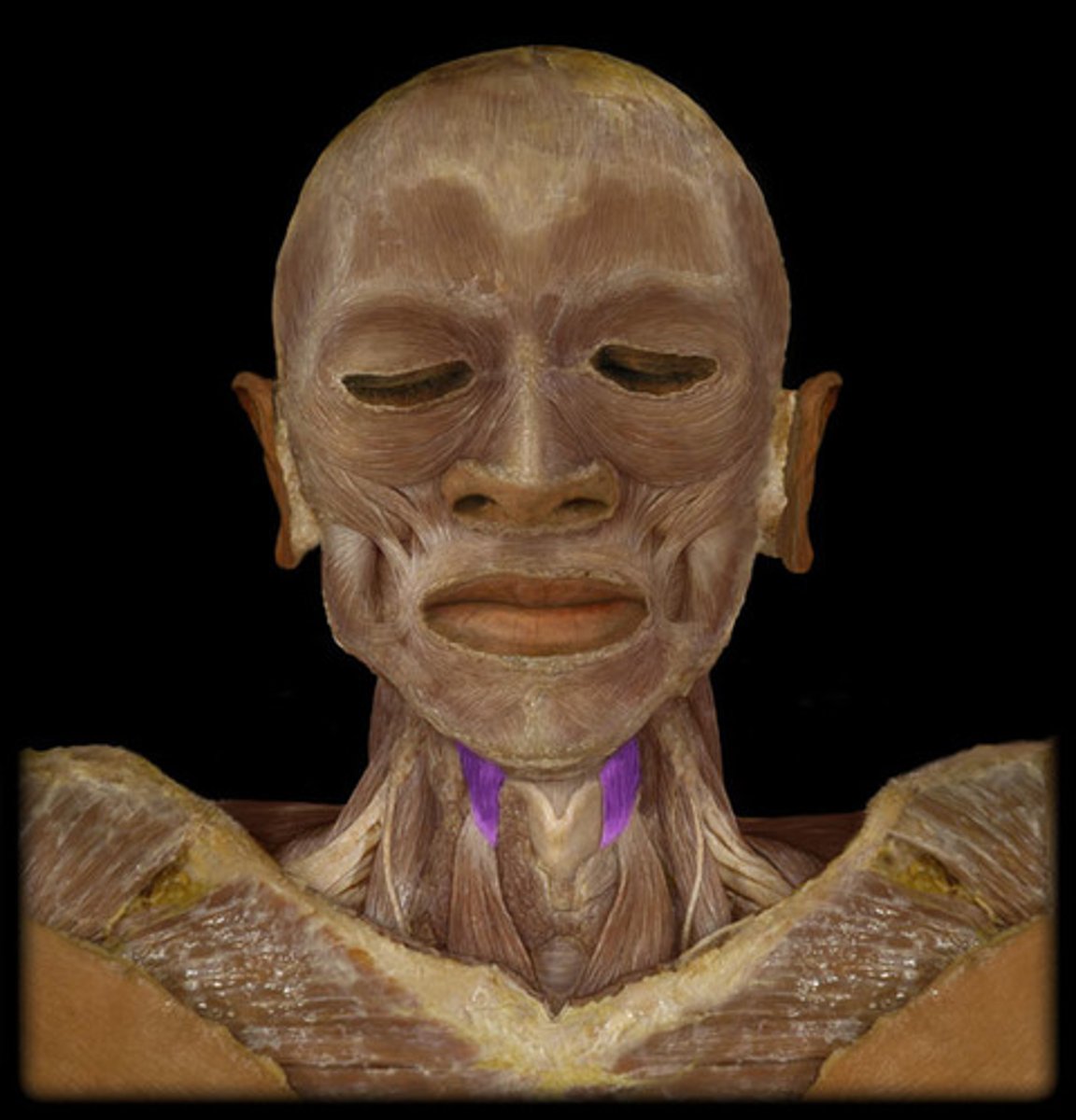
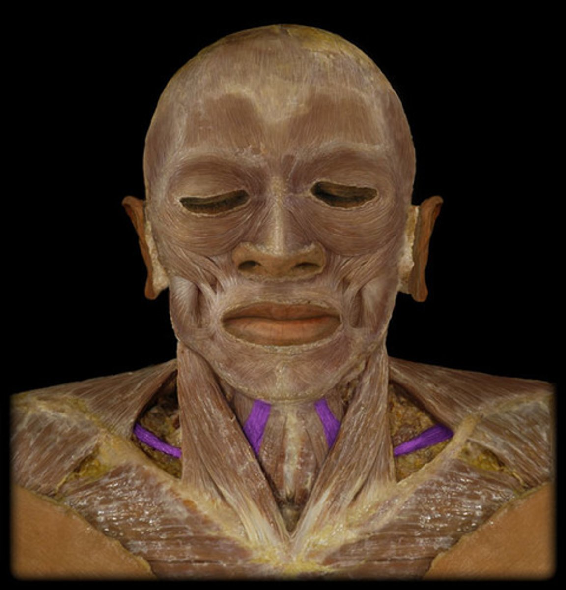
Sternothyroid Muscle
Red in the picture
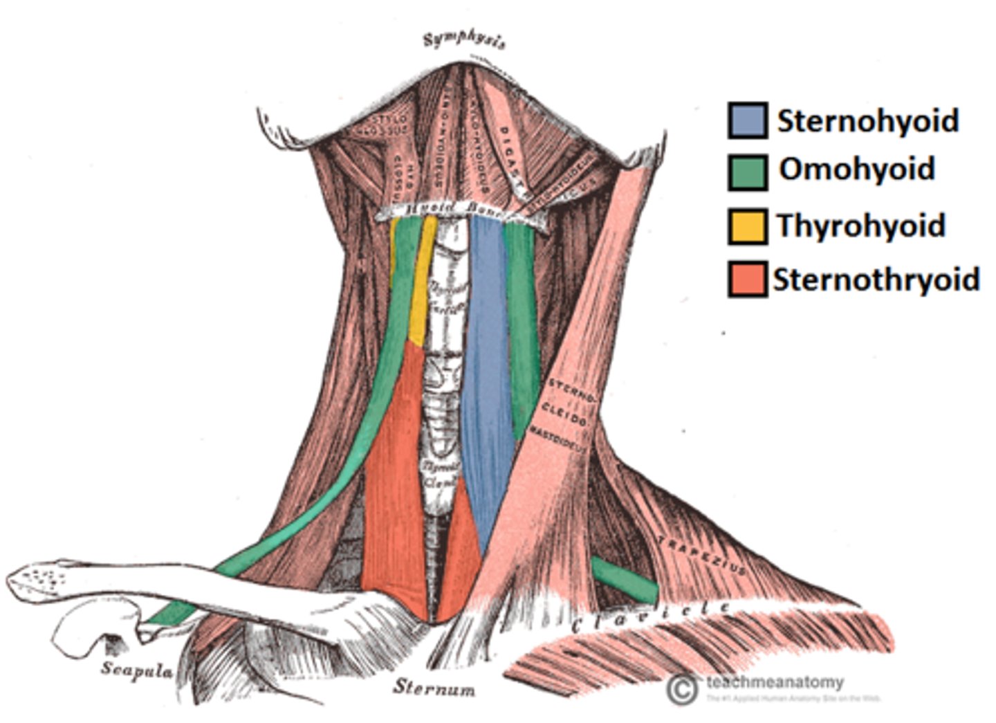
Thyrohyoid Muscle
Closest to the head of all the infrahyoid muscles
Omohyoid Muscle
Makes L shapes from neck to shoulders
Superior Belly of the Omohyoid Muscle
Thicker part of the Omohyoid Muscle
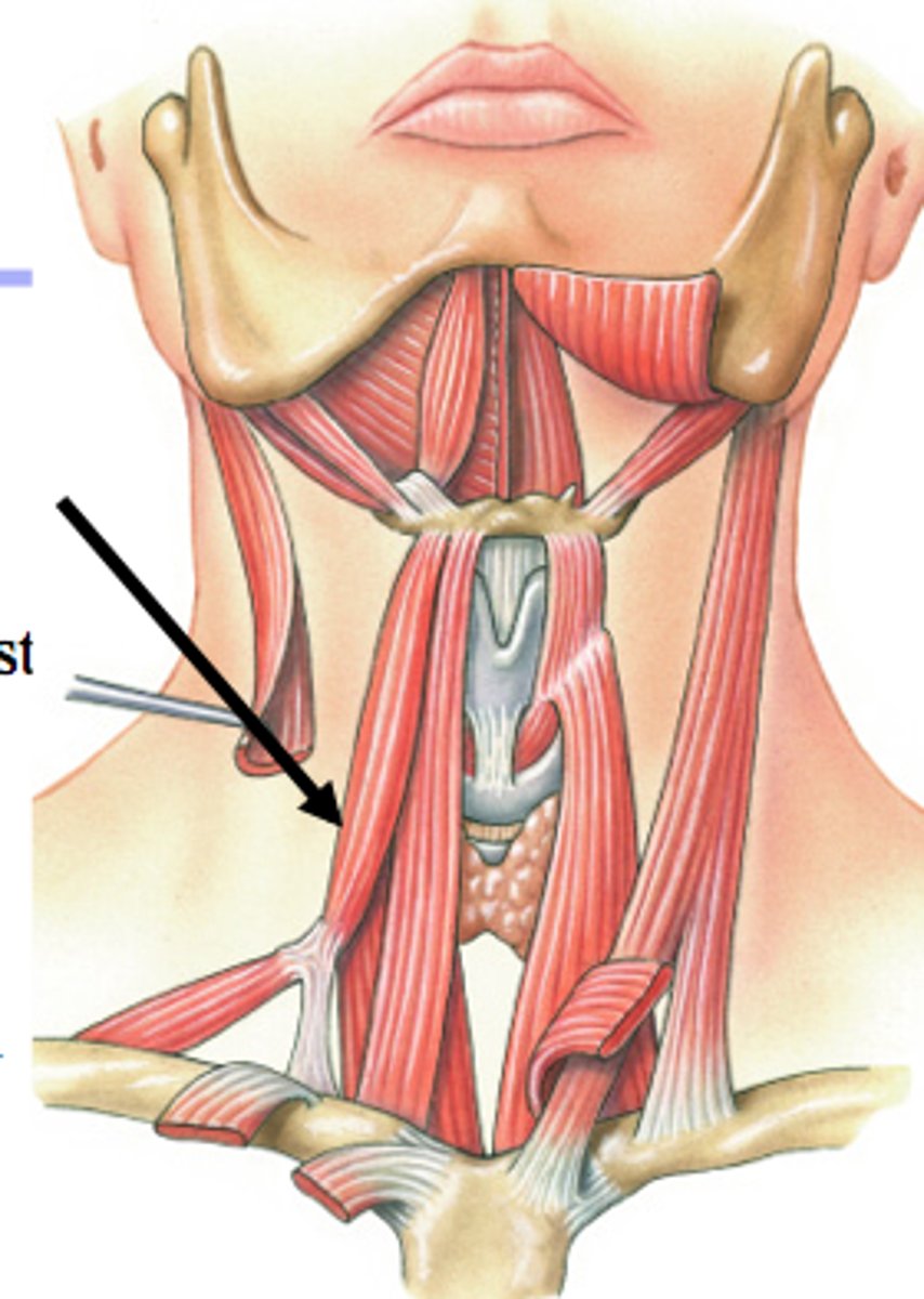
Inferior Belly of the Omohyoid Muscle
Thinner part of the Omohyoid Muscle
Occipitofrontalis Muscle
Attaches to and moves the scalp
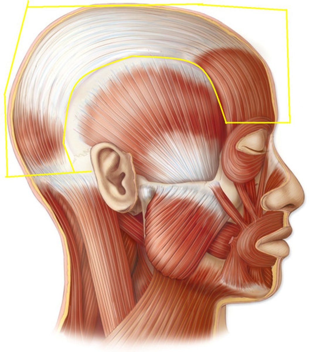
Frontal Belly of the Occipitofrontalis Muscle
Forehead
Occipital Belly of the Occipitofrontalis Muscle
In the posterior scalp
Orbicularis Oculi Muscle
Around the eyelids
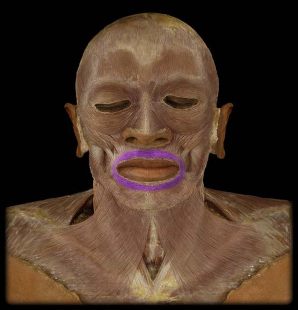
Orbicularis Oris Muscle
Around the lips
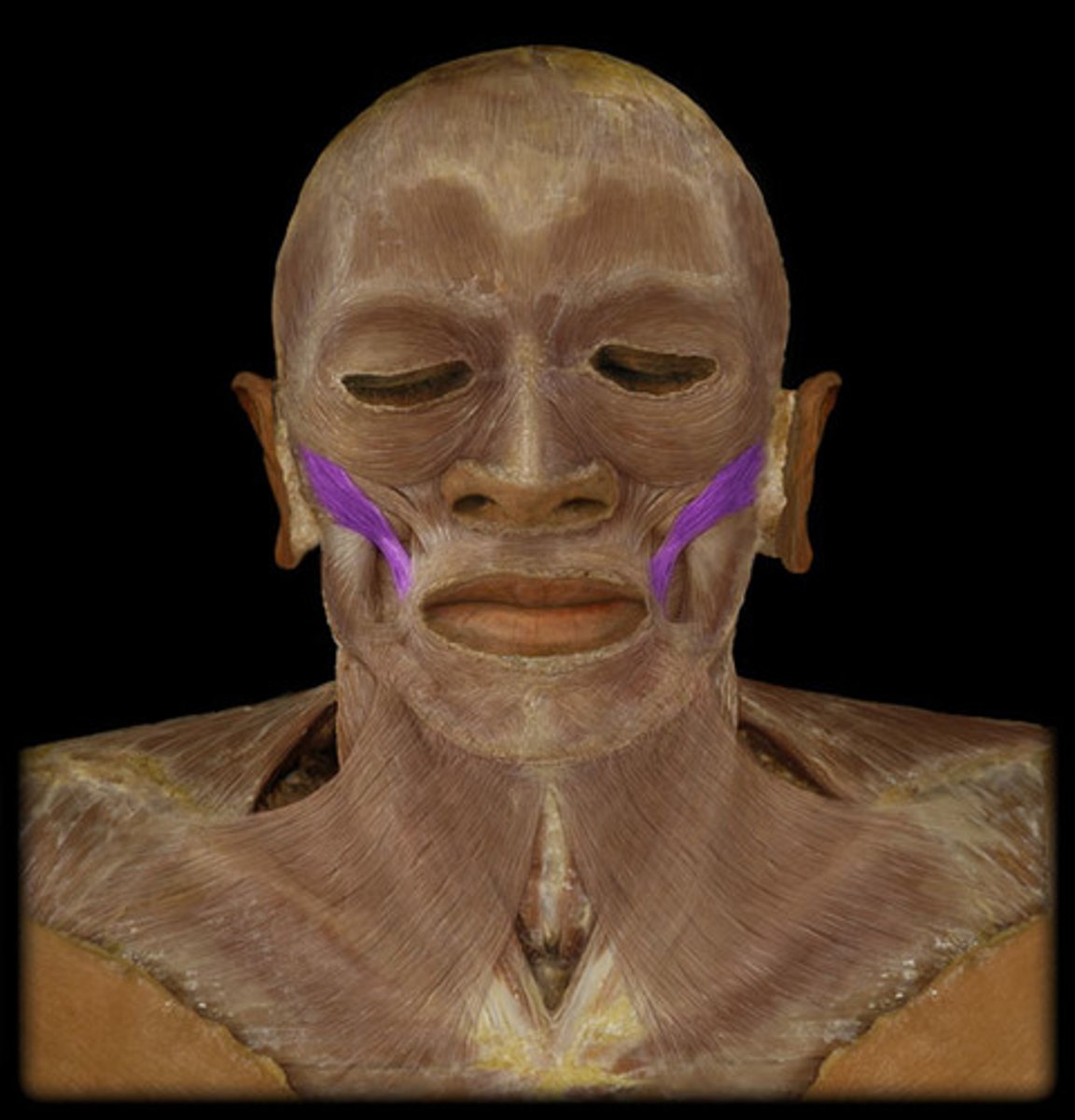
Buccinator Muscle
In the area of dimples

Zygomaticus Major Muscle
Covers zygomatic arch
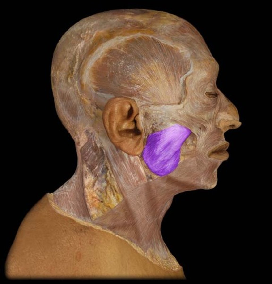
Masseter Muscle
Clench your teeth, and you can feel this muscle flex
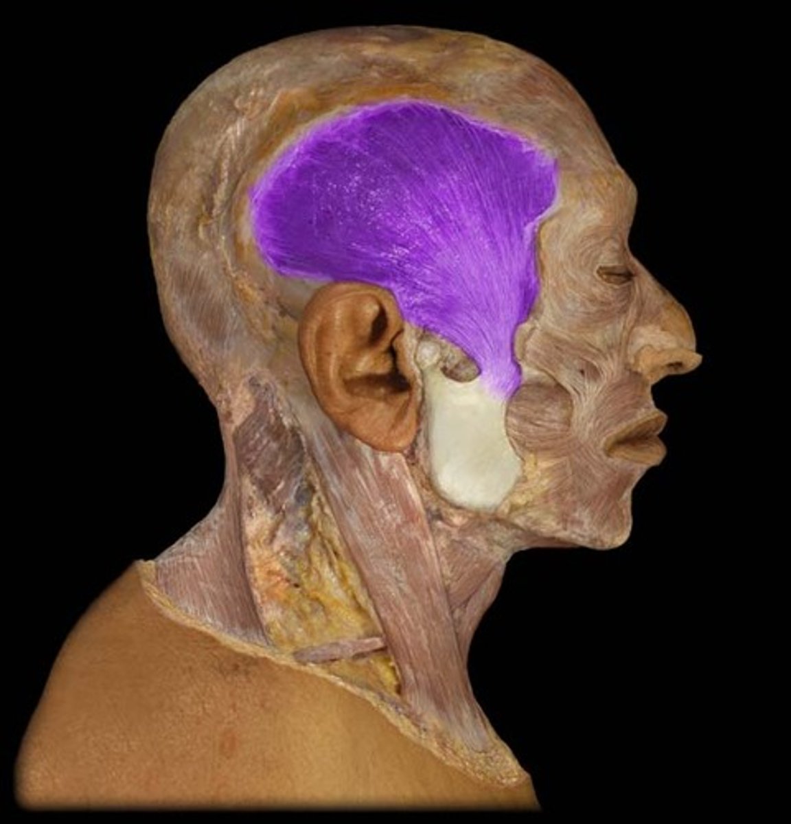
Temporalis Muscle
Temples, elevates and retracts the mandible
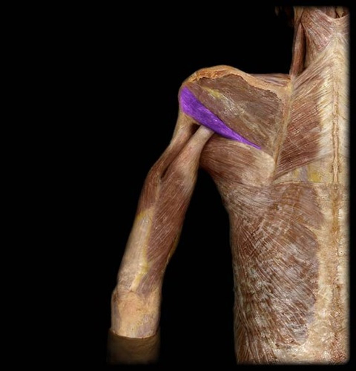
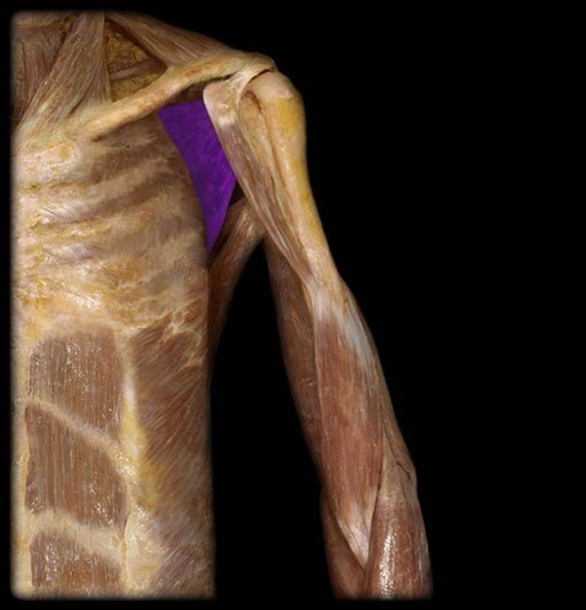
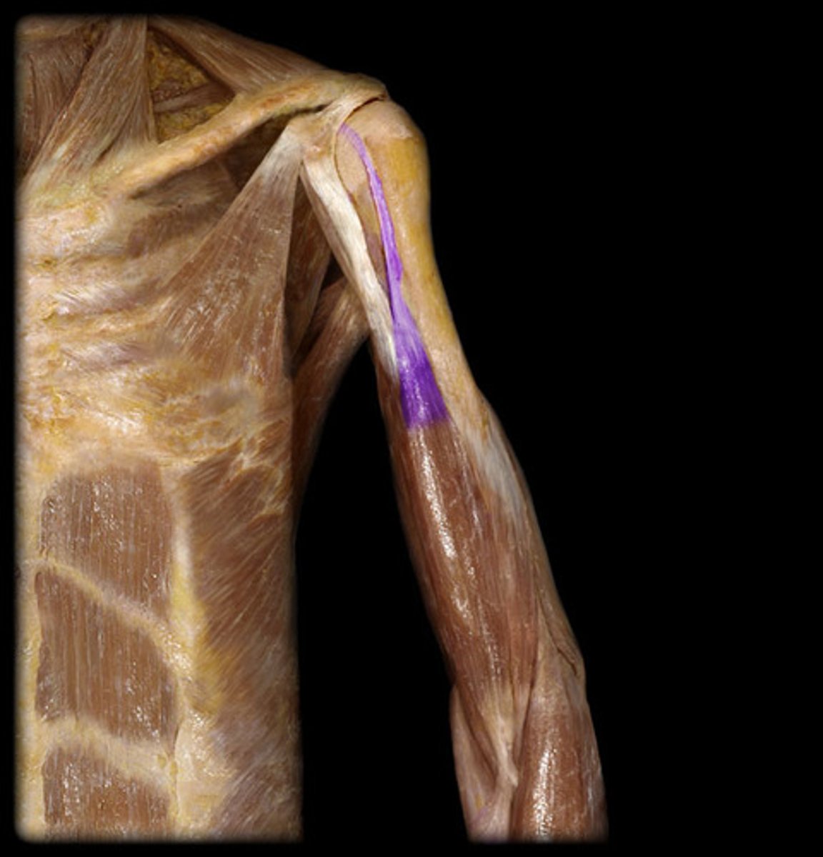
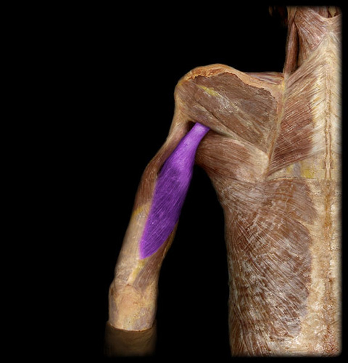
Coracobrachialis Muscles
BQ: Attachments are in the name, what connects the pectoral muscles to the biceps.
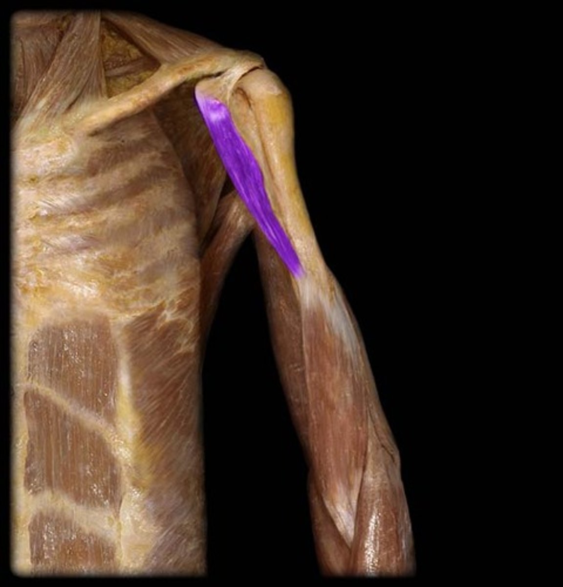
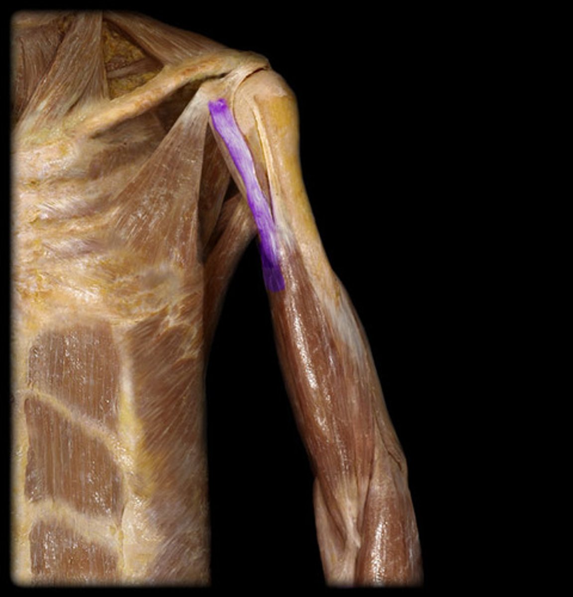
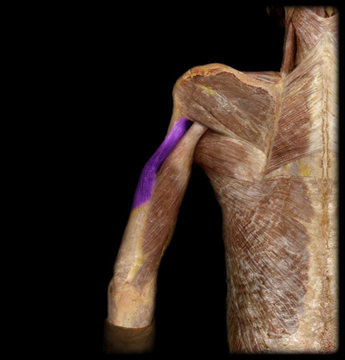
Biceps Brachii Muscles
Collective structure with two parts
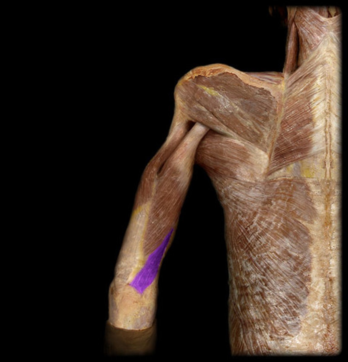
Long Head of the Biceps Brachii
Shorter head of the bicep
Short Head of the Biceps Brachii
Longer head of the bicep
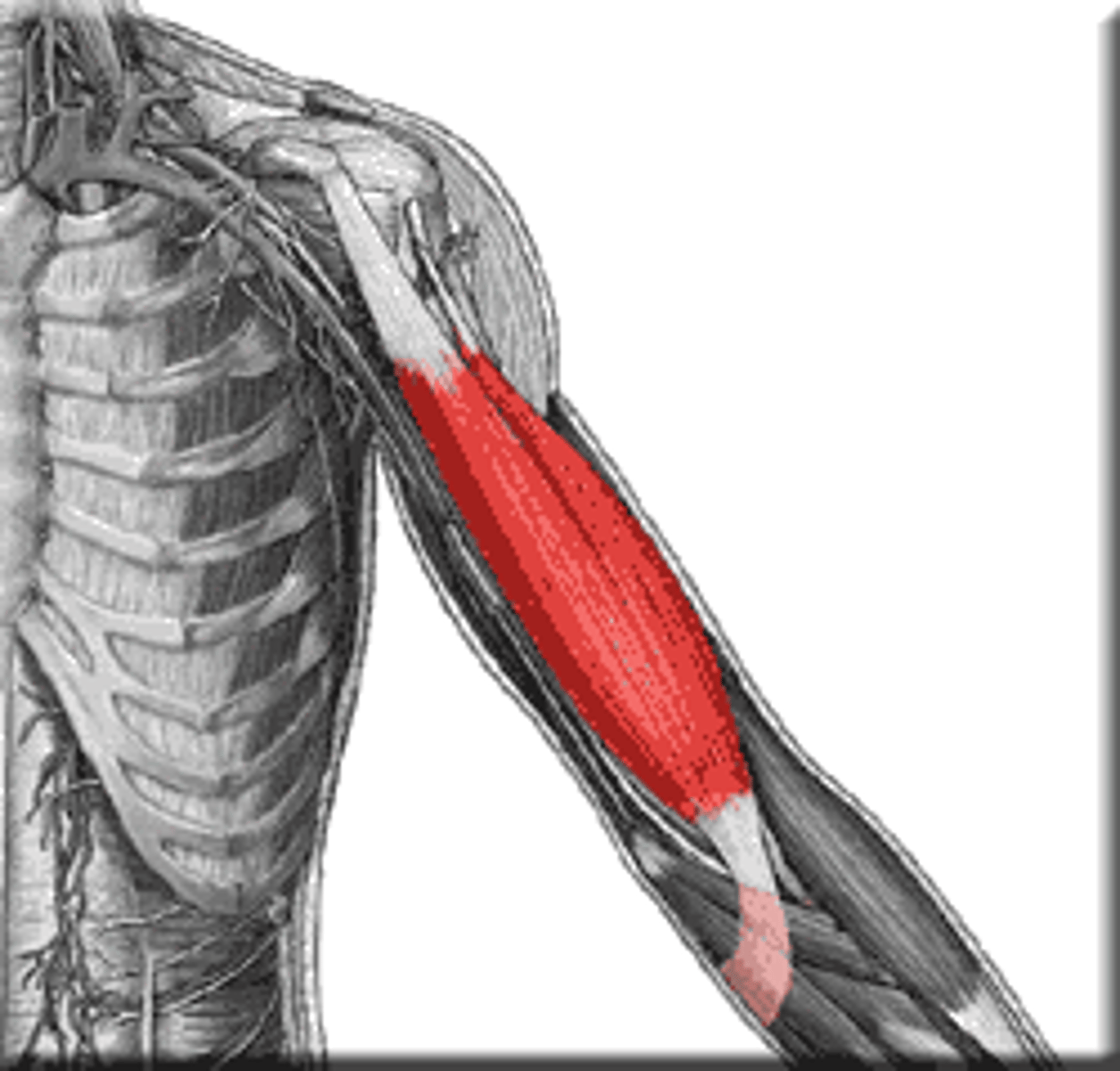
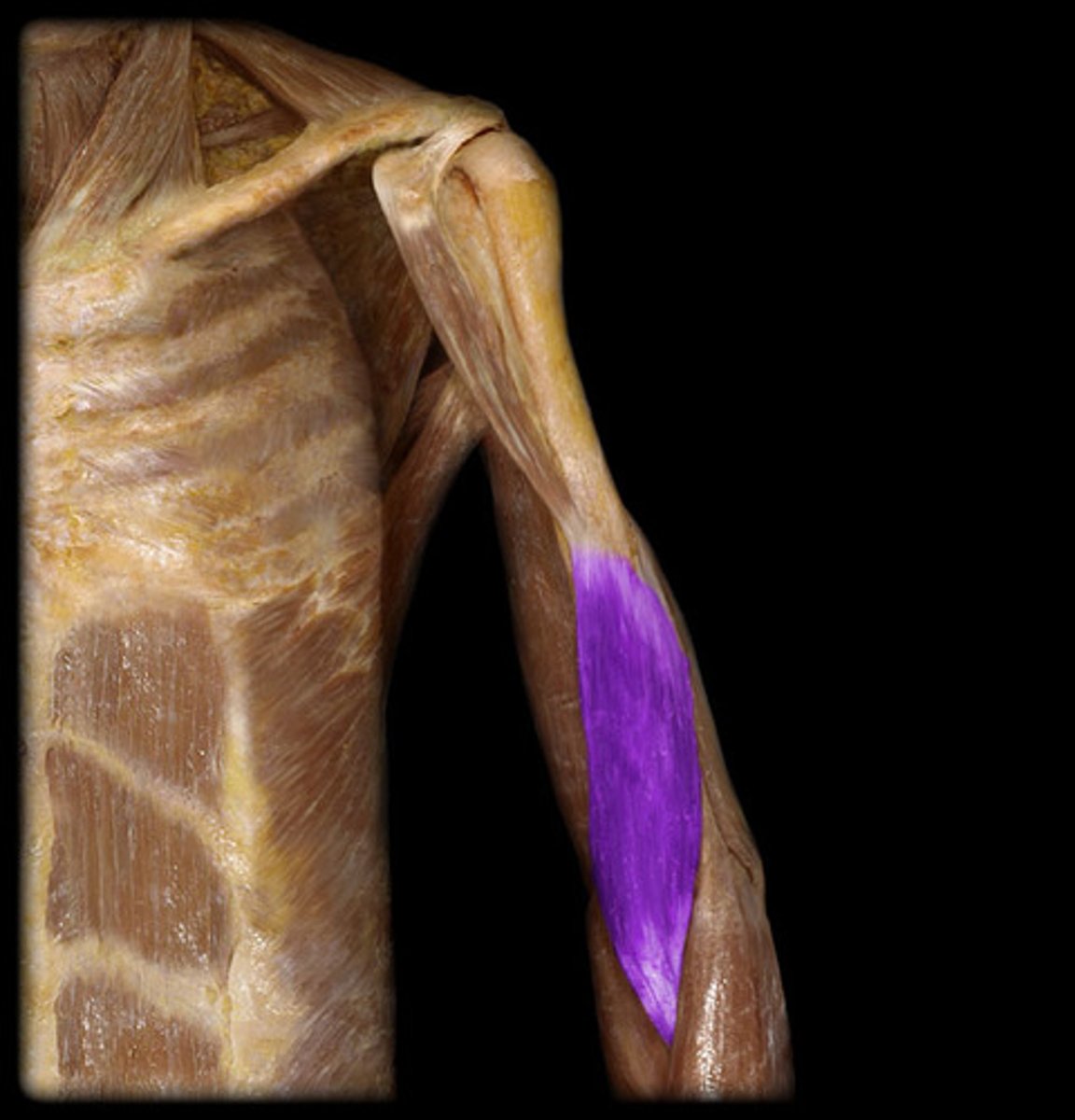
Brachialis Muscles
Flexes forearm at elbow joint
Triceps Brachii Muscle
Collective structure, three heads, back of the arm
Long Head of the Triceps Brachii
The only head that attaches to scapula, bottom, thick

Medial Head of the Triceps Brachii
Under the two other heads, in the middle
Lateral Head of the Triceps Brachii
BQ: Prominent in Body Builders.
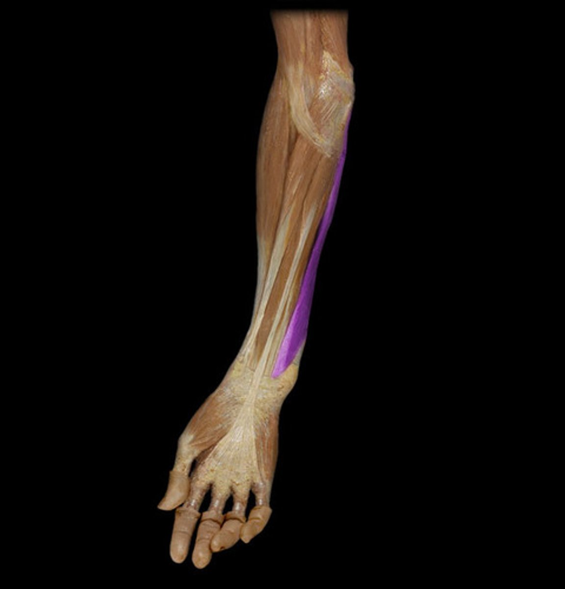
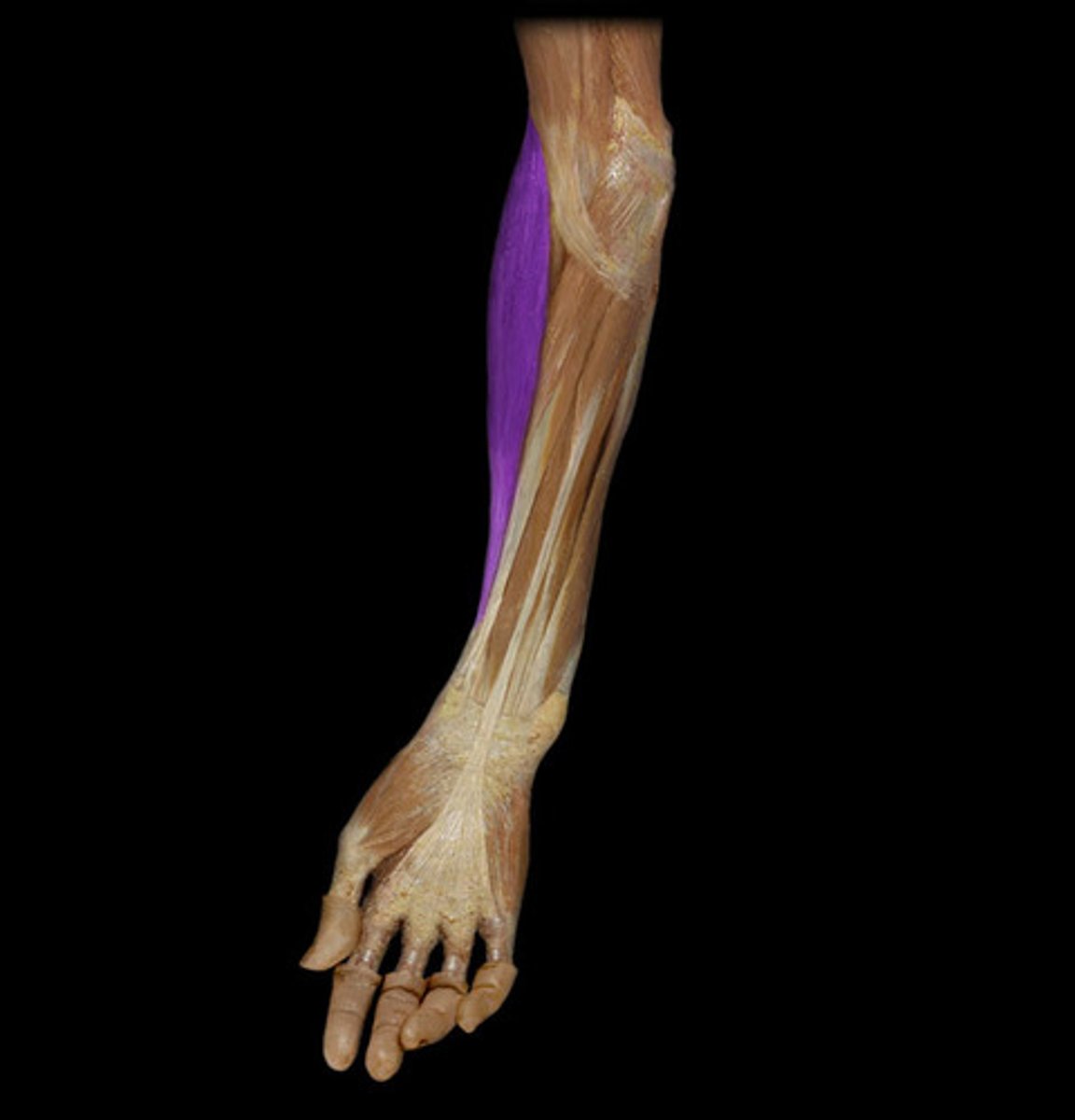
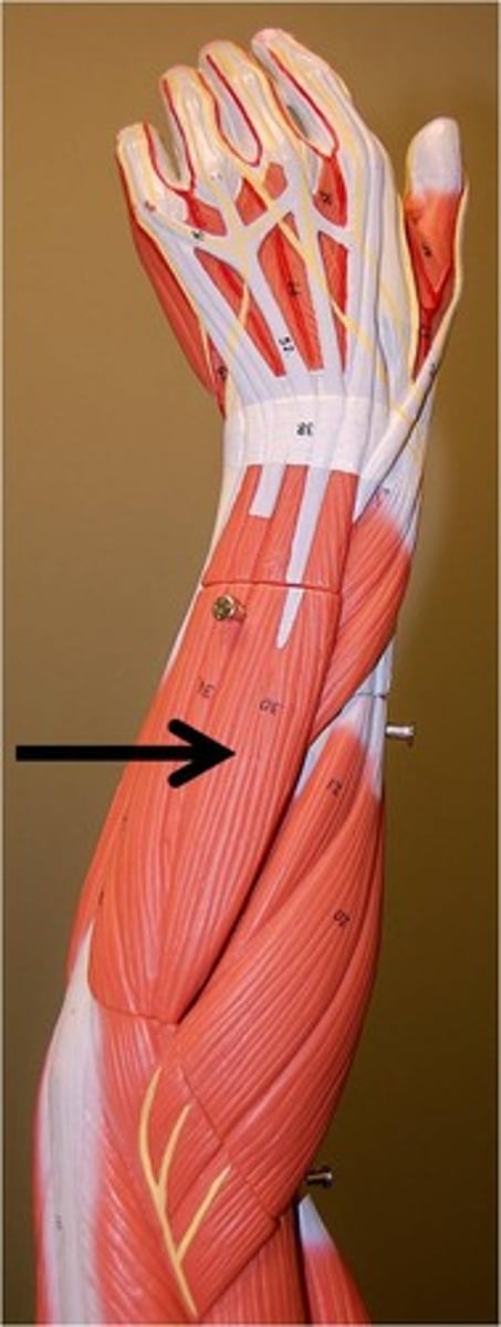
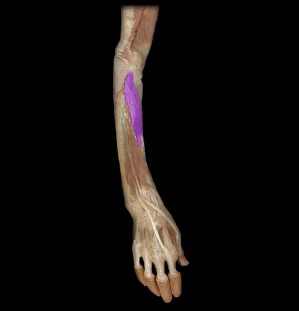
Pronator Teres Muscle
Forearm Muscles: 1
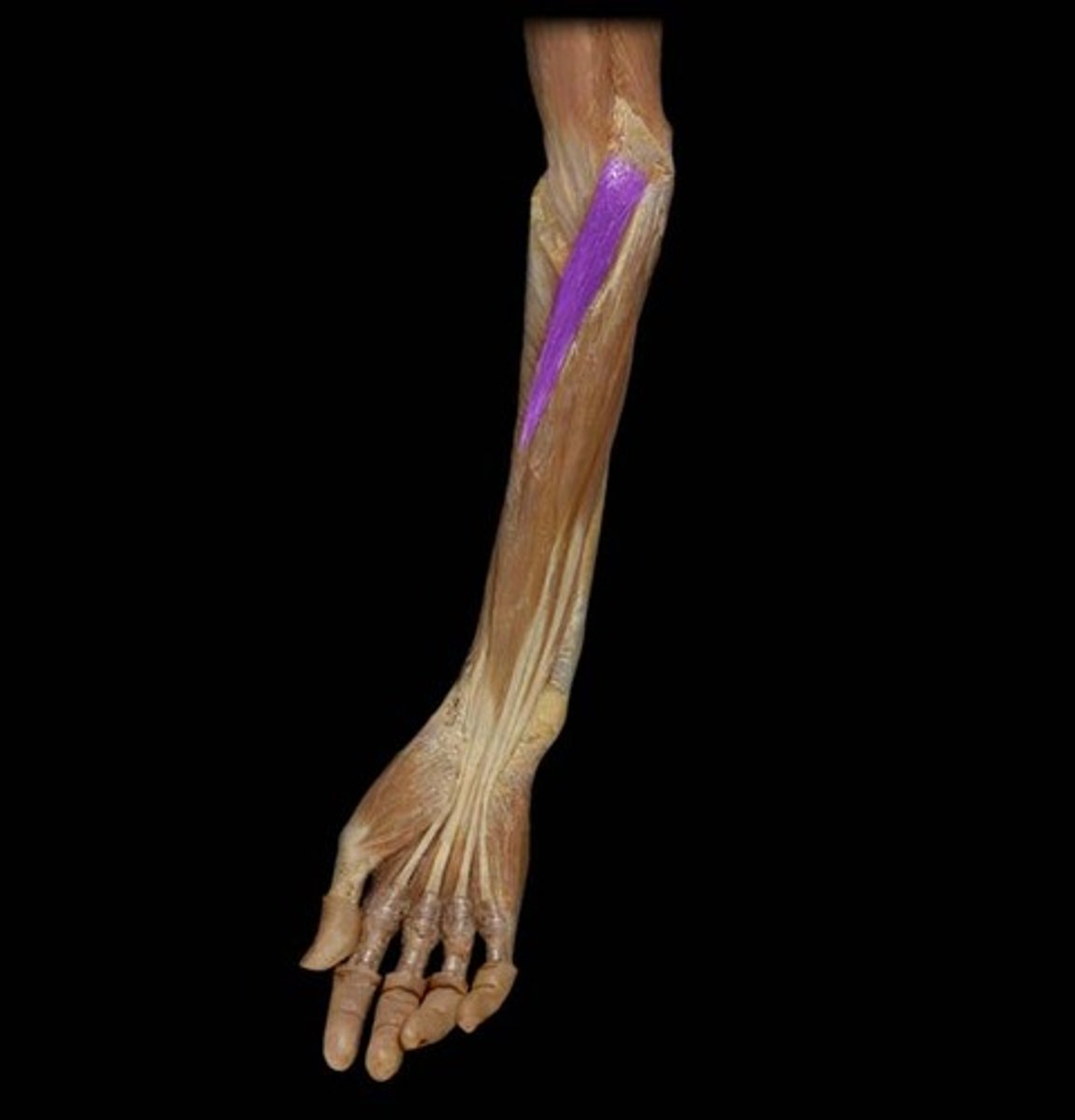
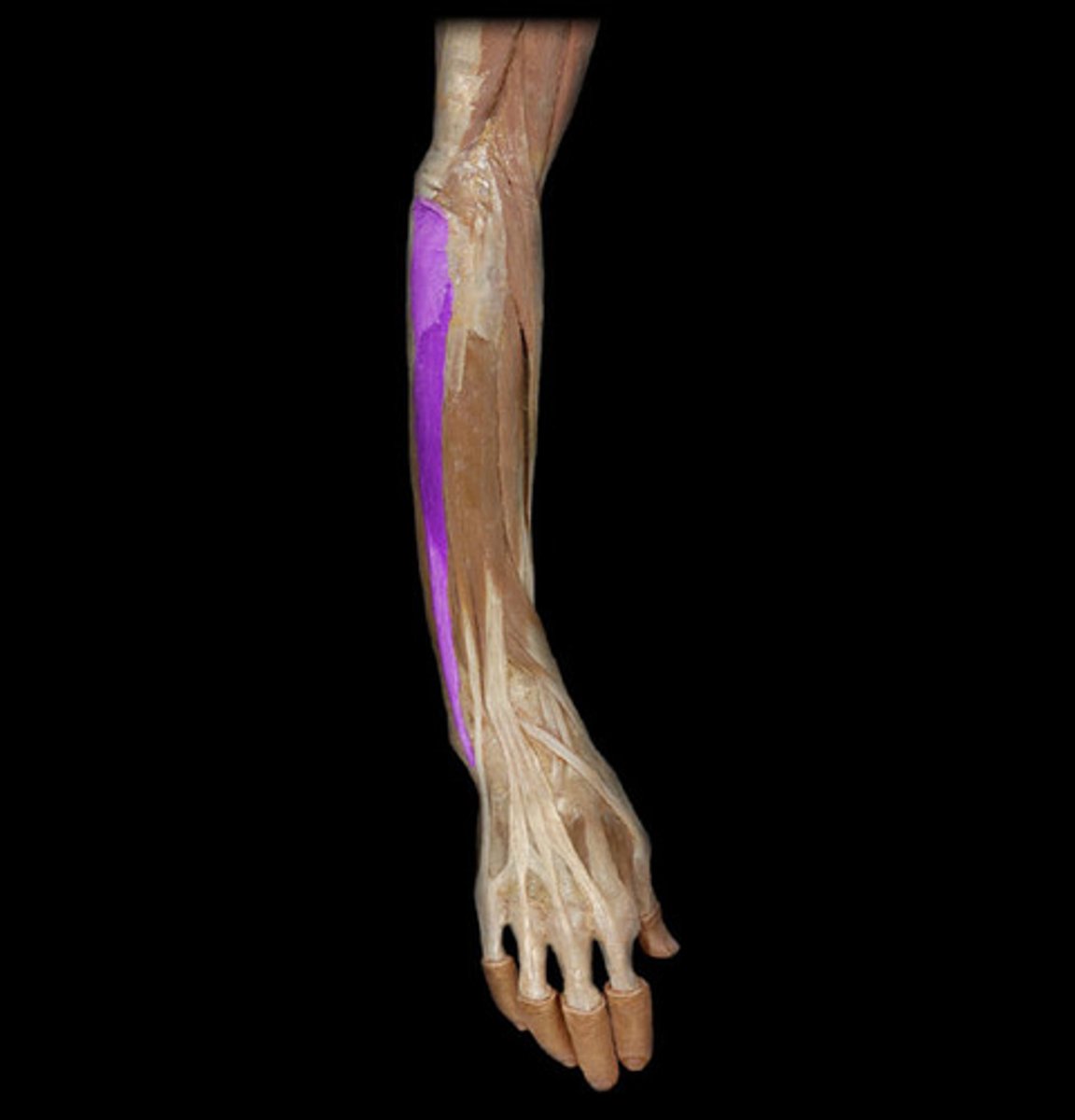
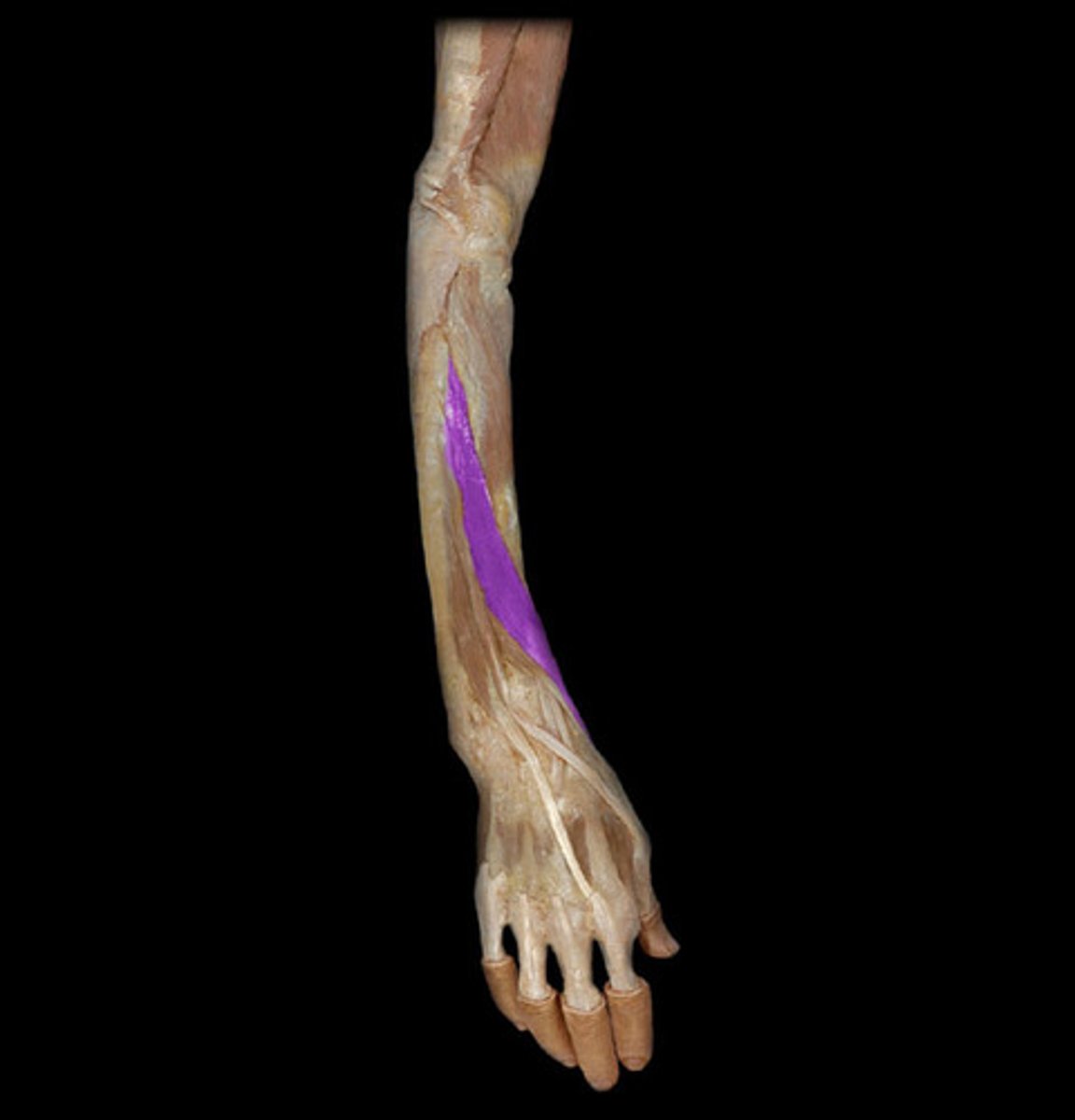
Flexor Carpi Radials Muscle
Forearm Muscles: 2
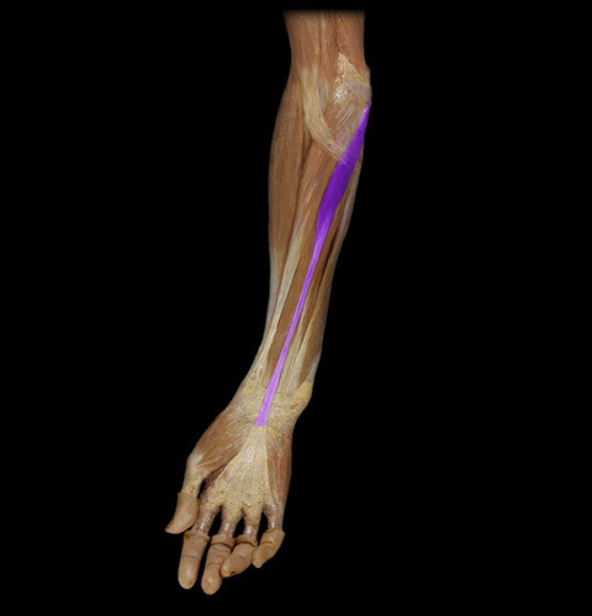
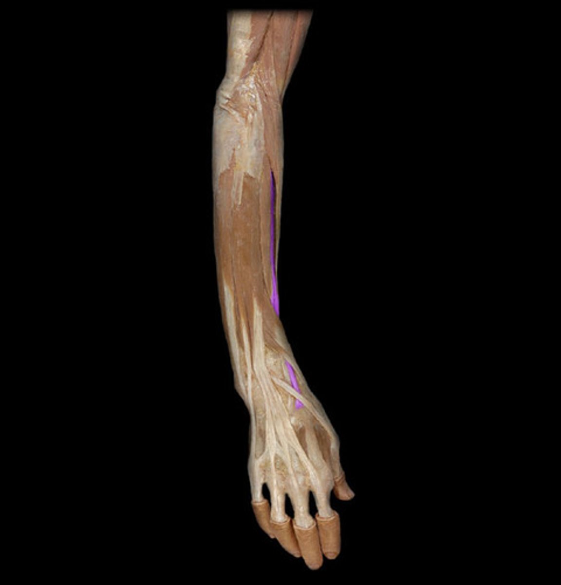
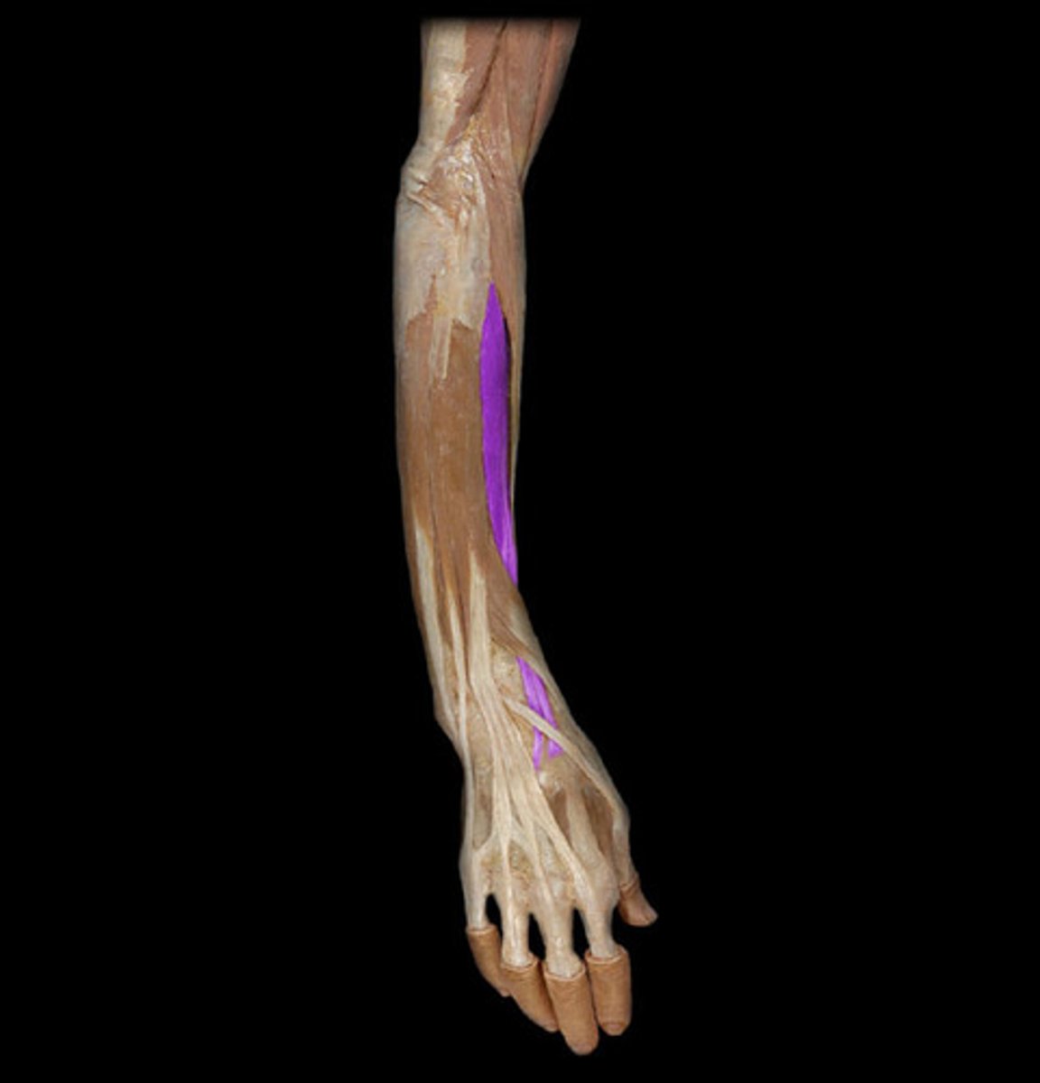
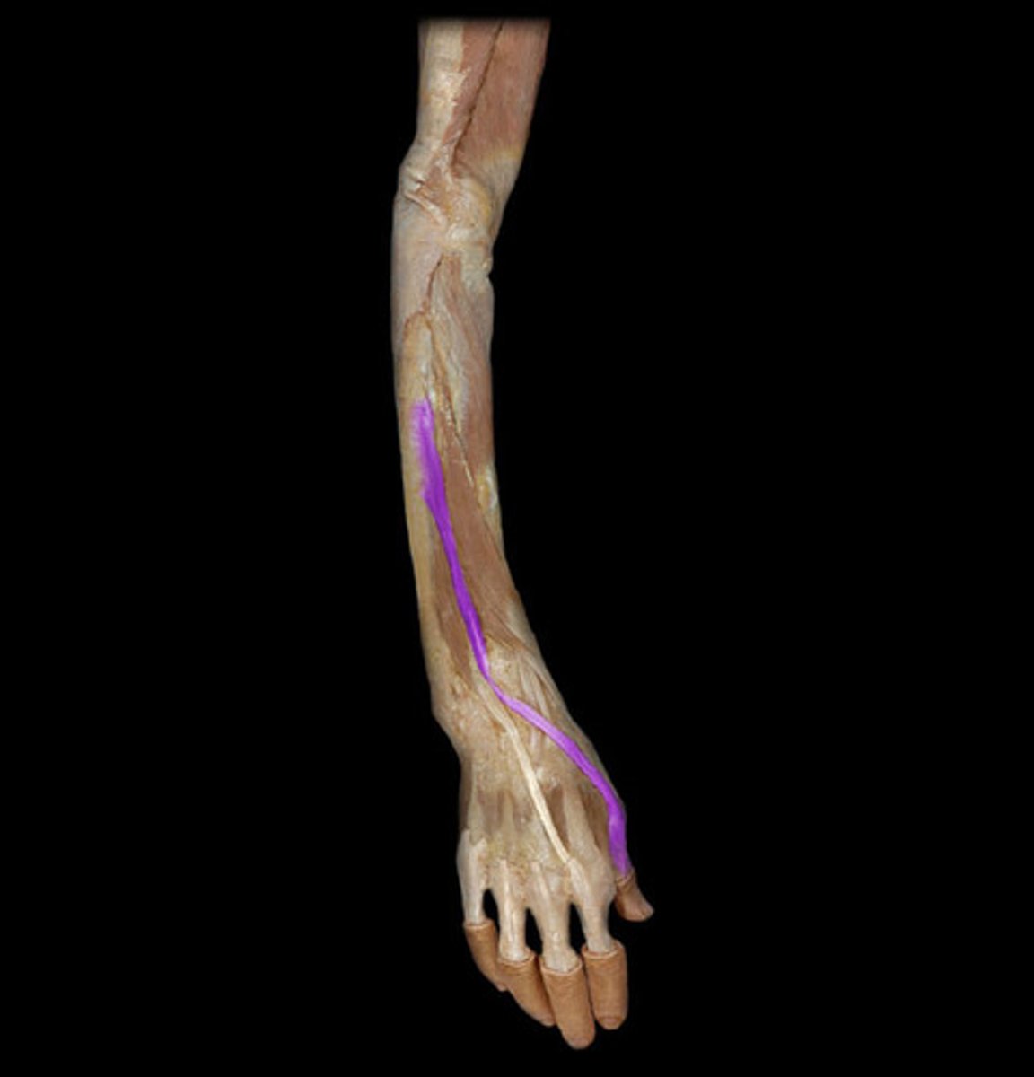
Palmaris Longis Muscle
BQ: About 20% of people don't have one.
Forearm Muscles: 3
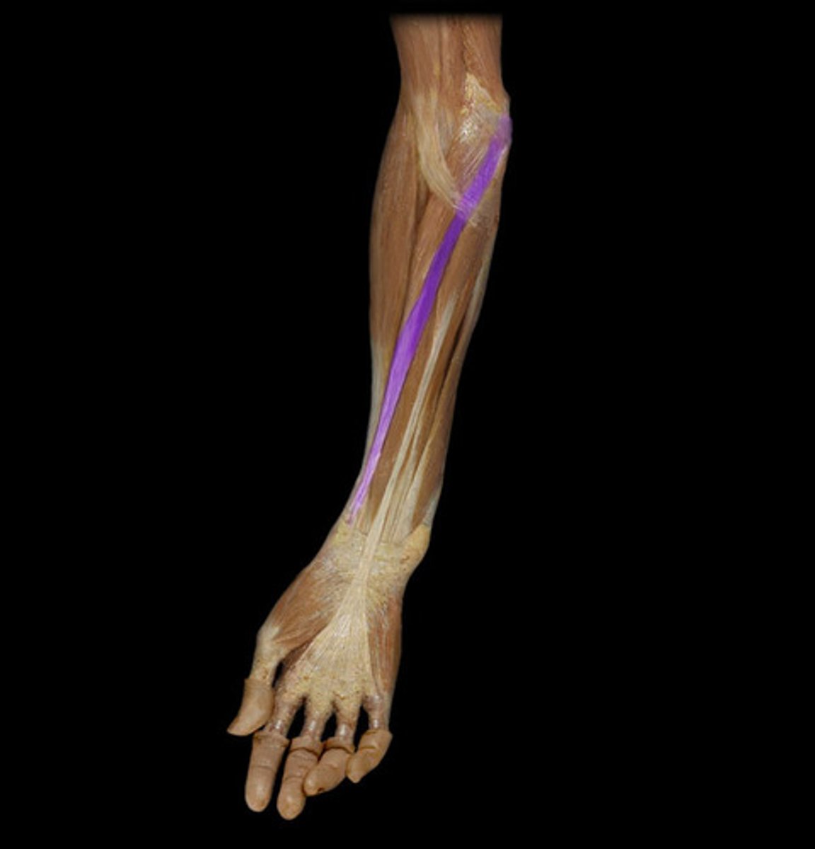
Flexor Carpi Ulnaris Muscle
Forearm Muscles: 4
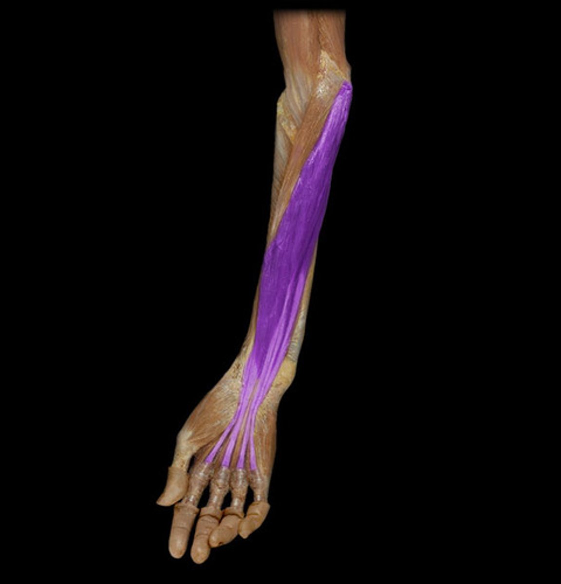
Flexor digitorum superficalis muscle
Thick slab, intermediate layer, inserts on medial phalanges
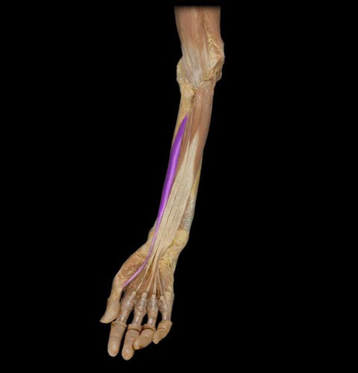
Flexor Pollicis Longus muscle
Flexes thumb, very deep inside the body
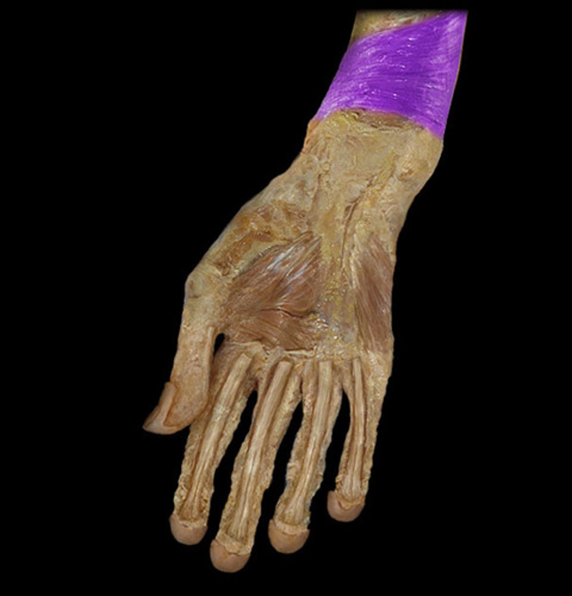
Pronator Quadratus Muscle
Pronates forearm, inside of the wrist
Brachioradialis Muscle
BQ: Attaches the humerus to the radius.
1
Extensor carpi radialis longus muscle
Extends wrist
2
Extensor carpi radialis brevis muscle
Extends wrist
3
Extensor Digitorum Muscle
In line with your middle finger, looks like guitar strings
4
Extensor Carpi Ulnaris muscle
Extension of wrist and adduction of hand, on pinky side
5
Supinator
Wraps around the top of the radius bone
Abductor Pollicis Longus Muscle
Takes thumb away from palm
1
Extensor Pollicis Brevis
Tendon right next to abductor
3

Extensor Pollicis Longus
Tendon next to EP brevis
2
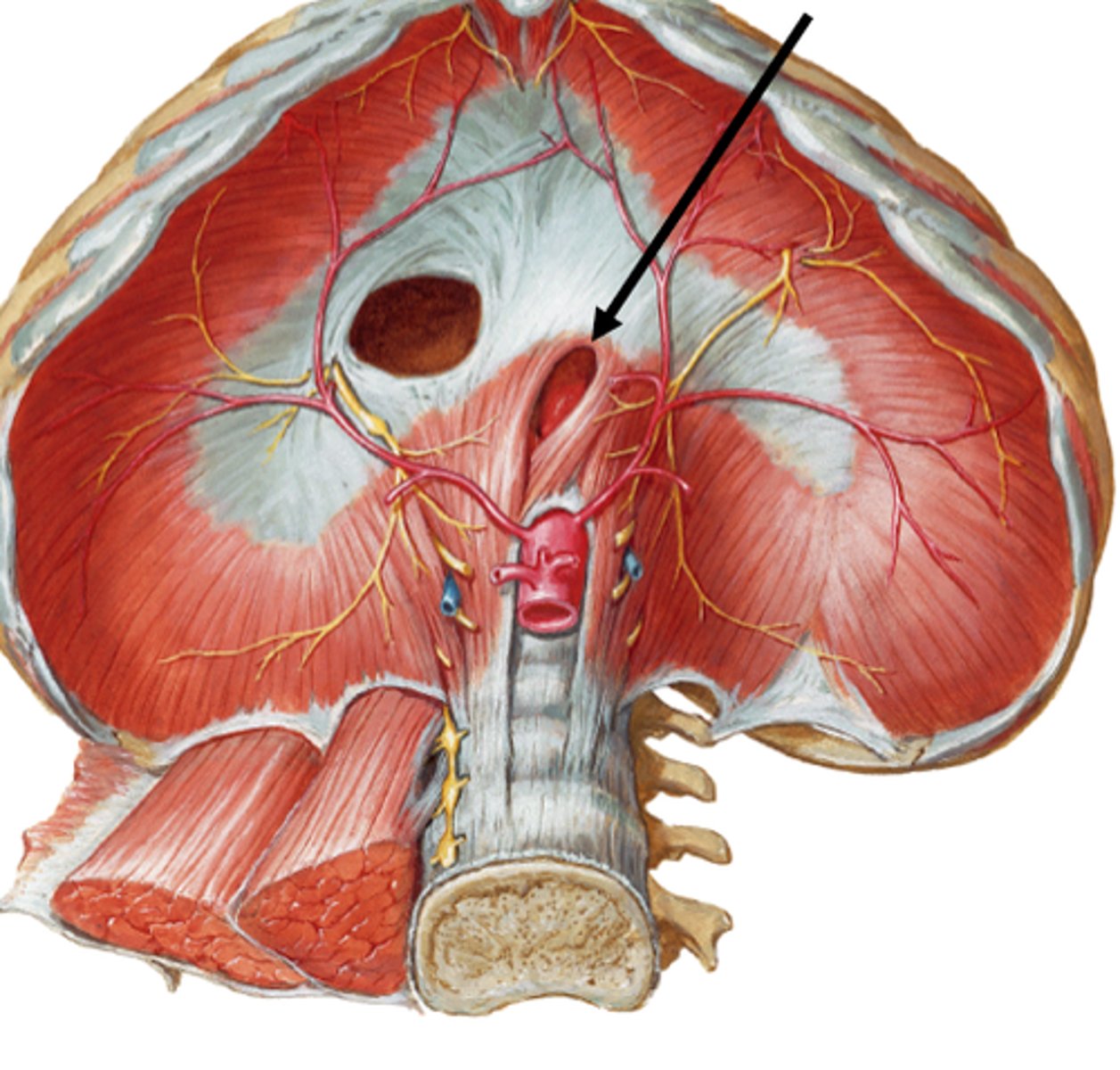
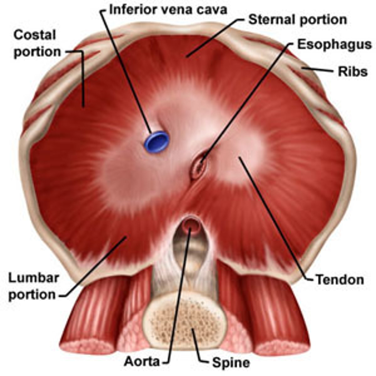
Diaphragm Muscles
A dome-shaped, muscular partition separating the thorax from the abdomen
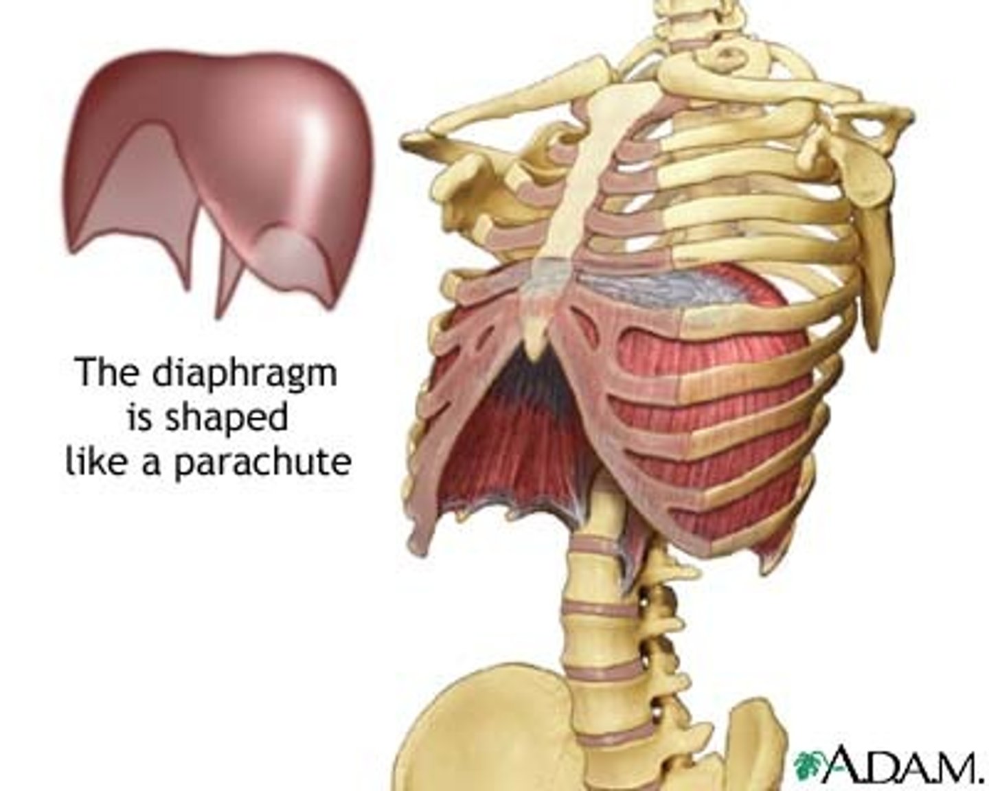
Sternocostal Part of the Diaphragm Muscle
Top edge of diaphragm
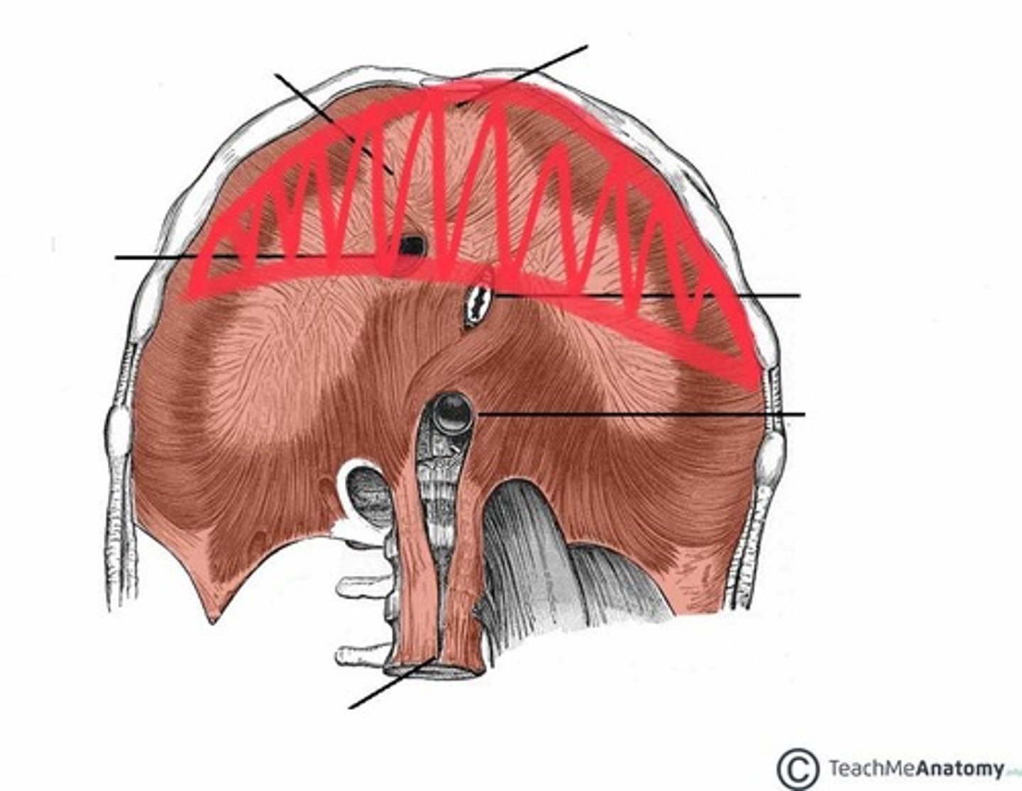
Lumbar Part of the Diaphragm Muscle
Bottom edge of diaphragm
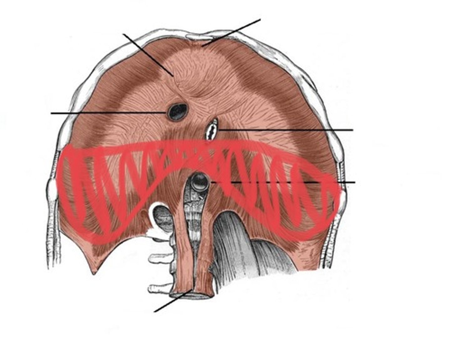
Central Tendon of the Diaphragm
Shiny line separating the two sections of the diaphragm
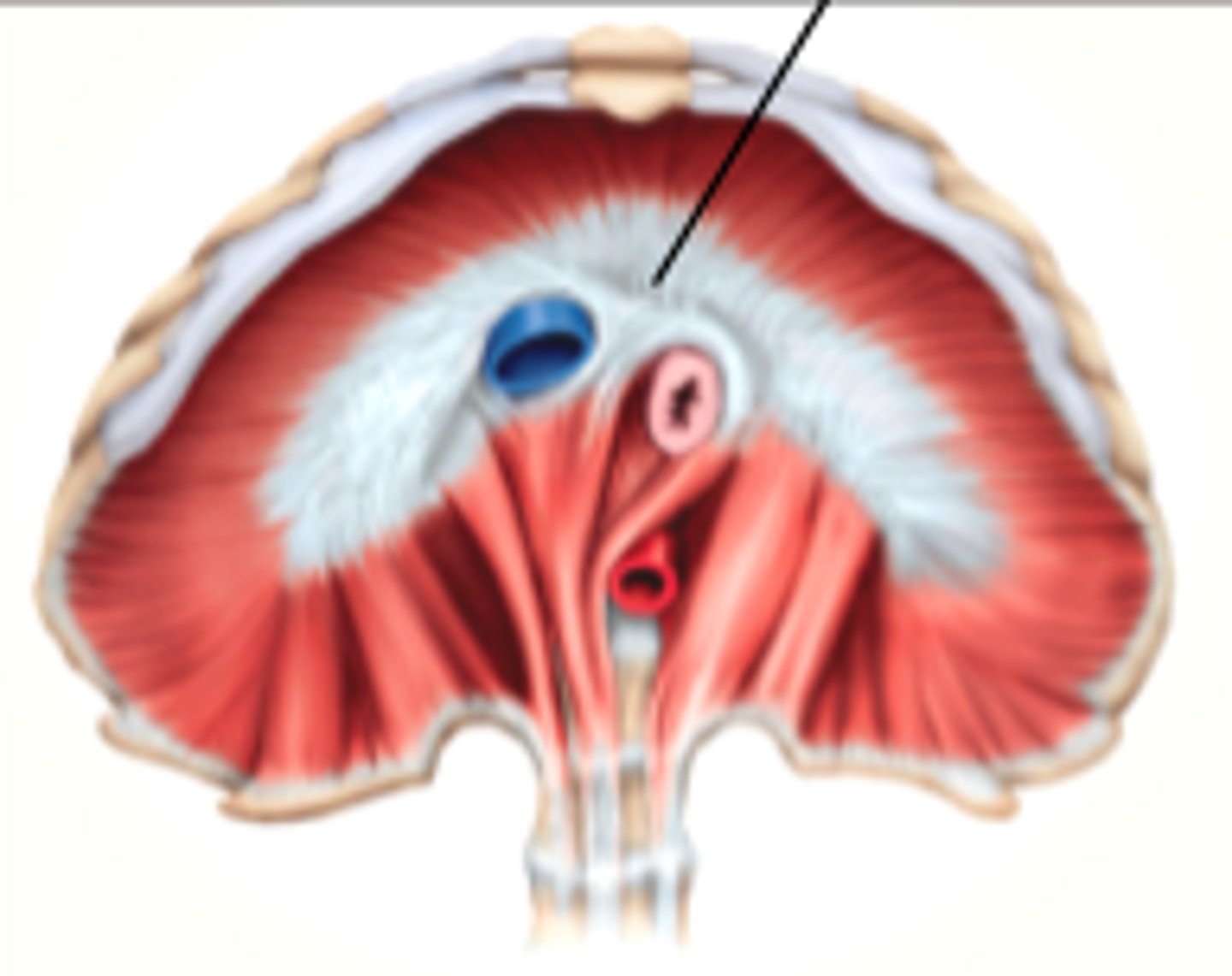
Esophageal Hiatus of the Diaphragm
Probe in the diaphragm from bottom to top
Caval Opening of the Diaphragm
Probe in the diaphragm from top to bottom, blue in the pic
Psoas Major Muscle
Flexes upper thigh
Psoas Minor Muscle
BQ: 40% of people do not have one.
Iliacus Muscle
Fills the iliac fossa

Iliopsoas Muscle
The psoas major and iliacus muscles fuse in the upper thigh
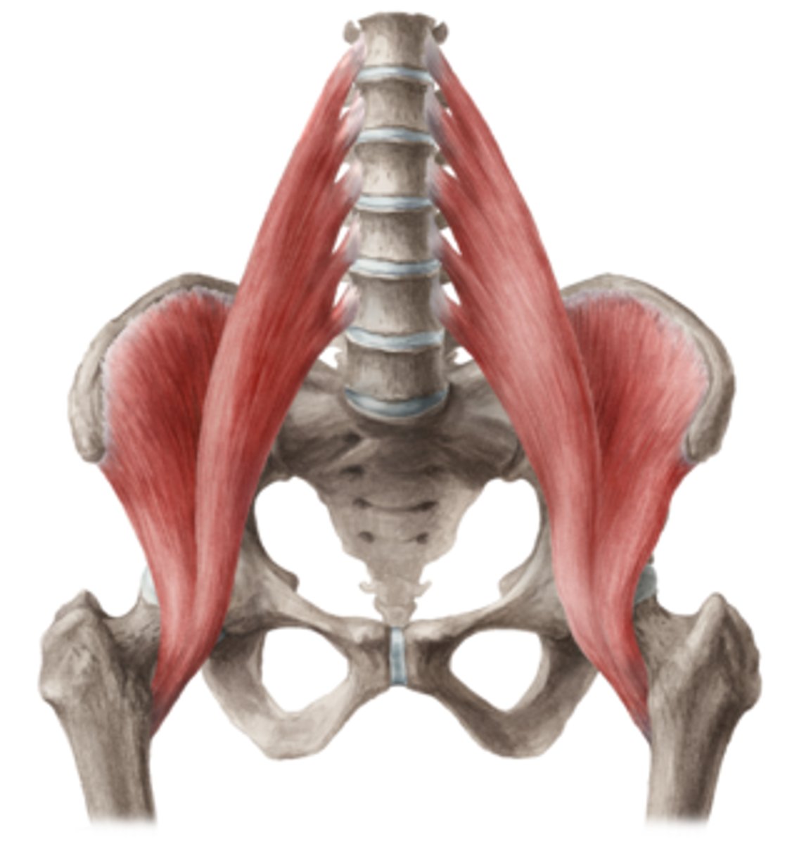
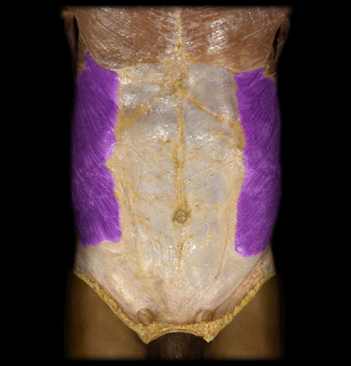
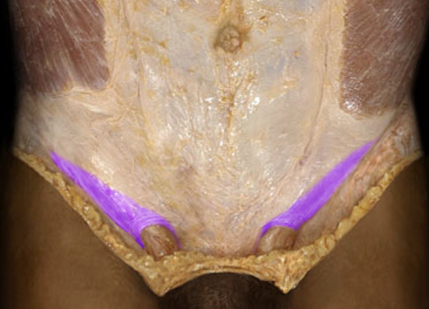
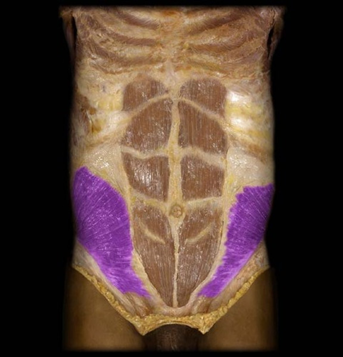
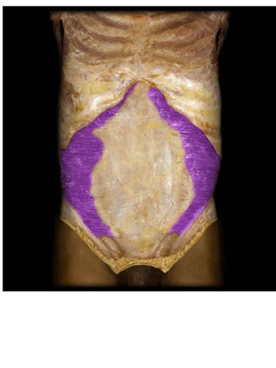
Quadratus Lumborum
Spans the gap between the 12th rib and iliac crest, behind kidneys
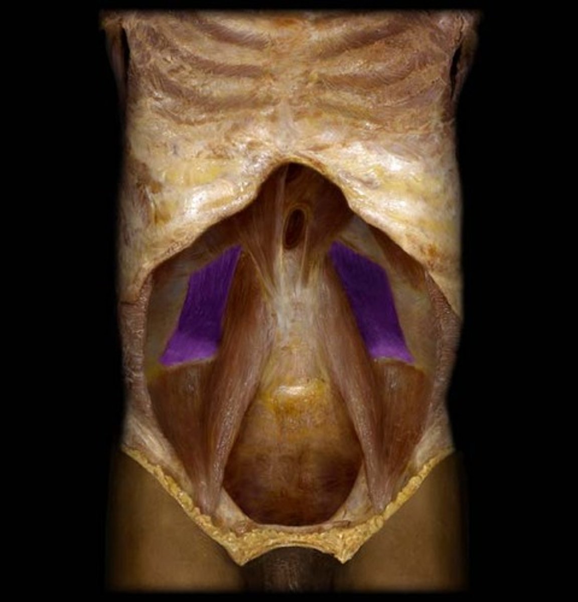
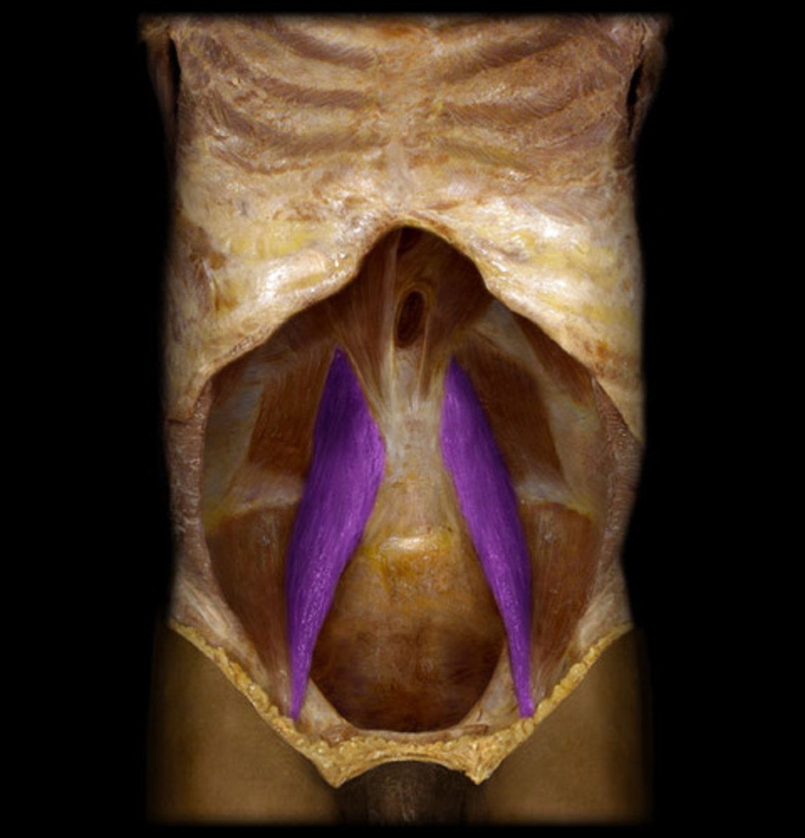
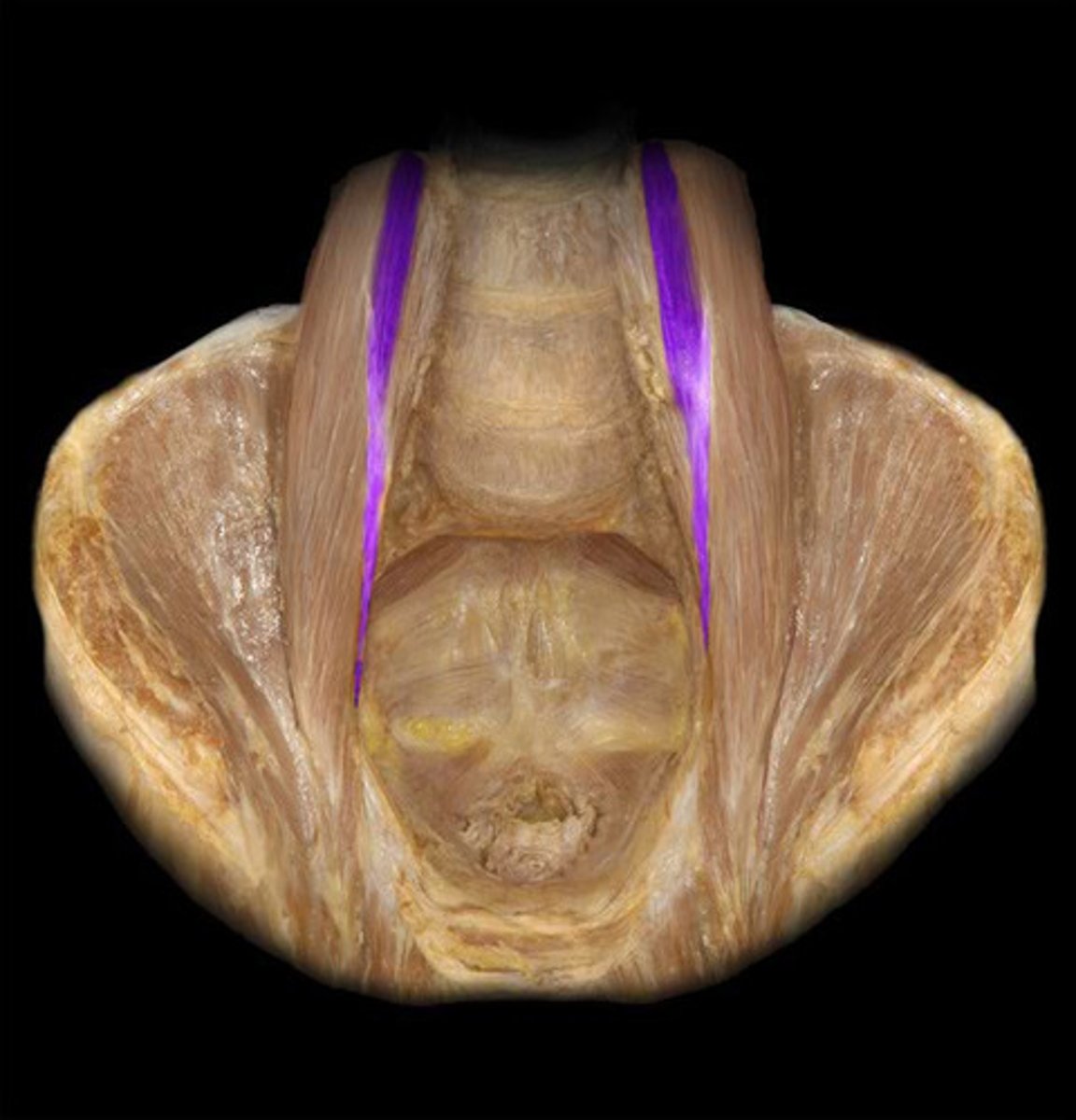
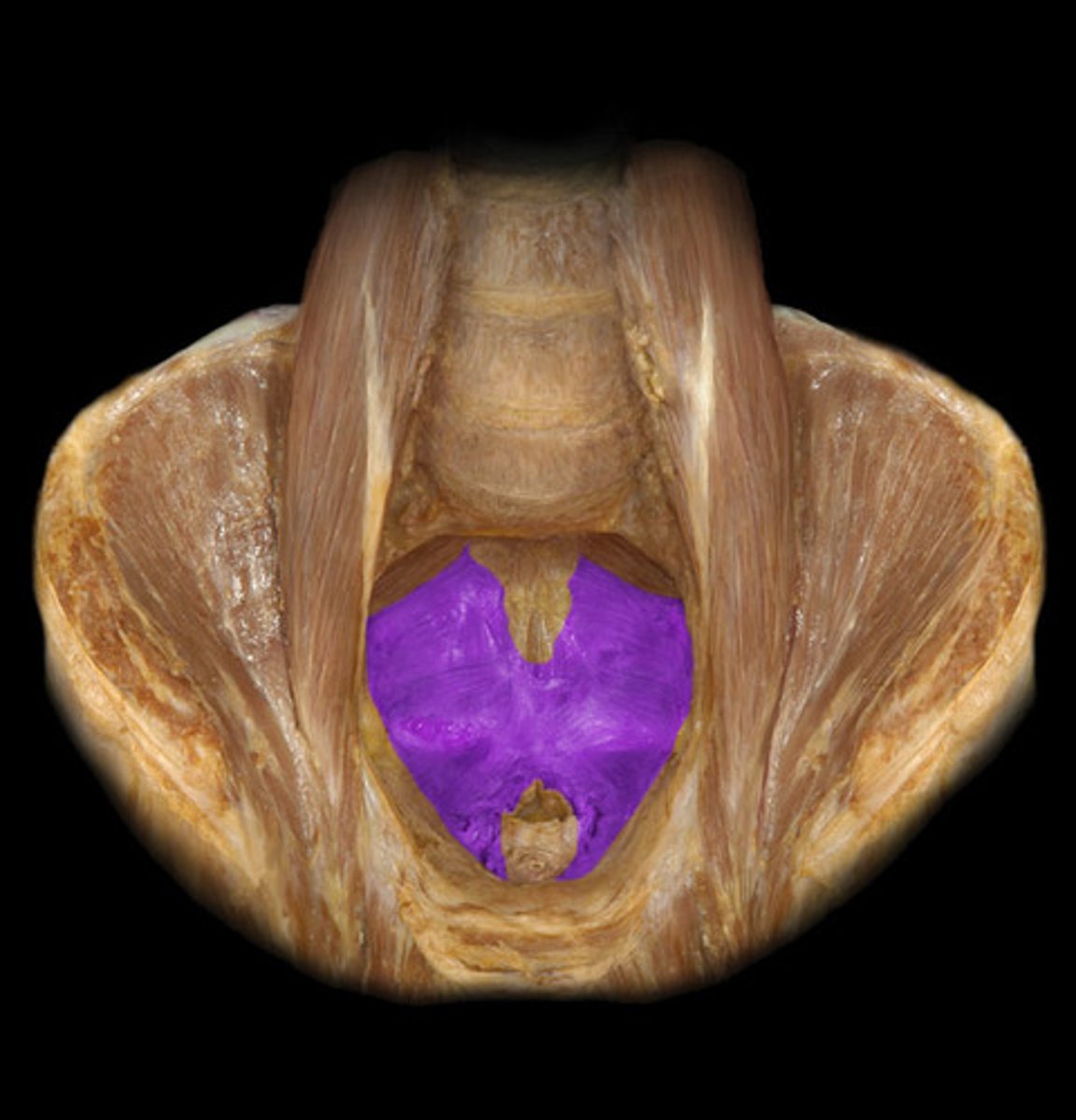
Pelvic Diaphragm Muscle
BQ: Forms the floor of the pelvic cavity. Very important muscle because it supports the pelvic organs.
External Anal Sphincter
Voluntary muscle to go #2
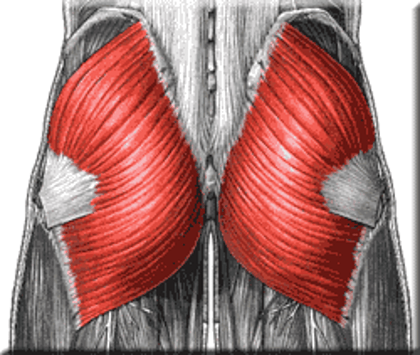
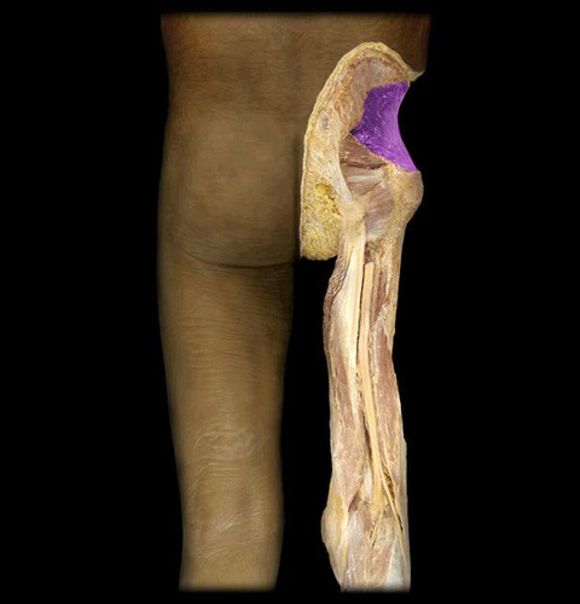
Gluteus Maximus Muscle
Largest single muscle in the body
-Big flap
Gluteus Medius Muscle
Small flap, middle gluteal
Gluteus Minimus Muscle
Most deep and smallest of gluteal
Tensor Fasciae Latae Muscle
Behind IT band
Iliotibial Tract
IT band
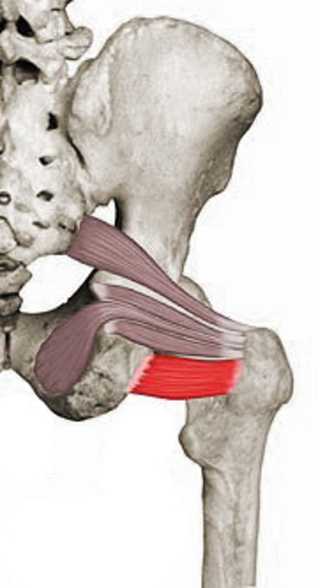
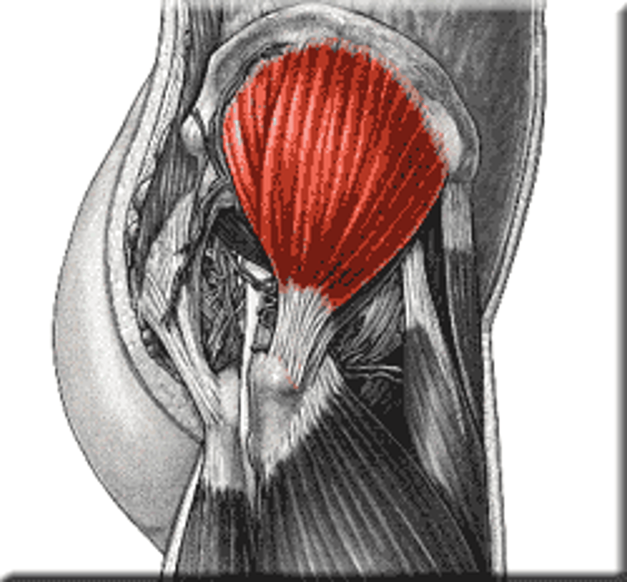
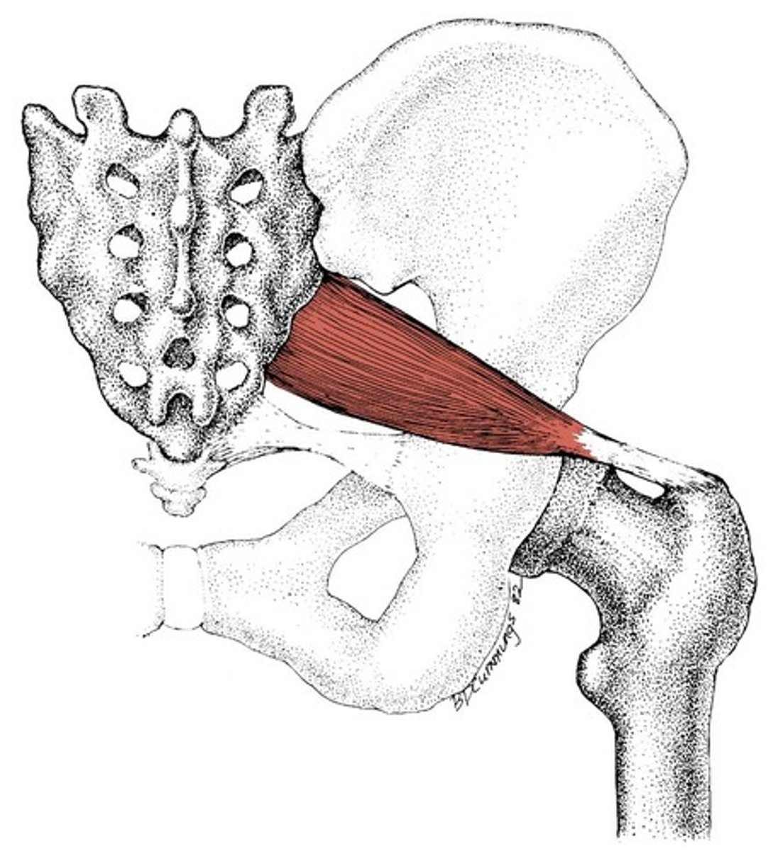
Piriformis Muscle
Pear shaped, passes through the greater sciatic notch to attach on the greater trochanter
Quadratus Femoris Muscle
Attaches to the ischial tuberosity and hip joint