Bio 240W Exam 2
5.0(1)
Card Sorting
1/72
Earn XP
Description and Tags
Last updated 10:19 PM on 2/27/23
Name | Mastery | Learn | Test | Matching | Spaced | Call with Kai |
|---|
No analytics yet
Send a link to your students to track their progress
73 Terms
1
New cards
Describe the three major types of nutrients: carbohydrates, fats/lipids, and proteins
Carbohydrates
* digested into small pieces usually glucose, sometimes other easily digestible sugars
* stored in limited quantities- body eager to use
* liver stores some excess also stored in muscles and nervous tissues
Fats
* broken down into fatty acids that can travel in the blood freely
* stored as triglycerides in fat cells
* typically provides more than half the body’s energy needs
* excess carbs stored as fatty acids
Proteins
* broken down into amino acids that are used to build new proteins
* can also yield energy if needed
2
New cards
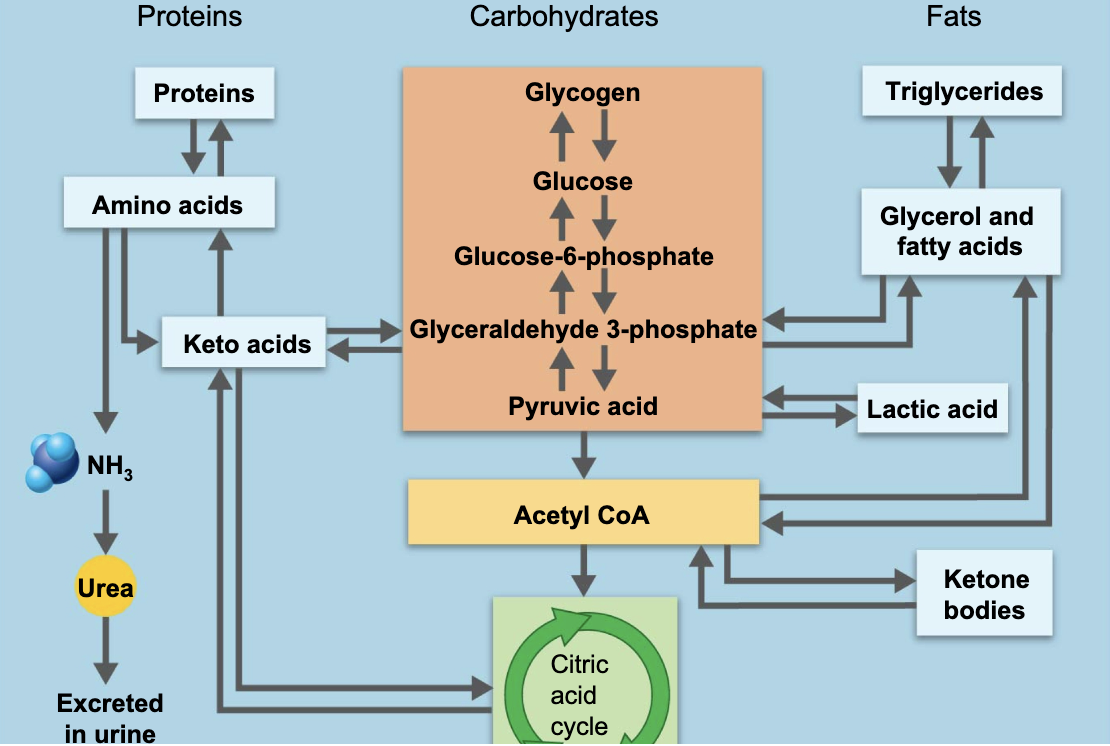
Discuss the ways in which carbohydrate metabolic pathways, glycolysis, and the citric acid cycle interrelate with protein and lipid metabolic pathways
\

3
New cards
Describe the absorptive state and the major events associated with this state
* called the fed state and lasts about 4 hours after eating
* anabolism exceeds catabolism
* excess nutrients stored as fats and sometimes carbs
* glucose → glycogen for storage
* triglycerides are used for energy by adipose tissue, the liver, and skeletal and cardiac muscles
* triglycerides → glycerol and fatty acids → triglycerides for storage
* removal of the amino group produces keto acids- energy in the citric acid cycle
* amino acids → fat in the liver
* amino acids are used for protein synthesis
* absorptive state controlled by insulin
4
New cards
Describe the post-absorptive state and the major events associated with this state
* fasting state
* gi tract is empty, energy sources are supplied by the breakdown of body reserves
* catabolism exceeds anabolism
* maintains blood glucose
* glucose sparing
* proteins → amino acids
* glycogen → glucose
* triglycerides → glycerol and fatty acids
* sympathetic nervous system- controlled by several hormones (more complex)
5
New cards
Sources of blood glucose in the post-absorptive state
* glycogenolysis in the liver- the first reserve used
* glycogenolysis in skeletal muscle- 2nd
* lipolysis in adipose tissue and liver- 3rd
* catabolism of cellular protein- not good, usually starving
* glycogenolysis in skeletal muscle- 2nd
* lipolysis in adipose tissue and liver- 3rd
* catabolism of cellular protein- not good, usually starving
6
New cards
Describe the process of insulin use during the absorptive state
* insulin secreted by beta cells in the pancreas
* insulin release is stimulated by elevated blood glucose levels and amino acids
* parasympathetic stimulation
* brings glucose into cells from the blood
* brain and liver take up glucose without insulin
* insulin is a hypoglycemic hormone
* inhibits glucose release from liver and gluconeogenesis
* insulin release is stimulated by elevated blood glucose levels and amino acids
* parasympathetic stimulation
* brings glucose into cells from the blood
* brain and liver take up glucose without insulin
* insulin is a hypoglycemic hormone
* inhibits glucose release from liver and gluconeogenesis
7
New cards
Describe how the catabolic-anabolic steady state is associated with these two metabolic states and how it helps to maintain homeostasis
* dynamic state in which organic molecules (except DNA) are continuously broken down and rebuilt
* the body uses nutrient pools available for immediate use
* anabolic- simple → complex, requires energy
* catabolic- complex → simple, releases energy
* the intermediates in the metabolic pathways are used for other processes
* keeps the energy needs in our body consistent as long as both processes occur
8
New cards
Describe glucose sparing especially regarding nutrients for the brain
* during prolonged periods of fasting the body uses more non-carbohydrate sources to conserve glucose
* the brain uses a bulk of glucose while other body cells switch to fatty acids as fuel
* glucose sparing saves glucose for organs such as the brain
* after 4-5 days ketone bodies are used by the brain if needed
* the brain uses a bulk of glucose while other body cells switch to fatty acids as fuel
* glucose sparing saves glucose for organs such as the brain
* after 4-5 days ketone bodies are used by the brain if needed
9
New cards
Discuss the roles of the nervous and endocrine systems in homeostasis
Nervous system—Autonomic system
* Sympathetic—arouses body for “fight or flight”
* Neurotransmitter released by sympathetic neurons is norepinephrine (similar to adrenaline)
* Parasympathetic—predominates during relaxation, “rest and digest”
* Neurotransmitter is acetylcholine—the same neurotransmitter used at the neuromuscular junction
Endocrine system
* Hormones involved in blood glucose regulation
* Insulin and glucagon
* Both are produced by the pancreas
\
10
New cards
Discuss homeostatic imbalances, specifically diabetes mellitus
Type 1
* inadequate insulin production
Type 2
* abnormal insulin receptors
\
both result in
* proteins and fats are used for energy
* unavailability of glucose to most body cells
* excessively high blood glucose levels (hyperglycemia)
* glucose loss in urine
* can lead to metabolic acidosis, protein wasting, weight loss
* may lead to coma and death
11
New cards
Why isn’t it sufficient to reduce **only dietary fat intake** to prevent **new fatty deposits from forming** in the body?
Acetyl CoA is a starting point for fatty acid synthesis.
12
New cards
TRUE OR FALSE. The preferred energy fuel for the brain is **fat**.
False
13
New cards
TRUE OR FALSE. **Carbohydrate and fat pools** are oxidized directly to produce cellular energy, but **amino acid pools** must first be converted to a **carbohydrate intermediate** before being sent through cellular respiration pathways.
True
14
New cards
When proteins undergo **deamination (removal of amino group**), the **waste substance found in the urine** is mostly ________.
urea
15
New cards
**Glycogen** is formed in the liver during the ________
absorptive state
16
New cards
**Lipogenesis** occurs when ________.
cellular ATP and glucose levels are high
17
New cards
Which of the following statements is FALSE?
* The amino acid pool is the body's total supply of amino acids in the body's proteins.
* Fats and carbohydrates are oxidized directly to produce cellular energy.
* Amino acids can be used to supply energy only after being converted to a citric acid cycle intermediate.
* Excess carbohydrate and fat can be stored as such, whereas excess amino acids are oxidized for energy or converted to fat or glycogen for storage.
The amino acid pool is the body's total supply of amino acids in the body's proteins.
18
New cards
If you were to **jog** one kilometer a **few hours after lunch**, which **stored fuel** would you probably tap?
liver glycogen and muscle glycogen
19
New cards
A **fasting** animal whose **energy needs exceed those provided in its diet** will draw on its **stored resources in which order?**
liver glycogen, then muscle glycogen, then fat
20
New cards
**Humans store glucose** in the form of ________________; **plants store glucose** in the form of ________________.
glycogen; starch
21
New cards
Which of the following **glucose storage forms** is the most **difficult for human digestive systems to break down**?
cellulose
22
New cards
In general metabolic terms, food digestion is a form of _______, while building new protein molecules is a form of _______.
catabolism; anabolism
23
New cards
The process whereby excess glucose is stored in cells is called _______.
glycogenesis
24
New cards
What is the primary process by which insulin is released after ingesting a meal?
Insulin is secreted in direct response to blood glucose.
25
New cards
What is the primary objective during the post-absorptive state?
To maintain blood glucose (typically around 70–110 mg/100 mL blood)
26
New cards
Describe the fibrous skeleton of the heart and its major functions.
* structural support for the heart
* gives the muscle cells something to pull against
* electrical insulation that helps regulate the heartbeat
* gives the muscle cells something to pull against
* electrical insulation that helps regulate the heartbeat
27
New cards
Describe intercalated discs and their components: desmosomes and gap junctions.
desmosomes
* provide a physical connection
* allows muscle cells to pull on each other without damaging membrane
gap junctions
* provide cytoplasmic connection between cells
* electrically connects the heart
* Allow waves of depolarization to spread rapidly from cell to cell—all heart muscle cells contract almost simultaneously
28
New cards

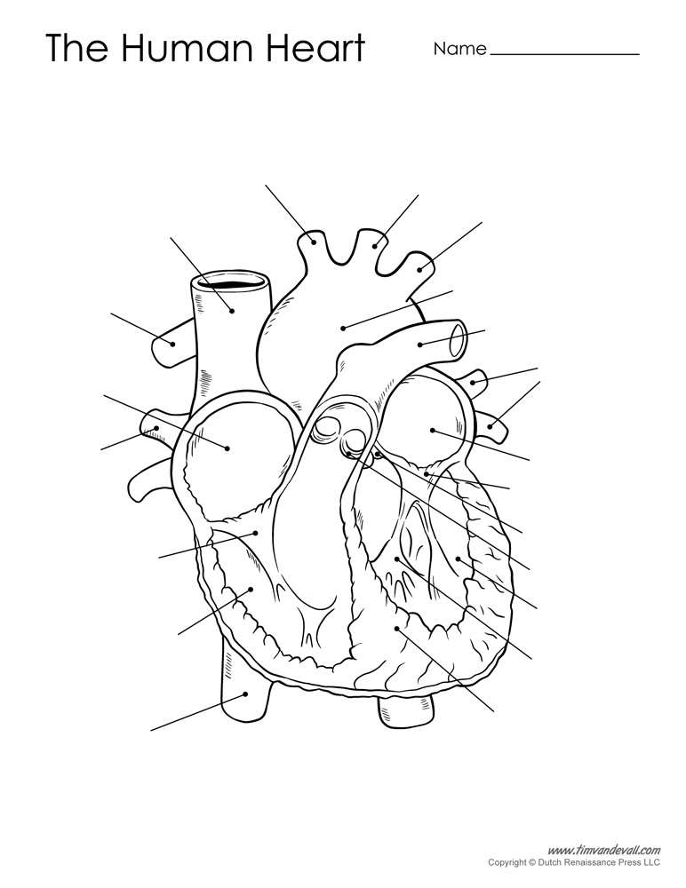
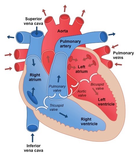
Identify the major anatomical features of the heart and major blood vessels.
\
\
Arteries carry blood away from the heart
Veins carry blood toward the heart
Tricuspid valve= right atrioventricular valve
pulmonary and aortic valves= semilunar valves
bicuspid valve= left atrioventricular valve
Veins carry blood toward the heart
Tricuspid valve= right atrioventricular valve
pulmonary and aortic valves= semilunar valves
bicuspid valve= left atrioventricular valve

29
New cards
Describe the blood flow through the heart and identify where oxygen and deoxygenated blood flows in the heart.
* start at right ventricle
* start at right ventricle
deoxygenated
* out of right ventricle through pulmonary semilunar valve
* through pulmonary arteries to the lung
* gas exchange in capillary beds of lungs
oxygenated
* return from lungs through pulmonary veins into left atria
* through bicuspid (left AV) valve into left ventricle
* out of the Aorta into the body
deoxygenated
* returns from the body through the superior and inferior vena cava into the right atria
* Through tricuspid (right AV) valve into right ventricle
repeat process
30
New cards
Which of the following structures is associated with oxygen-rich blood?
A. Right AV valve
B. Pulmonary vein
C. Right ventricle
D. Pulmonary artery
A. Right AV valve
B. Pulmonary vein
C. Right ventricle
D. Pulmonary artery
B. Pulmonary vein
31
New cards
Which of the following structures does blood flow into AFTER the left ventricle?
A. Right AV valve
B. Aorta
C. Right ventricle
D. Pulmonary artery
A. Right AV valve
B. Aorta
C. Right ventricle
D. Pulmonary artery
B. Aorta
32
New cards
Which of these vessels supplies blood to the heart (myocardium)?
A) Aorta
B) Vena cava
C) Pulmonary artery
D) Pulmonary vein
E) None of the above
A) Aorta
B) Vena cava
C) Pulmonary artery
D) Pulmonary vein
E) None of the above
E) None of the above
actually the coronary arteries
actually the coronary arteries
33
New cards
Why are the walls of the ventricles thicker than the walls of the atria?
Under higher pressure to get the blood farther away from the heart (either to lungs or the body)
34
New cards
Describe the four major structures of the conduction system and where they are related to in terms of anatomy of the heart.
SA node- Pacemaker cells coordinate the heartbeat, signal the atria to contract together first and then the ventricles contract together second
* the upper wall of the right atrium and the opening of the superior vena cava
* excited roughly 75 times per minute
AV node- conduct the action potential to additional conductive cells of the ventricles
* Without SA node, excited roughly 50 times per minute
AV bundle cells- conduct the action potential to the base of the ventricles
* Without SA node or AV node, excited roughly 40 times per minute
Purkinje fibers- conduct the action potential to muscle cells of the ventricle walls
* Without SA node, AV node, or bundle action potential, excited roughly 30 times per minute
35
New cards
How is the connection between heart muscle cells different than between two neurons or a neuron and a skeletal muscle cell?
A. All of the above have synapses between the cells but no need for neurotransmitters
B. All of the above have synapses and need neurotransmitters
C. Heart cells do not have synapses but still need neurotransmitters
D. Heart cells do not have synapses and no need for neurotransmitters
A. All of the above have synapses between the cells but no need for neurotransmitters
B. All of the above have synapses and need neurotransmitters
C. Heart cells do not have synapses but still need neurotransmitters
D. Heart cells do not have synapses and no need for neurotransmitters
D. Heart cells do not have synapses and no need for neurotransmitters
36
New cards
Describe the relationship between volume and pressure.
\-For a fixed volume of fluid, the pressure depends on the volume of the space it occupies
\-Pressure and volume are inversely proportional
* Large volume = lower pressure
* Small volume = high pressure
* As the size of the space changes, so does the pressure
\-Pressure and volume are inversely proportional
* Large volume = lower pressure
* Small volume = high pressure
* As the size of the space changes, so does the pressure
37
New cards
Define systole and diastole.
systole- contraction
diastole- relaxation
diastole- relaxation
38
New cards
During systole the space of the chamber decreases, the volume of fluid is the same – What happens to pressure?
A – Increases
B - Decreases
A – Increases
B - Decreases
A – Increases
39
New cards
During Diastole the space of the chamber increases, the volume of fluid is the same - What happens to pressure?
A – Increases
B - Decreases
A – Increases
B - Decreases
B - Decreases
40
New cards
Explain how contraction and relaxation of the chambers of the heart create pressure gradients and how these gradients cause the flow of blood in relation to anatomy.
1. All chambers in diastole
– Blood flows passively through atria and into ventricles
– Both AV valves open
– Note both atria and ventricles fill with blood
– But not all blood leaves atria
2. Atrial systole
– Ventricles (still in diastole) swell with extra blood that has been pumped in by atria
3. Ventricular systole (atrial diastole)
– Right and left AV valves pushed shut
– Blood forced into arteries through pulmonary and aortic semilunar valves
– ventricles contract from the bottom up
4. Now back to all chambers in diastole
– as ventricles enter diastole, a little blood sucked back in
– however right and left semilunar valves snap shut
41
New cards
Explain what the first and second heart sounds are.
1. Bicuspid and tricuspid valves are pushed shut during ventricular systole (first heart sound—“lub”)
2. Pulmonary and aortic semilunar valves snap shut when reentering all chambers in diastole (second heart sound—“dub”)
42
New cards
Where would pressure be greater?
A) coming out of the right ventricle (pulmonary circuit—heading to lungs)
B) coming out of the left ventricle (systemic circuit—to the rest of the body)
A) coming out of the right ventricle (pulmonary circuit—heading to lungs)
B) coming out of the left ventricle (systemic circuit—to the rest of the body)
B) coming out of the left ventricle (systemic circuit—to the rest of the body)
43
New cards
Which set of valves are involved with the second heart sound?
A. Pulmonary and right AV valves
B. Pulmonary and aortic valves
C. Right and left AV valves
D. Aortic valve and left AV valve
A. Pulmonary and right AV valves
B. Pulmonary and aortic valves
C. Right and left AV valves
D. Aortic valve and left AV valve
B. Pulmonary and aortic valves
44
New cards
Define a brain center.
a collection of interneurons that receive sensory input about a specific function and create motor output to alter that function
“Cardiovascular control centers” include:
* Cardiac control (heart)
* Cardioacceleratory neurons
* Cardioinhibitory neurons
* Vasomotor control (vessels)
Together they regulate blood pressure and heart function
“Cardiovascular control centers” include:
* Cardiac control (heart)
* Cardioacceleratory neurons
* Cardioinhibitory neurons
* Vasomotor control (vessels)
Together they regulate blood pressure and heart function
45
New cards
Describe the action potential in the pacemaker cells/SA node and how it contributes to the autorhythmicity of the heart.
Have leak-like channels that allow cations to diffuse into the cell
Channels result in depolarization to threshold **without** neuronal excitation
no Na+/K+ pump needed to return to Resting Membrane potential
Channels result in depolarization to threshold **without** neuronal excitation
no Na+/K+ pump needed to return to Resting Membrane potential
46
New cards
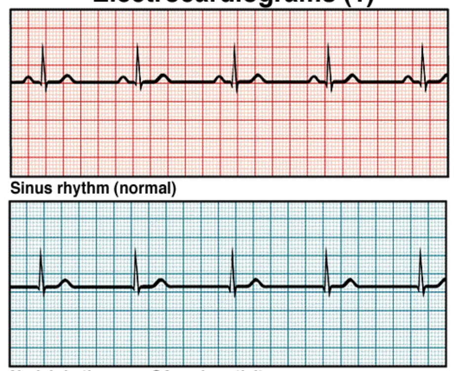
Analyze how the major regions of an EKG correlate with the conduction system function and contraction
P wave- SA node excitement and atrial depolarization
PR segment- atrial depolarization is complete, the impulse is delayed at the AV node
QRS complex- ventricular depolarization, atrial repolarization
ST segment- ventricular depolarization is complete
T wave- ventricular repolarization begins at the apex
after the T wave- ventricular repolarization is complete
47
New cards
A patient visits a physician for a second opinion about a diagnosed heart murmur affecting the right AV valve. Heart murmurs are caused by valve prolapse (the valve turns inside out). Which of the following symptoms could the physician check to confirm the heart murmur?
A. A missing QRS complex on an EKG
B. A weak/missing second heart sound
C. A missing T wave on an EKG
D. A weak/missing first heart sound
A. A missing QRS complex on an EKG
B. A weak/missing second heart sound
C. A missing T wave on an EKG
D. A weak/missing first heart sound
D. A weak/missing first heart sound
48
New cards

What is happening in the lower EKG?
A. No atrial contraction
B. No ventricle depolarization
C. No ventricle contraction
D. No AV node activity
A. No atrial contraction
B. No ventricle depolarization
C. No ventricle contraction
D. No AV node activity
A. No atrial contraction
49
New cards
Define stroke volume and cardiac output.
stroke volume- the amount of blood pumped out of each ventricle in one pump (both ventricles pump the same amount). normally between 70-80mL
cardiac output- the amount of blood the heart pumps per minute INTO ARTERIES
* heart rate (beats/min) x stroke volume (mL/beat)= cardiac output (mL/min)
cardiac output- the amount of blood the heart pumps per minute INTO ARTERIES
* heart rate (beats/min) x stroke volume (mL/beat)= cardiac output (mL/min)
50
New cards
Describe blood vessel anatomy, especially the differences seen in arteries, veins, and capillaries.
Arteries are thicker than veins because they carry blood under a higher pressure
Arteries transport blood away from the heart
Veins return the blood back to the heart
Capillaries surround body cells and tissues to deliver oxygen, amino acids and glucose, and return carbon dioxide and wastes
The capillaries also connect the branches of arteries to the branches of veins.
In capillaries, the arterial end blood pressure is higher than the osmotic pressure, and net pressure out
The venous end has higher osmotic pressure than blood pressure, net pressure in.
51
New cards
Describe blood pressure and how it is controlled.
\-the force blood exerts against an arterial wall (most commonly the brachial artery)
\-Neural circuits in the brain also regulate heart rate, the strength of contraction, vasoconstriction, and dilation throughout the entire body
\-Neural circuits in the brain also regulate heart rate, the strength of contraction, vasoconstriction, and dilation throughout the entire body
52
New cards
Three main variables that affect blood pressure? what do they do?
Cardiac output
* Amount of blood the heart pumps per minute into the arteries
* arterial volume is constant but the amount of fluid in arteries is altered
Resistance of vessels
* the combined effect of blood composition, vessel diameter, and vessel length
* vessel diameter can change (vasomotion)
Blood volume
* controlled by the kidneys and hormones
53
New cards
Describe the afferent signals to the brain, including three types of receptors, and how they affect heart rate.
Propriocenters- sensory input from muscles and tendons
* informs the brain on changes in physical activity
Baroreceptors- sensory input from blood vessels
* informs the brain on changes to pressure in vessels
Chemoreceptors- sensory input from blood vessels
* informs the brain on changes in carbon dioxide or oxygen levels in the blood
* important in respiratory system but has some affect on heart rate
54
New cards
Compare and contrast the efferent responses that affect heart rate and how they alter the SA node activity including the neurotransmitters associated with these responses.
Sympathetic system
* cardioacceleratory system- increased heart rate
* Postganglionic neuron secretes norepinephrine (NE)
* Adrenergic receptors on cells of the SA node bind to NE
* Cause an increased rate of action potentials of SA node
* Maximum heart rate is 230 beats/minute
* Limit of SA node excitation
Parasympathetic system
* cardioinhibitory system- decreased heart rate
* Postganglionic neuron secretes acetylcholine (ACh)
* Cholinergic receptors on cells of the SA node bind ACh
* Allow potassium to leave the cell → hyperpolarizing reaction
* The rate of action potentials decrease
* Normal heart rate is about 75 beats/minute
* Without nervous control, the heart rate would be about 100 beats/minute
55
New cards
Describe the roles of the sympathetic and parasympathetic nervous systems in controlling heart rate.
Sympathetic
* blood pressure is too low
* visceral motor neurons cause
* increase heart rate by activating cardioacceleratory neurons
* vasoconstriction which is the constriction of blood vessels regulated by the sympathetic release of norepinephrine or epinephrine
Parasympathetic
* blood pressure is too high
* visceral motor neurons cause
* decrease heart rate by activation of cardioinhibitory neurons
* vasodilation which is the dilation of blood vessels due to a decrease in norepinephrine
56
New cards
If the right AV valve does not close completely and allows blood to pass through when it should be shut, then you could expect:
the output of the right ventricle to be decreased.
57
New cards
The **semilunar valves** prevent blood from **flowing backwards into** the...
ventricles
58
New cards
**Normal heart sounds** are caused by which of the following events?
closing of the heart valves
59
New cards
Without **SA node activity**, what type of **heart surgery would be indicated**?
inserting an artificial pacemaker
60
New cards
Which of the following develops the **greatest pressure** on the blood in the **aorta**?
systole of the left atrium
diastole of the right ventricle
systole of the left ventricle
diastole of the right atrium
systole of the left atrium
diastole of the right ventricle
systole of the left ventricle
diastole of the right atrium
systole of the left ventricle
61
New cards
Among the following choices, which organism likely has the **highest systolic pressure**?
mouse
human
hippopotamus
giraffe
mouse
human
hippopotamus
giraffe
giraffe
62
New cards
The heart muscle (myocardium) receives its nutrients from the blood moving through **the left atrium and ventricle.**
True
False
True
False
False
63
New cards
The **left side** of the heart pumps the **same volume of blood as the right.**
True
False
True
False
True
64
New cards
The **atrioventricular (AV) valves** are **closed** ________.
when the ventricles are in systole
65
New cards
**During exercise**, which of the following would occur on an **electrocardiogram (ECG/EKG) compared to an individual at rest**?
the T wave would decrease
the P-R interval would decrease
the time from one R to the R of the next heartbeat would decrease
the S-T segment would decrease
the T wave would decrease
the P-R interval would decrease
the time from one R to the R of the next heartbeat would decrease
the S-T segment would decrease
the time from one R to the R of the next heartbeat would decrease
66
New cards
Ultimate goal of plant nutrient uptake
sucrose biosynthesis
67
New cards
During the day what happens to the CO2 in the carbon reactions
what happens at night?
what happens at night?
* stored as starch in chloroplast or sucrose in the cytosol
* at night, co2 assimilation stops, and starch in chloroplast broken down for transport
* at night, co2 assimilation stops, and starch in chloroplast broken down for transport
68
New cards
what is sucrose transported through?
Sucrose transported into vascular tissue to sinks throughout plant
69
New cards
What are sinks and sources?
\-Sources synthesize sugars \n –Sinks use sugars
70
New cards
What do the xylem and the phloem do?
* Xylem conducts water and dissolved minerals \n upward from roots into the shoots (“xylem up”)
* Phloem transports sugars to roots and other parts of \n plant as needed (“phloem down”?)
* Phloem transports sugars to roots and other parts of \n plant as needed (“phloem down”?)
71
New cards
Where do plants get sucrose from and in which direction can it move?
* Plants can use sucrose locally within the leaf or transport it to another part of the plant that needs it
* Sucrose can move up OR down in in the phloem, only up in the xylem
* Sucrose can move up OR down in in the phloem, only up in the xylem
72
New cards
Which of the following is most likely to be a source in plants? \n a) immature leaves \n b) developing fruits \n c) roots in the early spring \n d) roots in late summer
d) roots in late summer
73
New cards
Why is sucrose used for carbon transport through the plant rather than glucose?
* glucose is smaller than sucrose
* need pressure gradient
* if glucose builds up there is ketoacidosis so sucrose is better because it is reactive
* need pressure gradient
* if glucose builds up there is ketoacidosis so sucrose is better because it is reactive