Ruminant Oral Facial, Esophagus, Gastric Disorders
1/239
There's no tags or description
Looks like no tags are added yet.
Name | Mastery | Learn | Test | Matching | Spaced | Call with Kai |
|---|
No study sessions yet.
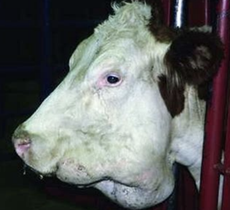
240 Terms
what is bovine papular stomatitis?
(zoonotic) disease causing proliferative and erosive stomatitis
what causes bovine papular stomatitis?
parapox virus
what do lesions caused by bovine papular stomatitis look like?
epidermitis causes small, red raised nodules
nodules may have a depressed or eroded center
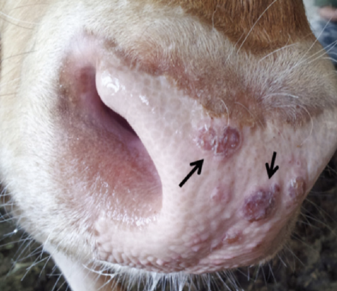
where are lesions caused by bovine papular stomatitis typically found?
-the muzzle
-oral mucosa
-rarely esophagus
-rumen
-teats
-feet
how do young cattle present with bovine papular stomatitis?
usually non-clinical to mild infections
when are cases of bovine papular stomatitis seen?
tends to occur in outbreaks, and usually regresses within a month
reinfections can occur (immunity is short lived)
what are severe forms of bovine papular stomatitis associated with?
weight loss, diarrhea, and will rarely have ulcerative lesions
what can worsen stomatitis in cattle with bovine papular stomatitis?
ulcerations and secondary infections
how is bovine papular stomatitis diagnosed?
clinical signs
biopsy
virus isolation
EM
PCR
what is seen on histopathology that can confirm diagnosis of bovine papular stomatitis?
eosinophilic intracytoplasmic inclusion bodies
which species get contagious ecthyma? what animals have more severe disease?
sheep and goats
most severe disease is seen in the young animals (but all ages are susceptible)
what is contagious ecthyma?
aka orf/soremouth
(zoonotic) disease caused by a parapox virus that is related to bovine papular stomatitis virus, causing similar lesions
what lesions are caused by contagious ecthyma?
causes proliferative lesions, but the surface is often eroded and covered by a pustule/scab/crust
what is the disease course of contagious ecthyma?
disease is self-limiting with total course of 1-2 months
chronic infections, reinfections, and secondary infections can occur
when do outbreaks of contagious ecthyma occur?
usually occur every lambing/kidding season in infected flocks
where are lesions caused by contagious ecthyma commonly found?
-mucocutaneous junction of face
-coronary band
-vulvar/teat leasions
what is most morbidity of contagious ecthyma due to?
morbidity due to decreased nursing due to painful mouth and/or udder
in severe cases, mucosa of mouth, esophagus, and rumen may be infected
what parts of the lesions caused by contagious ecthyma are contagious?
crusts and secretions are infective, and the virus survives a long time (6+ months) in the environemnt
how is contagious ecthyma treated?
antibiotics can be used to control secondary infections and supplemental feeding for animals that are not nursing
but, most animals are not treated
where are vaccines given for contagious ecthyma?
given at non-mucocutaneous sites (live vaccine)
what is the purpose of the contagious ecthyma vaccine?
may reduce morbidity, but do not use in orf-free flocks
how is contagious ecthyma diagnosed?
can use the following tests on fluids/crusts of lesions:
-histopath lesions (intracytoplasmic inclusions)
-electron microscopy
-immunofluorescent antibody
can also use serology/PCR
what lesions are seen in humans due to contagious ecthyma infections?
painful, proliferative and ulcerative lesions
usually regress within 2-5 weeks
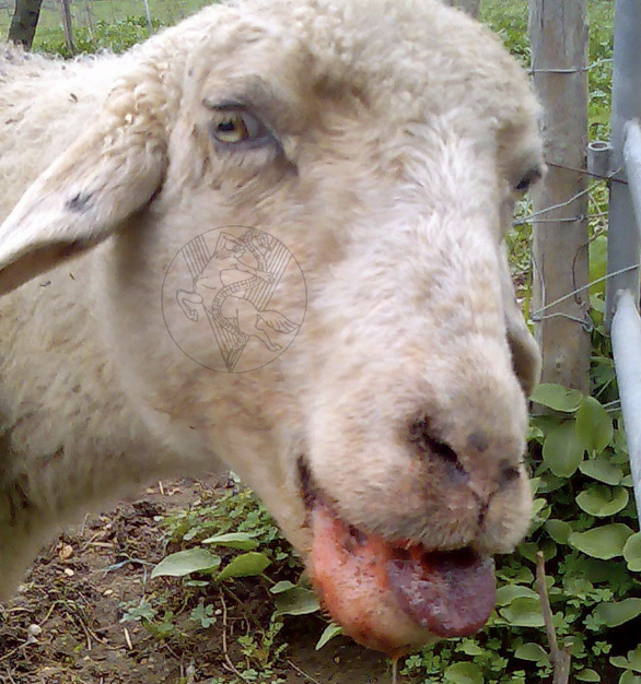
what is vesicular stomatitis?
reportable disease characterized by vesicular/erosive/ulcerative stomatitis (non-proliferative)
caused by a rhabdovirus
which animals are affected by vesicular stomatitis?
cattle
small ruminants
camelids
horses
pigs
what lesions are seen with vesicular stomatitis?
epidermitis leads to transient vesicles which will then rupture (vesicles are rarely witnessed)
non-proliferative disease!
how long after infection with vesicular stomatitis will lesions appear?
after 3-14 day incubation period, epidermitis leads to transient vesicles
the vesicles form and rupture, then the animal becomes febrile (vesicles are rarely witnessed)
where are lesions caused by vesicular stomatitis found?
most common in the mouth, but can also be found on:
-coronary bands
-interdigital space
-teats
what are clinical signs of cows with vesicular stomatitis?
-salivation
-dysphagia
-lameness
-anorexia
-severe weight loss
-decreased milk production
what age of cows tend to have more severe disease caused by vesicular stomatitis?
older cows are affected more severely
cows <1yr usually show no signs
when will signs of vesicular stomatitis regress?
in 3-6 weeks (high morbidity, low mortality)
when are outbreaks of vesicular stomatitis typically seen?
usually in the summer or fall that end with the first killing frost
how is vesicular stomatitis transmitted?
insect vectors may play a role in transmission (biting midge, black flies) or pollens
people, milking equipment, and direct contact are also important in disease spread
why are outbreaks of vesicular stomatitis worse in herds with coarse feed, poor foot care, and poor teat care?
since the virus is thought to not break intact skin
if feeding course feed or improper husbandry, the virus can enter the body easier through damaged skin/membranes
how is vesicular stomatitis diagnosed?
state vet- CF or FA to identify virus
-maybe PCR
-SN to show rising titer
how long is the quarantine period for farms with confirmed cases of vesicular stomatitis?
21 days after last lesion heals
how do horses with vesicular stomatitis present?
horses usually get a mild, usually oral, form of the disease
is vesicular stomatitis in small ruminants/llamas common?
these animals are susceptible, but are not commonly affected
what is the major differential for vesicular stomatitis?
foot and mouth disease
what animals are affected by foot and mouth disease?
all animals but the horse (horses are resistant)
what is foot and mouth disease (FMD)?
reportable disease caused by a picornavirus
how can foot and mouth disease be distinguished from vesicular stomatitis?
clinical disease of FMD is basically indistinguishable from vesicular stomatitis, except FMD is more persistent and spreads more easily
all ages are affected, small ruminants and llamas are more commonly affected
how severe is FMD is sheep and goats?
sheep and goats get little disease (mild, usually lameness), but still shed (important in spread of dz)
is abortion more common in FMD or vesicular stomatitis?
FMD
how long is the disease course of FMD?
most animals get better in 2-3 weeks
what is the 'malignant' form of FMD?
characterized by myocardial necrosis (tiger heart) and death (usually in calves)
how long can affected cattle, sheep, and goats shed the FMD virus?
cattle: up to 3 years
sheep and goats: up to 9 months
which part of animals infected with FMD is considered contagious?
all body secretions, tissues, and animal products, including:
-milk
-semen
-meat
-leather
-cheeses
-offal
how is FMD transmitted?
flies, ticks, and birds
how is FMD diagnosed?
by state vet via combo of:
-virus isolation (from vesicles)
-IHC
-serology
what is the treatment for animals infected with FMD?
slaughter or quarantine (quarantine is not beneficial tho)
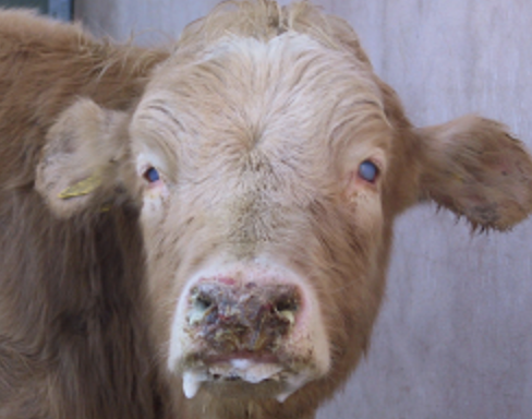
what is bluetongue?
notifiable disease in sheep caused by the virus orbivirus, causing widespread vasculitis
what are the clinical signs of bluetongue?
-transient high fever
-oral and nasal discharges with cyanosis (bluetongue)
-erosions of oral MM
-facial edema
-foot lesions
-myopathy
-pulmonary edema w/ secondary bacterial pneumonia
-diarrhea in lambs (not adults)
how do sheep infected with bluetongue present compared to infections with orf?
sheep infected with bluetongue appear much more depressed and clinically ill than with orf
when do outbreaks of bluetongue usually occur?
during warm months
outbreaks stop with the first killing frost
how is bluetongue spread?
by the bites of culicoides (midges)--> fly control important for prevention!
can also be spread in semen and transplacentally
which animals may serve as reservoir hosts for bluetongue?
cattle and goats can be unapparent carriers, and rarely get clinical disease
what can occur with in-utero infections of bluetongue in cattle?
can lead to abortion, birth defects (arthrogryposis), and persistent viremia
abortion is also common in sheep
how is bluetongue diagnosed?
PCR, viral isolation, or serology
what is the mortality rate of bluetongue?
5-30%
what is death usually due to in animals infected with bluetongue?
cardiomyopathy, secondary infections, or severe vasculitis
are there vaccines available for bluetongue?
yes, modified-live vaccines available in some areas, but should be used before fly season
must be used with caution near pregnant sheep (can abort)
what is epizootic hemorrhagic disease?
disease in cattle caused by a virus related to bluetongue virus
how is epizootic hemorrhagic disease spread?
primarily by culicoides
which animals is epizootic hemorrhagic disease enzootic in?
white tailed deer
what is bovine viral diarrhea?
notifiable disease caused by a pestivirus in cattle, affects rapidly dividing cells
what are the important GI-tract syndromes of disease of BVD?
acute BVD and mucosal disease
how does infection with acute BVD occur?
occurs with a new infection of immunocompetent cattle with either cytopathic or non-cytopathic virus
what are the clinical signs of cattle with acute BVD?
-transient high fever
-diarrhea
-leukopenia
-oral ulcers
-oculonasal discharge
what cells does acute BVD virus multiply in?
multiplies and kills:
-epithelial cells (GI ulcers, necrosis of crypt cells)
-WBCs (neutropenia)
-necrosis of peyer's patches
where are lesions caused by acute BVD seen?
causes ulcers in:
-from mouth to colon
-nasal mucosa
-ocular mucosa
-coronary band
-respiratory tract
why is the acute form of BVD rare currently?
due to good vaccination strategies (relatively effective vaccine available)
however, when new strains emerge, will make these vaccines less effective
how is acute BVD diagnosed?
-PCR (blood or lesion)
-virus isolation (whole blood or lesions); short viremic phase, collect between 3-12 days post-infection
-serology (acute and convalescent, 21-30 days apart)
which animals is mucosal disease (BVD) seen in?
occurs in persistently infected animals that subsequently becomes exposed to a cytopathic virus
what is a persistently infected (PI) animal (BVD)?
PI refers to cattle exposed to non-cytopathic virus in utero between days 42-125 of gestation
PI calves develop immunotolerance and lifelong BVD infections
how may PI BVD calves present clinically?
PI calves may have birth defects, be a poor-doer, or appear clinically normal
PI calves have poor immune responses and sheds virus into the environment (has persistent viremia)
how does exposure to a cytopathic strain of BVD most commonly occur?
through mutation
cytopathic and non-cytopathic viruses differ by one amino acid
which disease is BVD mucosal disease similar to?
malignant catarrhal fever
how do BVD mucosal disease and malignant catarrhal fever differ?
-mucosal disease course is longer
-bladder and brain infections are rare in mucosal dz
-lymphadenopathy does not occur in mucosal dz
what are the typical findings/lesions of mucosal disease (BVD)?
-ill thrift
-oral erosions
-blunting of buccal papillae
-peyer's patch necrosis
how is BVD mucosal disease diagnosed?
-IHC via ear notch
-PCR
-virus isolation (whole blood or serum)
-antigen capture ELISA
-not serologic tests
what is border disease?
variant strain of the same virus causing BVD in cattle, but in sheep and goats
what is malignant catarrhal fever (MCF)?
notifiable disease of wildebeest and sheep caused by a herpesvirus :
-alcelaphine herpes virus-1 and 2
-ovine herpesvirus 2
do sheep and wildebeest show disease when infected with MCF?
no, these animals do not show disease
but, they shed the virus in secretions, especially around lambing
which animals are susceptible to MCF?
cattle, bison, and more exotic species
pigs have recently been identified as
when can animals show signs of MCF after infection with the virus?
since it's a herpesvirus, animals can become infected once and not show signs of MCF until later (up to 6 months) due to periods of latency
what is the importance of the latency period of MCF in cattle?
infected cattle can serve as the source of infection for herds w/o recent exposure to wildebeest or sheep
what is the basic mechanism of MCF?
the virus causes vasculitis affecting most epithelial surfaces
mediated thru cytotoxic T-cell activity
what are the clinical signs of MCF?
-crusting oral and nasal ulcers
-conjunctivitis with corneal edema and uveitis
-high fever
-diarrhea
-hematuria
-coronitis
-encephalitis
-lymphadenopathy
what occurs once animals show clinical signs of MCF?
almost all animals with clinical signs die within 96 hours
how is MCF diagnosed?
serologic tests
what is lumpy jaw?
chronic osteomyelitis caused by the gram+ anaerobe actinomyces bovis
leads to lysis and proliferation of bone of the maxilla or mandible (results in a hard, bony swelling)
how does lumpy jaw occur?
the bacteria is an oropharyngeal commensal and is thought to invade the bone thru mucosal lesions or erupting teeth (in young animals)
which animals is lumpy jaw common in?
more common in cattle than small ruminants
what are consequences of lumpy jaw?
weight loss can occur as the swelling disrupts dental alignment or adjacent soft tissue becomes inflamed
how do lesions caused by lumpy jaw appear?
lesions often have intermittent drainage thru fistulous tracts
the drainage contains 2-5mm yellow 'sulfur' granules, clumped aggregations of organisms and debris
what will gram-stain of drainage from lumpy jaw lesions show?
gram-positive, filamentous organisms that vary in shape
how is lumpy jaw diagnosed?
culture and radiographs (to diff. osteomyelitis from tooth root abscess)
how is lumpy jaw treated?
long term (30+ days) of antibiotics may arrest growth of lesion, but are rarely curative unless lesion is caught early
iodides have been recommended but have minimal efficacy against bony lesions
what is wooden tongue?
cellulitis or lymphadenitis caused by gram- anaerobe actinobacillus lignieresii
leads to acute diffuse soft tissue swelling, followed by chronic focal, soft tissue masses