L4: Integration of signals
1/23
Earn XP
Description and Tags
Name | Mastery | Learn | Test | Matching | Spaced | Call with Kai |
|---|
No study sessions yet.
24 Terms
Difference between GP and AP
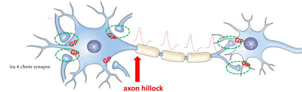
GP: in dendrites n the cell body, signals decay w/ distance, short-lived n temporary, local signals, amplitude depends on the intensity of stimulus
AP: in axon hillock n axons, dont undergo decay, self-propagating, all-or-none, consistent shape n magnitude, long-distance communication
How GP formed? how it help fire AP at axon hillock?
triggered by stimulus → open ligand/mechanically-gated ion chan, Multiple GPs from different dendrites/cell body areas travel to the axon hillock and sum together EPSP n IPSP (spatial/temporal). If the combined depolarization reaches the threshold potential (-55mV), it determines whether an AP will be produced successfully at the axon hillock
How GPs be converted to an AP that is triggered only at the axon hillock?
graded potential (GP) can be summed (postsynaptic potential)
Excitable membranes of dendrites and cell bodies are communicating with the axon terminals (chemical synapse) of another neuron.
Presynaptic n Postsynaptic neurons
Pre: conducts impulses toward a synapse, sends info
Post: transmit an electrical signal away from a synapse, receives the info.
## a single neuron post to one cell can be pre to another cell.
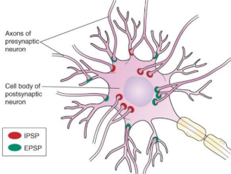
In summation, what’s EPSP and IPSP
EPSP: generated by EXCITATORY chem synapse, cause depolarisation
IPSP: generated by INHIBITORY chem synapse, hyperpolarisation
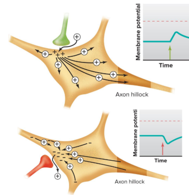
Spatial summation? how the mechanism only excitatory neurons
= summation of postsynaptic potentials that occurs when several presynaptic axon terminals produce graded potentials at the same time.
Summed potential created by more than one EPSP and/or IPSP arriving together at different synapses on a postsynaptic cell membrane.
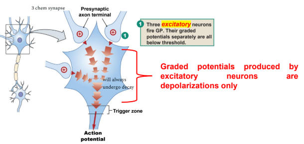
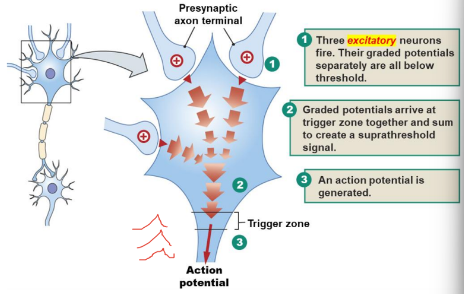
1. excitatory neurons fire, GP all separately below threshold
2. GP arrive at trigger zone, sum & create suprathreshold
3. AP fired
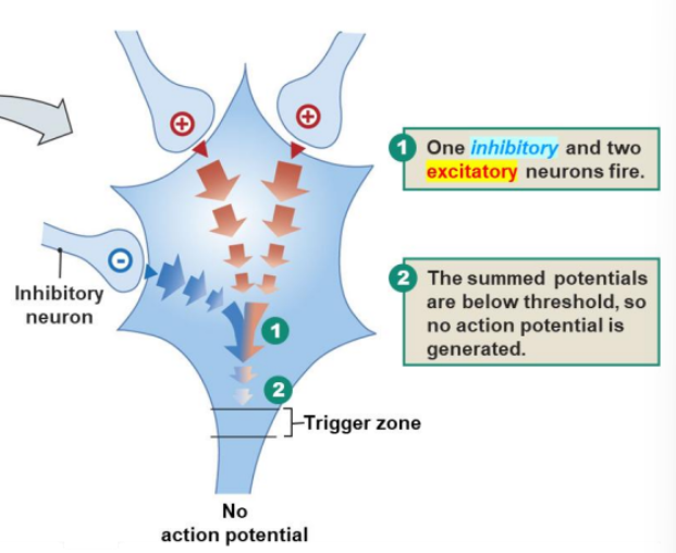
Spatial summation when excitatory n inhibitory involved
strength of a depolarization produced by one excitatory postsynaptic neuron can be reduced or cancelled out by a hyperpolarization produced by another postsynaptic neuron
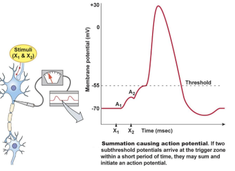
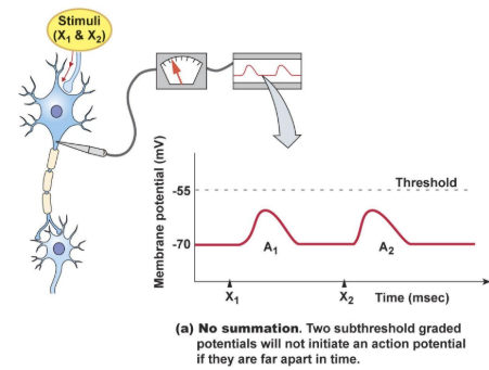
pic: so what’s temporal summation? can it failed?
= summation of postsynaptic potentials occurs when a pre-synaptic neuron fires repeatedly at a high rate (close in time). will fail if the two graded potentials are too distant from each other
what does it mean
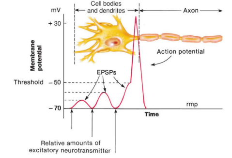
Excitatory postsynaptic potential (EPSP) increases possibility of forming an action potential in the axon hillock via summations
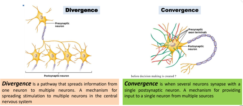
Types of neural circuit (the complex grouping of neural pathways)
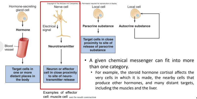
categories of chem messengers
Post-synaptic potential is produced by a chemical synapse. what’s synapes? what r the types?
Synapses are interaction points or junction within the nervous system and transmit electrical signals between cells: chemical synapse and electrical synapse
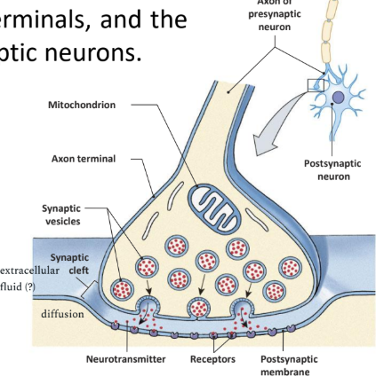
Chem synapses?
releases chemicals (neurotransmitters) between presynaptic and postsynaptic neuron. Presynaptic neurons release NT from their axon terminals → NT bind to specific receptors on post-synaptic neurons.
##Axon terminals of the presynaptic neuron hold the synaptic vesicles that contain neurotransmitter molecules.
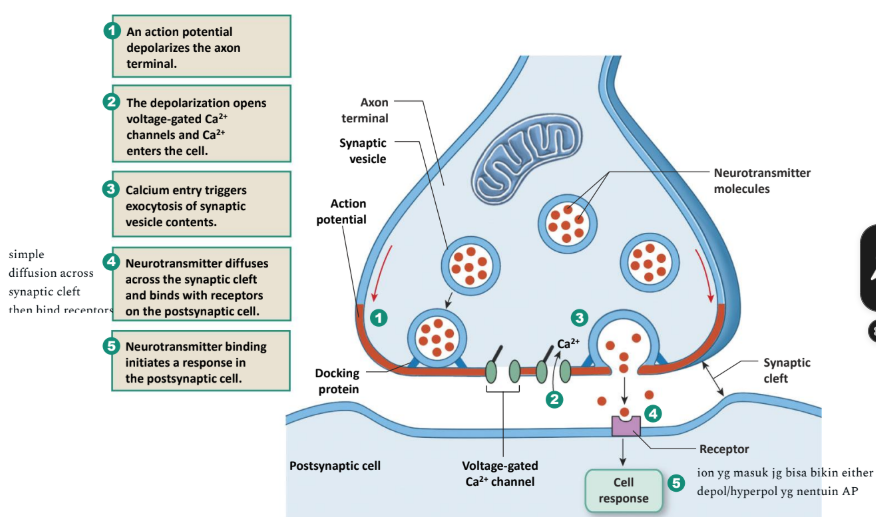
Sequence of events at a chemical synapse
#Synaptic cleft: narrow space between the pre and postsynaptic
#Exocytosis is a process in which intracellular vesicle fuses with plasma membrane, the vesicle opens, and its contents are released into the extracellular fluid (i.e., the synaptic cleft)
#receptor step4 usually ligand-gated
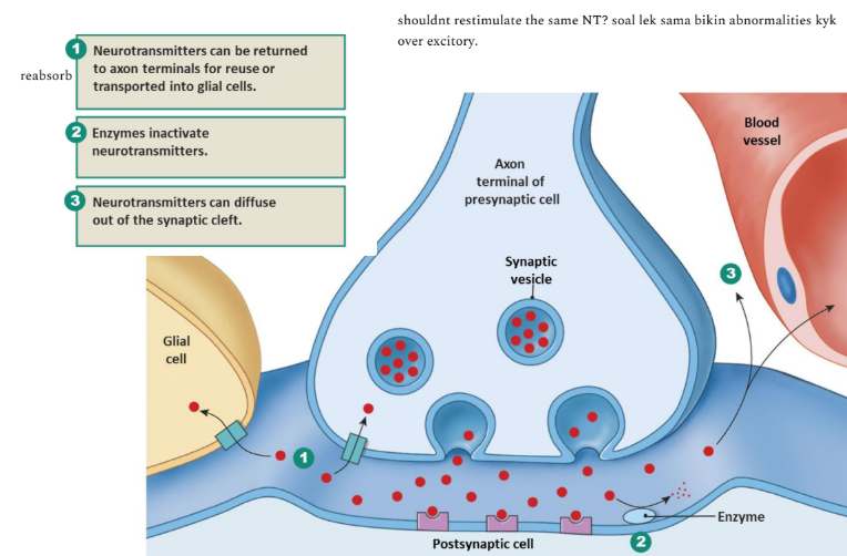
3 methods effecting termination of NT
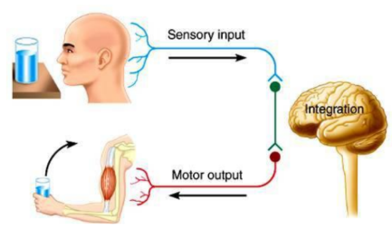
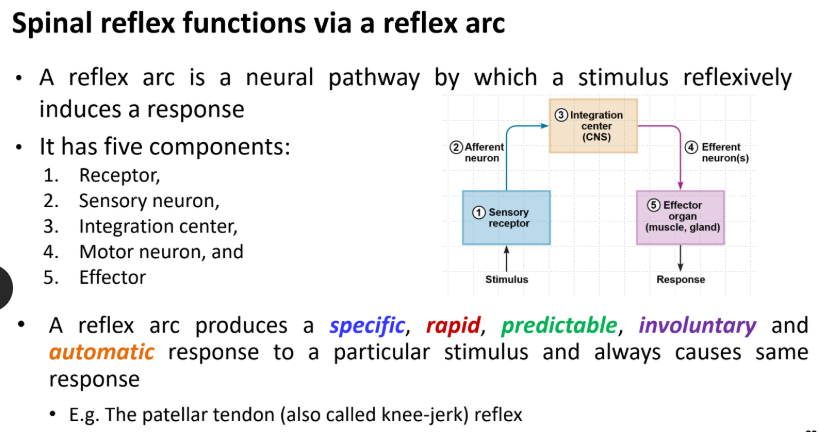
The central nervous system comprises the brain and the spinal cord. define 3 of em
Sensory input: provides CNS w/ info abt the internal n external envi
Integration: CNS takes all info → interprets → determine an appropriate effector response
Motor output: executes CNS commands to cause appropriate effector response
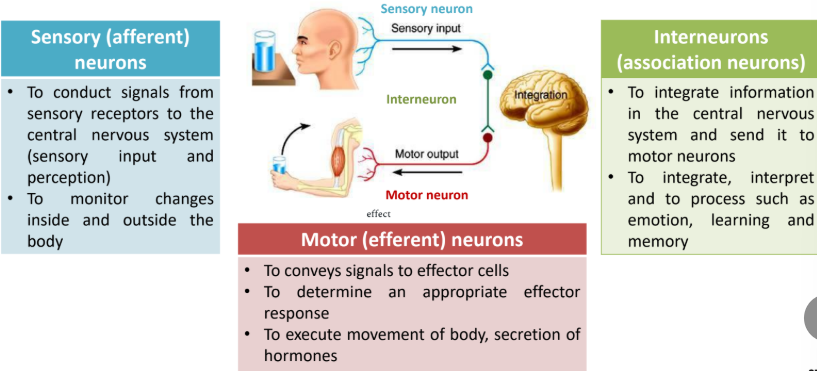
3 types of neurons
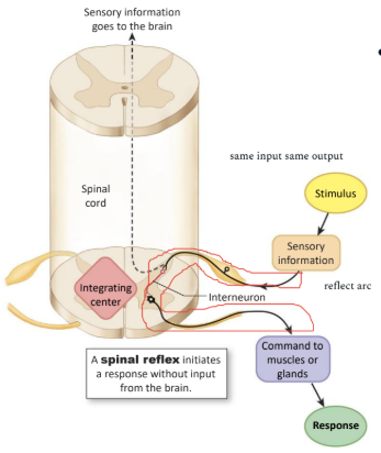
Spinal reflexes are simple behaviors produced by CNS pathways that lie entirely within the spinal cord.
Flexion: bending a joint, extension: straightening a joint
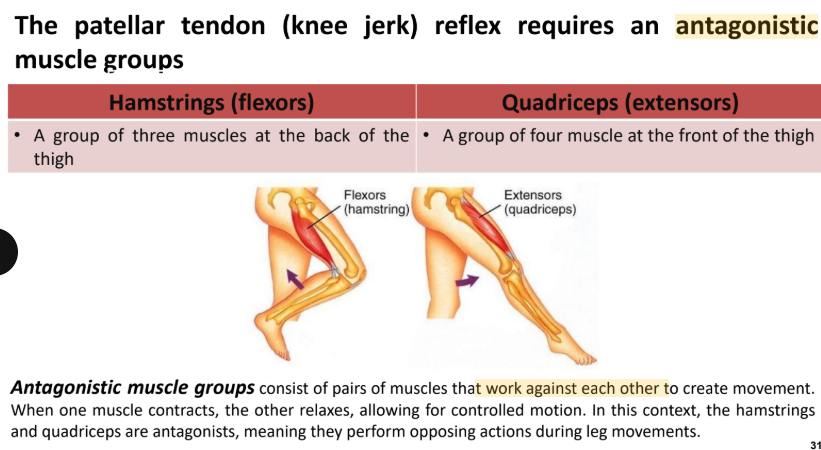
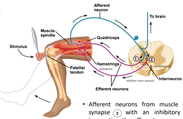
The patellar tendon (knee-jerk) reflex is a mono-synaptic reflex
Step 1: tapping the PT below the kneecap, muscle spindle is the receptor in this reflex; when stretched, action potentials are triggered and travel in afferent neurons to the spinal cord which is the integration center
Step 2: In the spinal cord, the afferent neuron synapses with efferent neuron that innervate the quadriceps to contract, and the leg ‘kick’ forward (extension).
Step 3: Afferent neurons from muscle spindle also synapse with an inhibitory interneuron innervating the efferent neurons going to the hamstrings causing it to relax.
Step 4: Afferent neurons also ascend to the brain, forming synapses with various interneurons. Simultaneous excitation of the quadriceps and inhibition of the hamstrings causes leg extension during the knee jerk reflex.
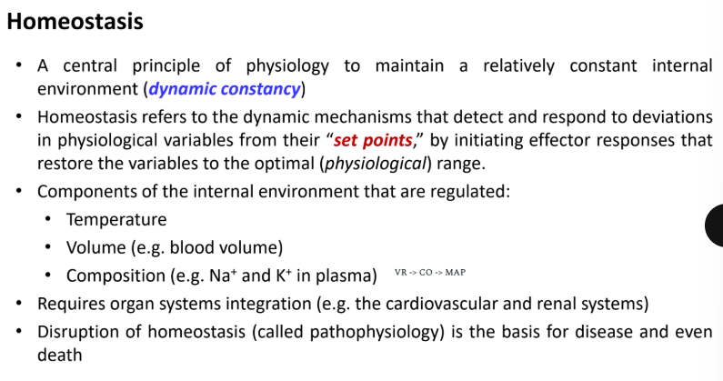
Homeostatis
#VR (venous return) -> CO (cardiac output) ->MAP
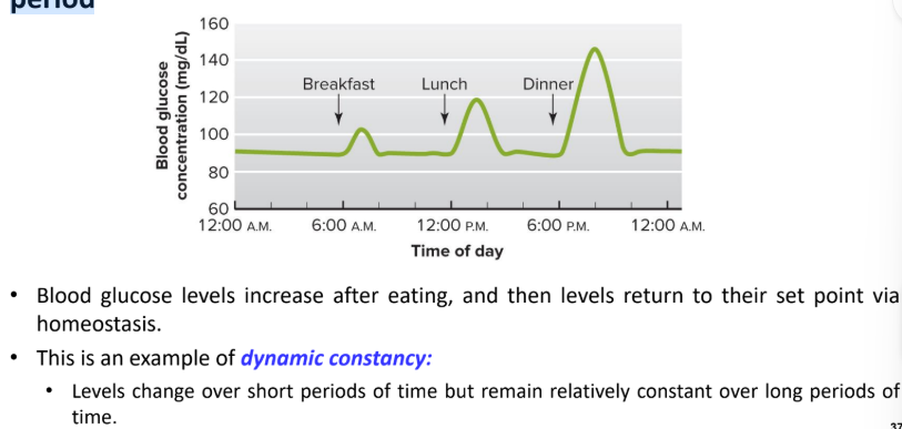
Changes in blood glucose concentration during a typical 24-hour period
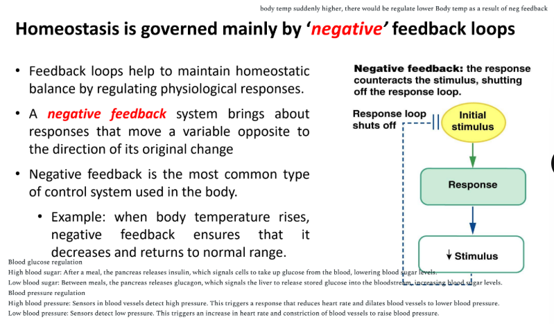
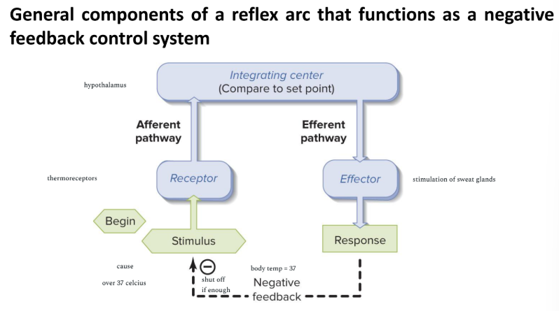
negative feedback in homeostatis
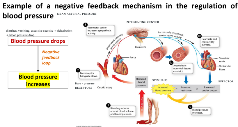
neg feedback in blood pressure
homeostatis imbalanced: