Biology 315 Wk 7 - Circulatory System I
1/129
There's no tags or description
Looks like no tags are added yet.
Name | Mastery | Learn | Test | Matching | Spaced | Call with Kai |
|---|
No analytics yet
Send a link to your students to track their progress
130 Terms
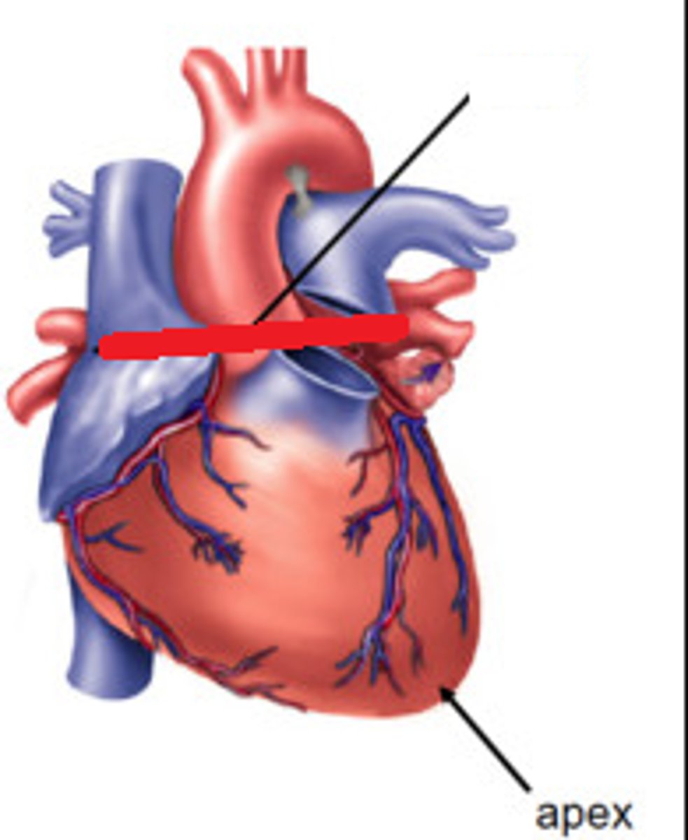
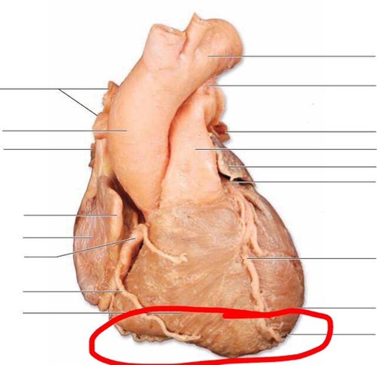
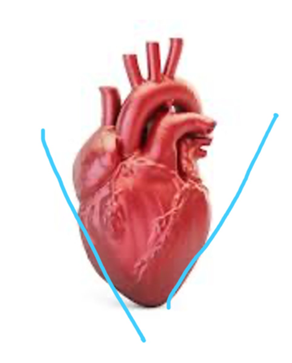
Apex of heart
feature. tip of the heart pointing down toward the 5th left intercostal space
Base of heart
feature. Circumference, TA circles top of the heart.
Right atrium
Chambers. TA pinches top right of the heart
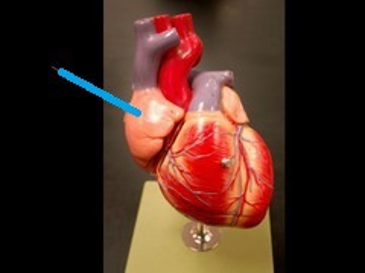
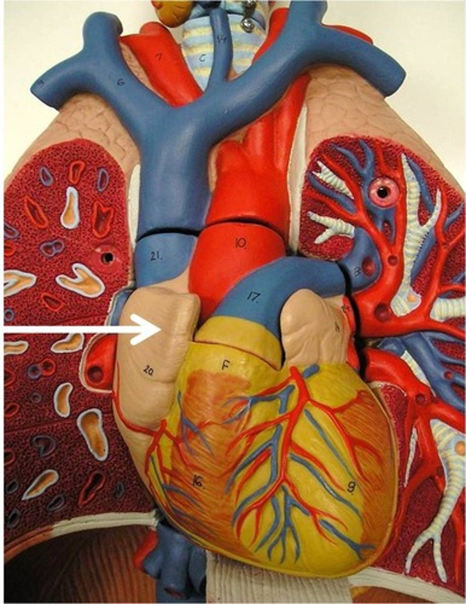
Right auricle
Chamber. Identify the flap.
Left Atrium
Chamber. TA pinches top left of the heart
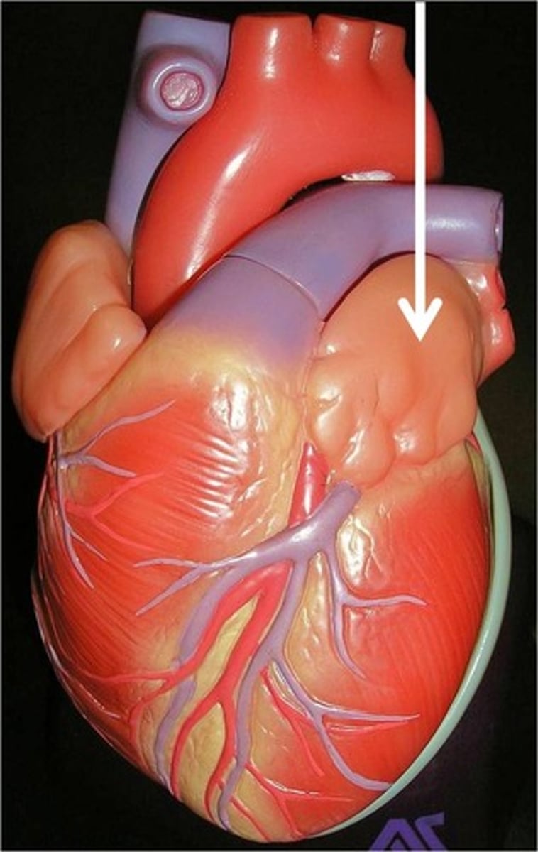
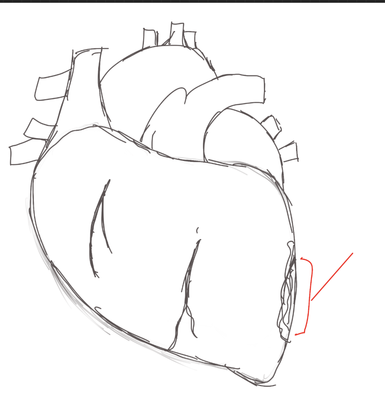
Left auricle
Chamber. Identify the flap.
Right ventricle
Chamber. TA pinches bottom right.
Left ventricle
Chamber. TA pinches bottom left.
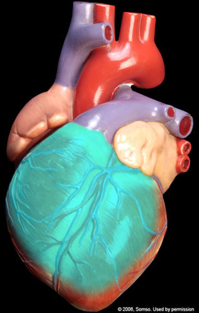
Anterior Surface of the heart
"region", front side of the heart
diaphragmatic surface of the heart
Region
Right pulmonary surface of the heart
Region. Right side.

Left pulmonary surface of the heart
Region. Left side.
Right border of the heart
left border of the heart
Inferior border of the heart
Superior border of the heart
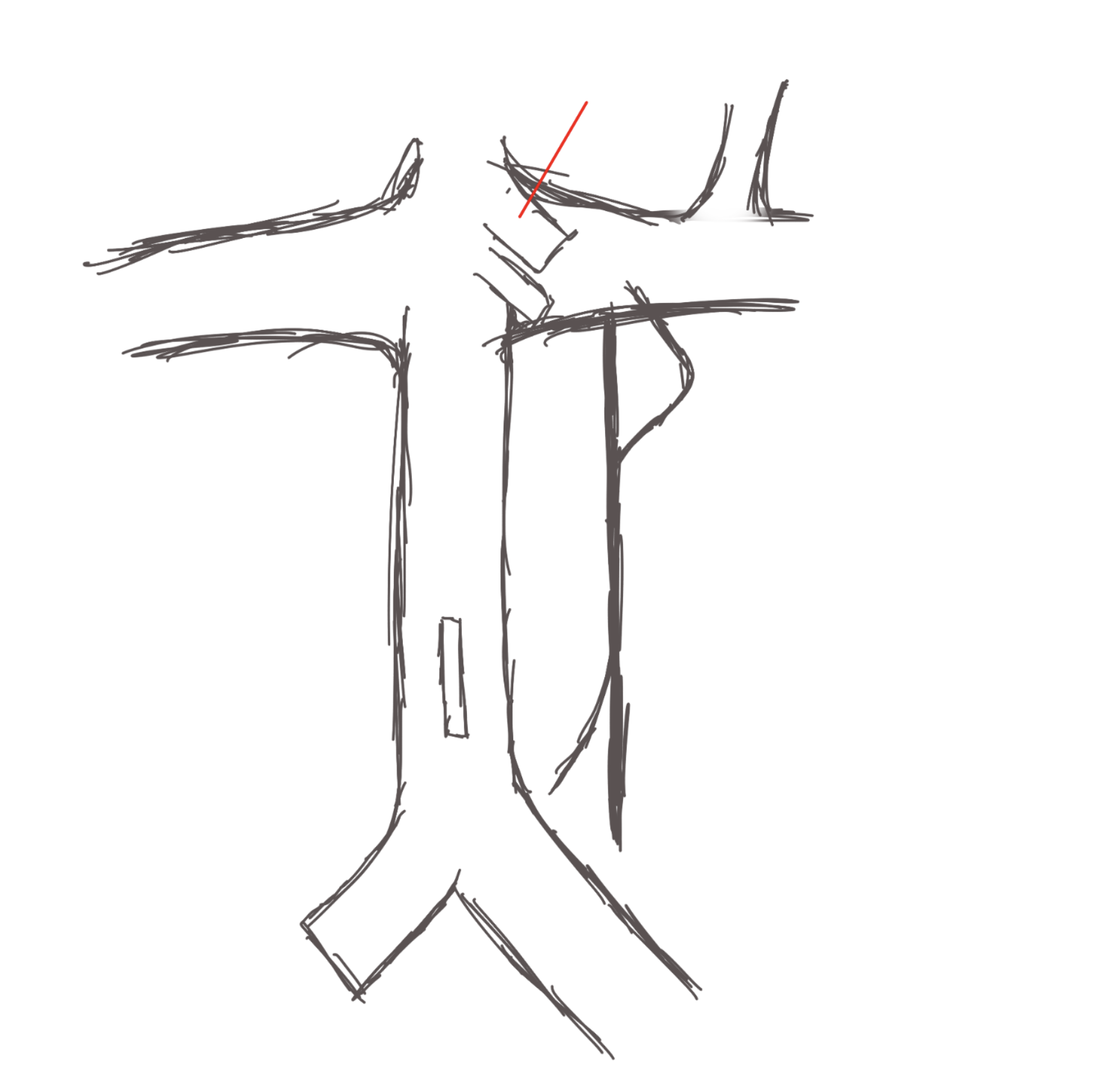
Coronary sulcus
Depression, on top of the heart, inside.
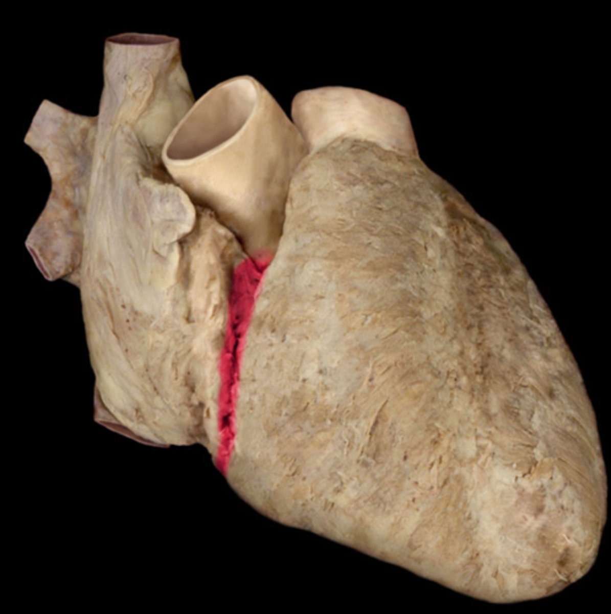
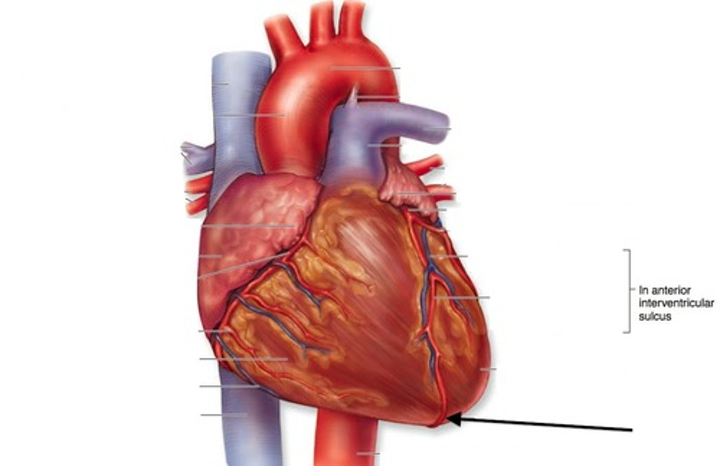
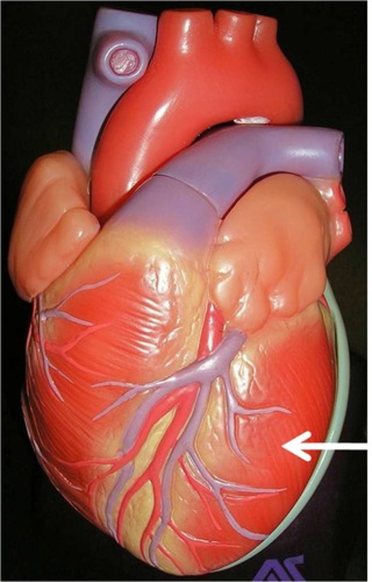
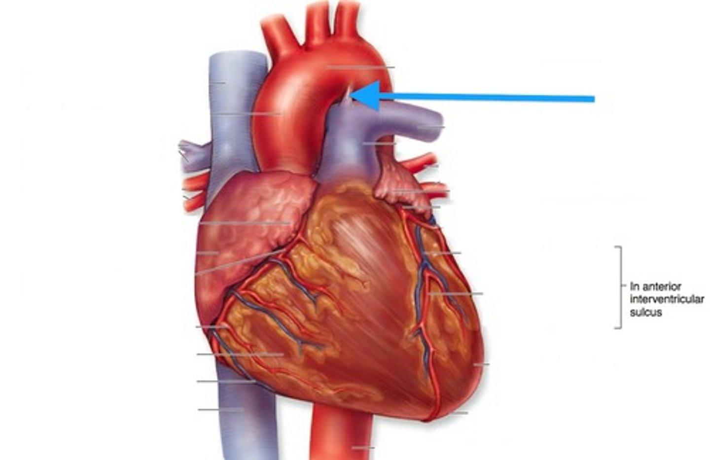
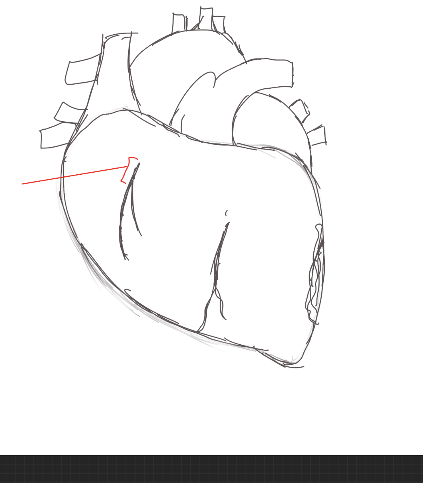
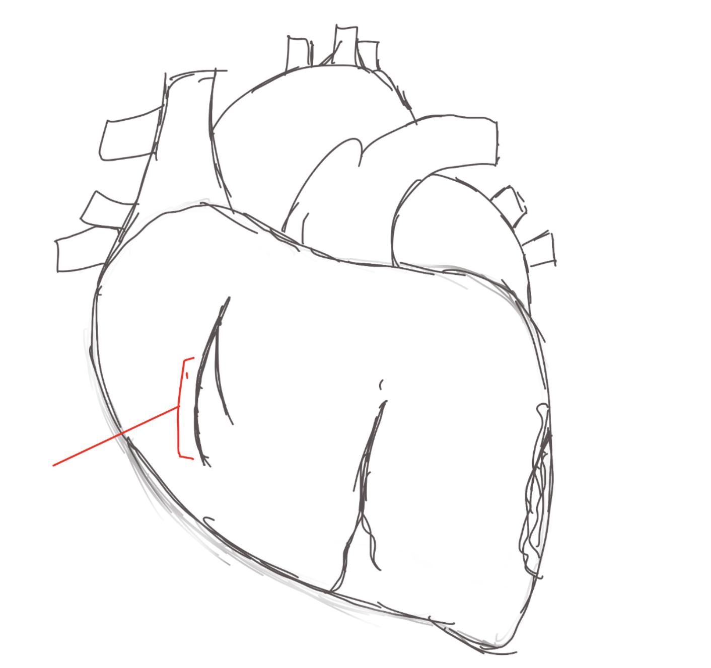
anterior interventricular sulcus
depression. anterior of the heart.
posterior interventricular sulcus
depression. posterior of the heart
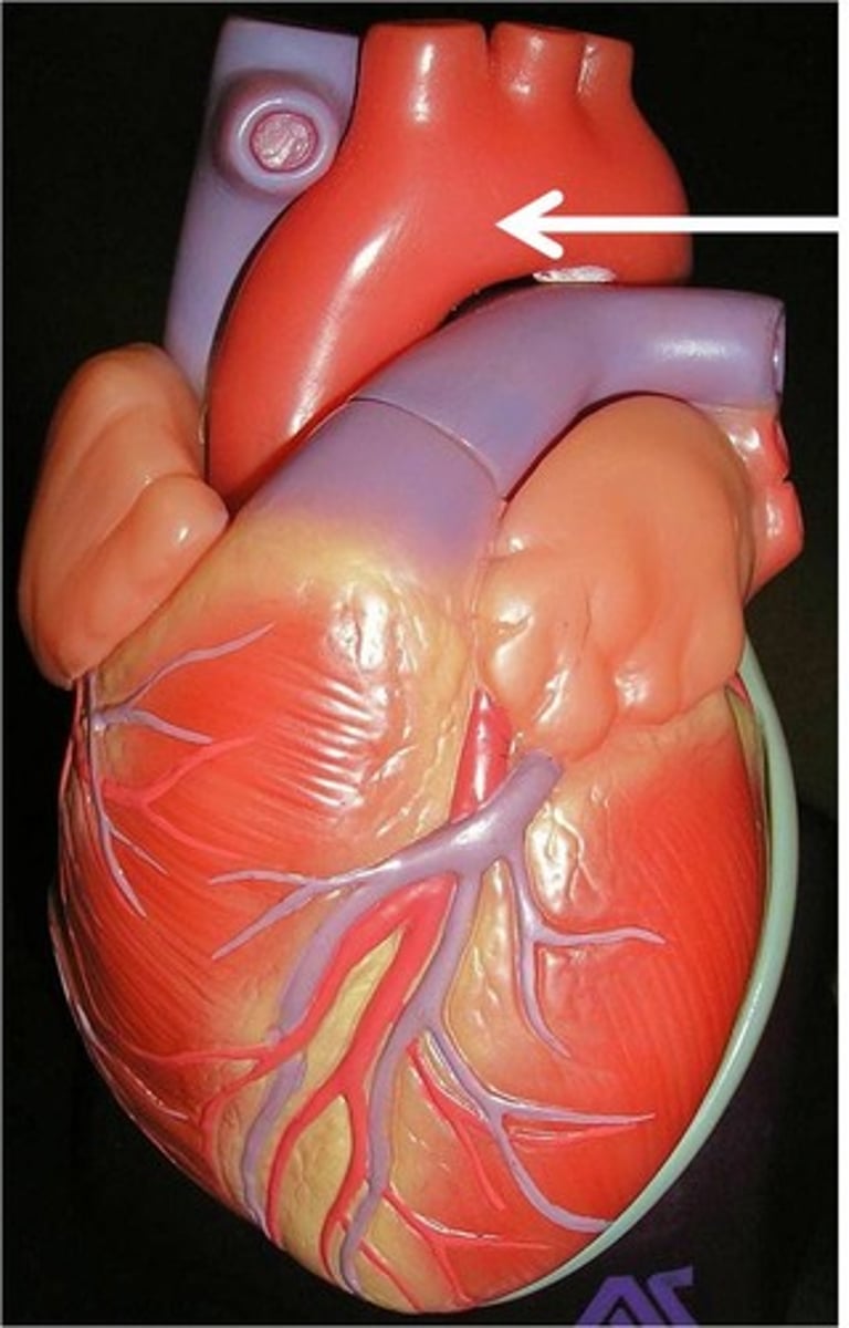
Aorta
structure. Leaves the left ventricle

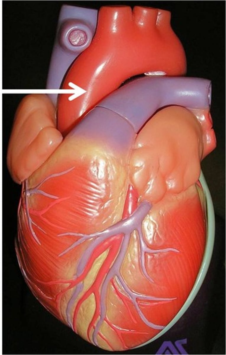
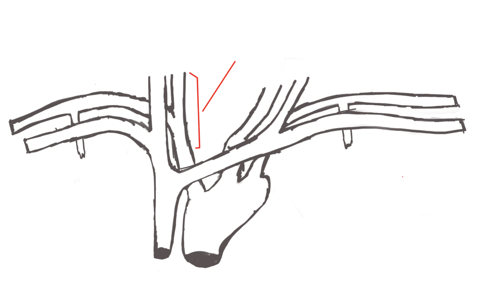
Ascending aorta
Portion. The portion of the aorta that rises from the heart.
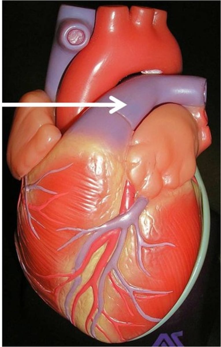
Arch of aorta
Portion. TA makes a rainbow (This is the only time)
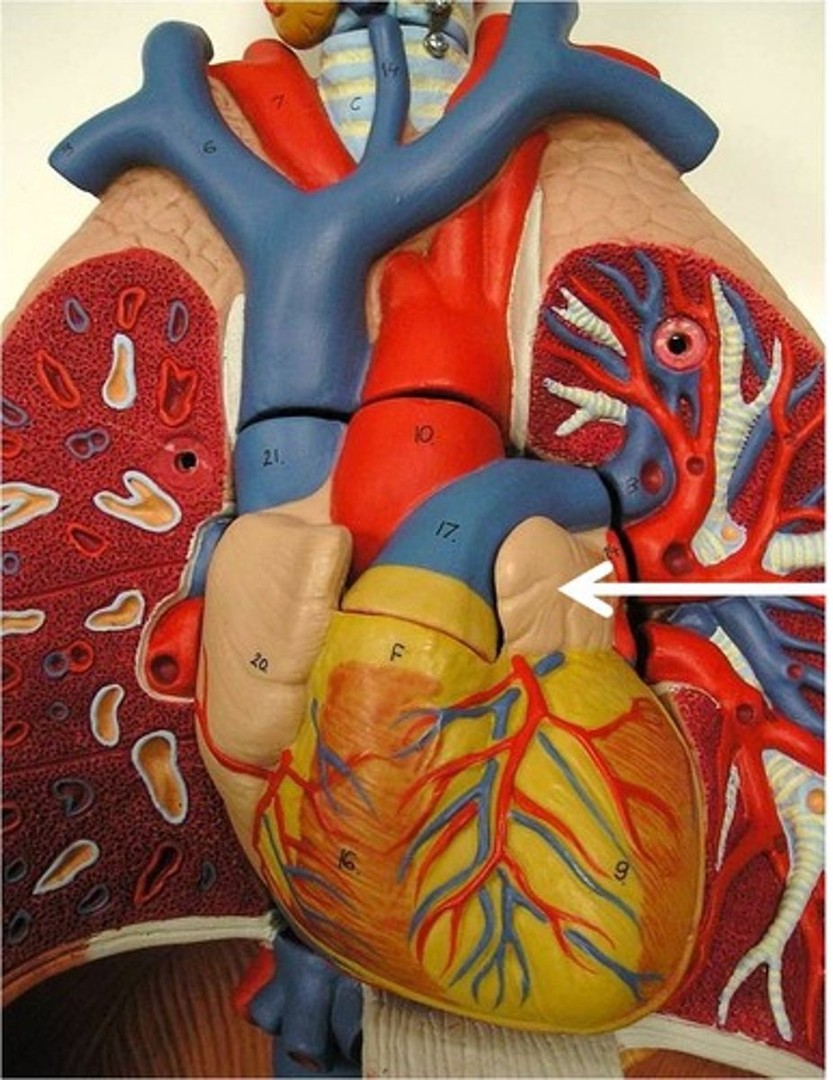
Pulmonary trunk
Collective structure. Leaves the right ventricle.
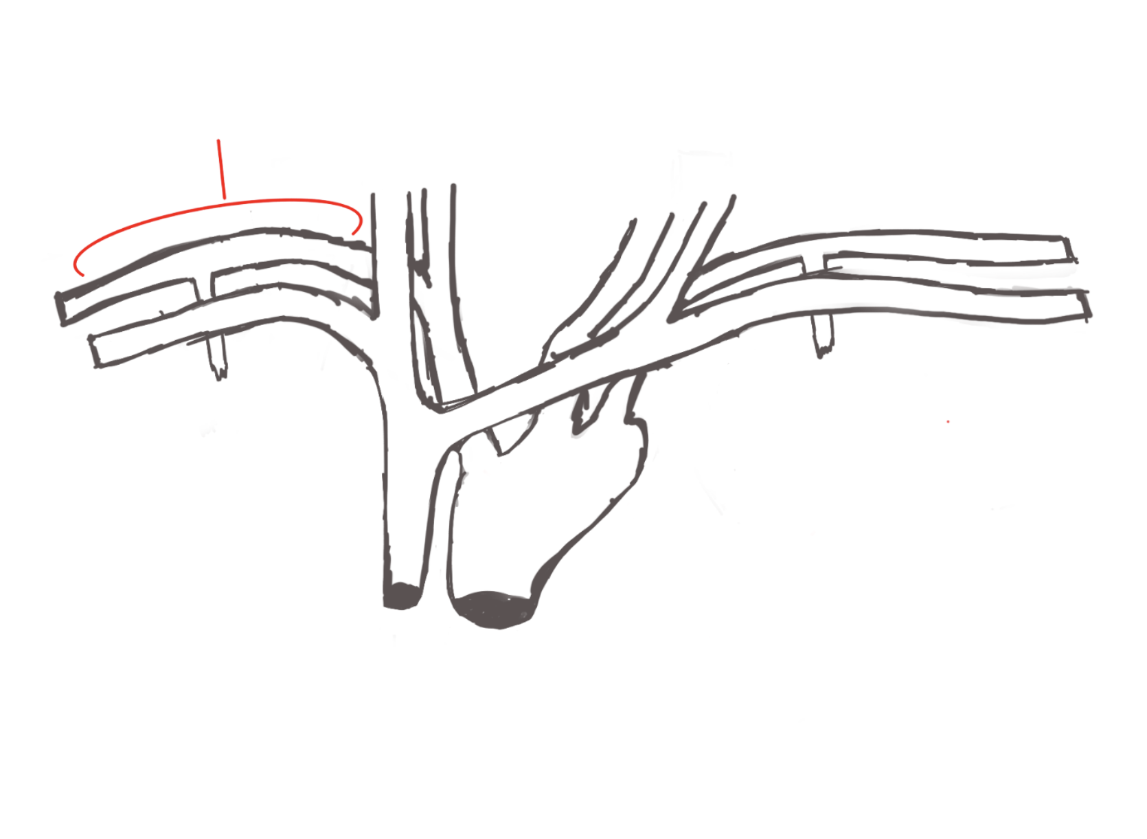
left pulmonary arteries
structures.
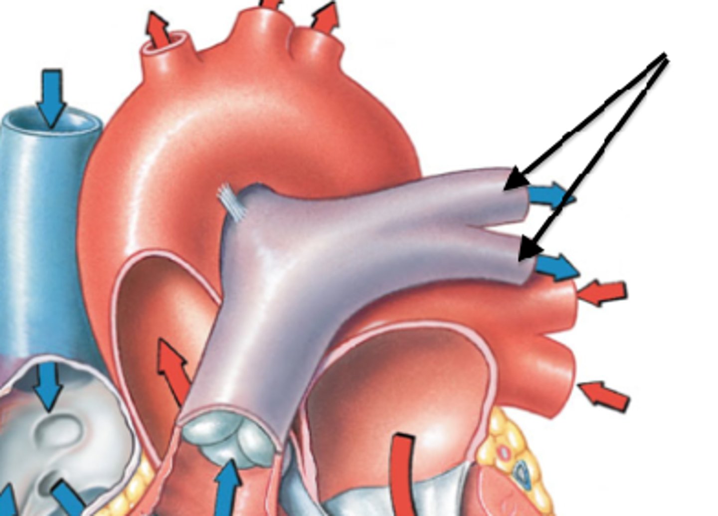
right pulmonary arteries
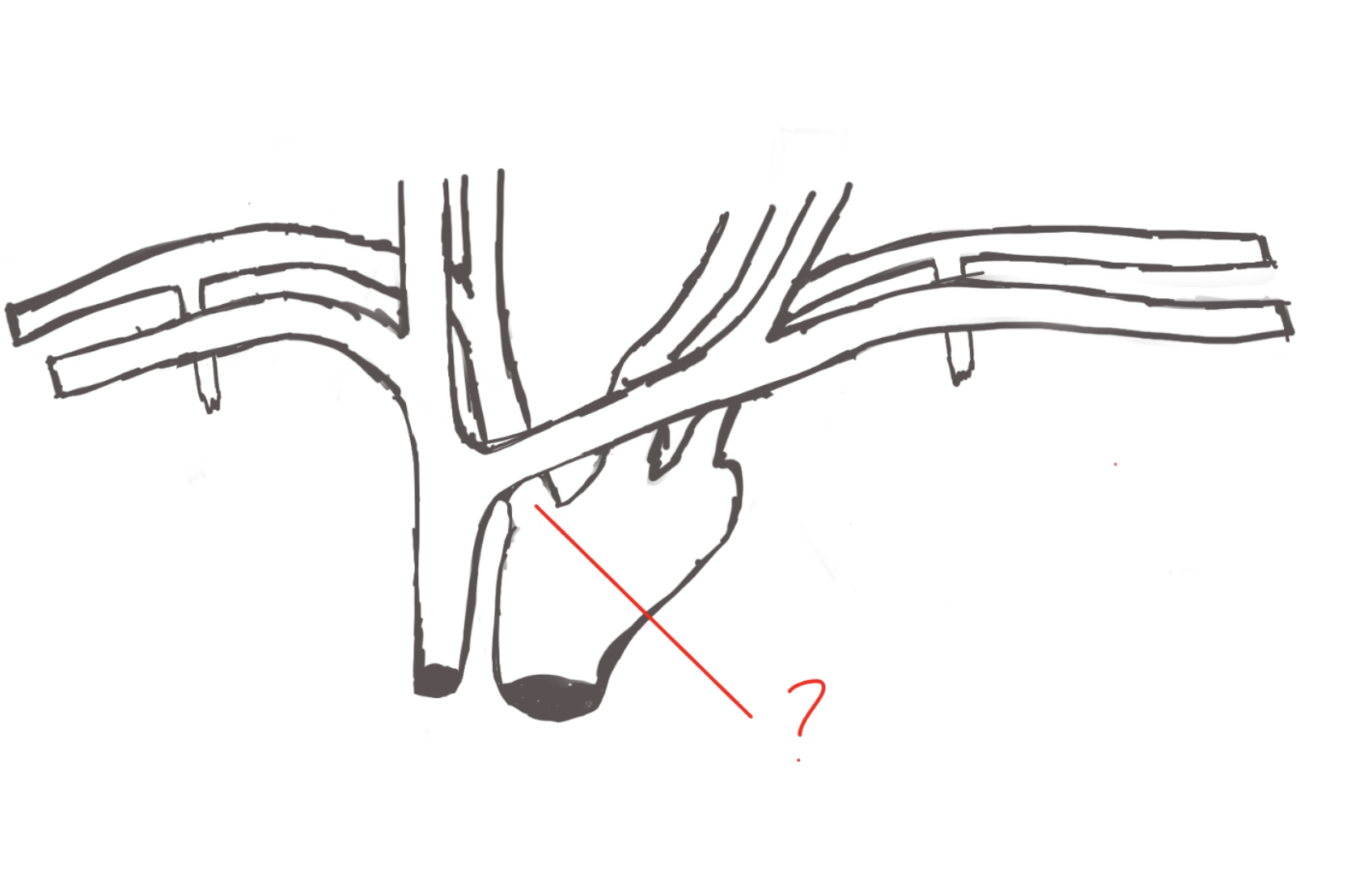
ligamentum arteriosum
feature. Has blood clot, also right next to black color.
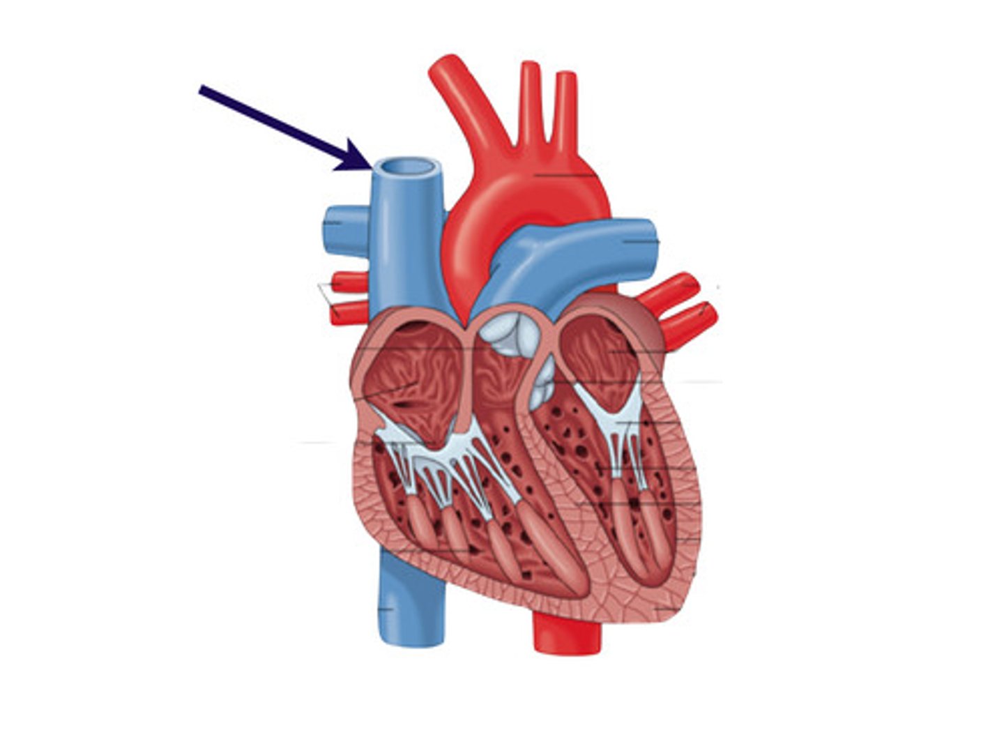
superior vena cava
feature.
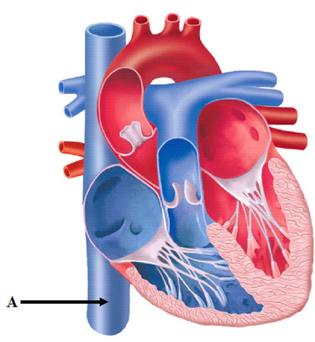
inferior vena cava
feature.
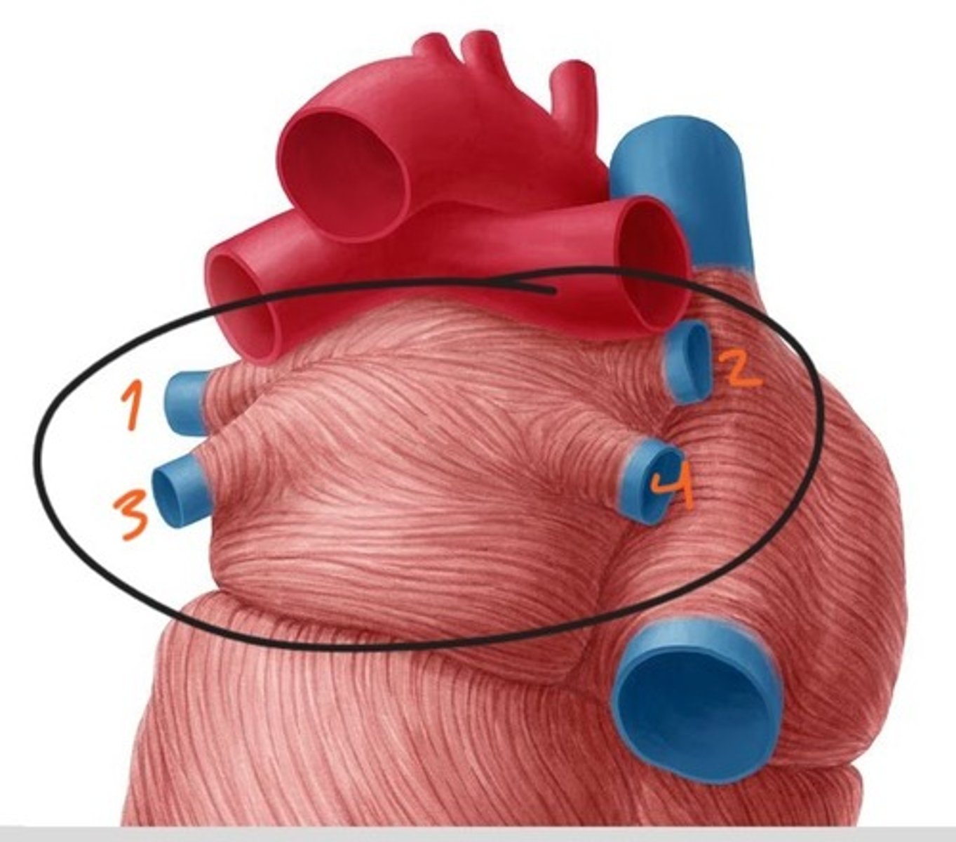
Pulmonary veins
Collective structure. Both side of this picture.
Left superior pulmonary veins
structure
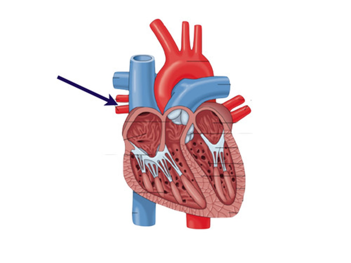
Right superior pulmonary veins
structure
Left inferior pulmonary veins
structure
right inferior pulmonary veins
structure
Right coronary artery
structure. below the right auricle.
Right marginal artery
structure. to lateral wall of right ventricle
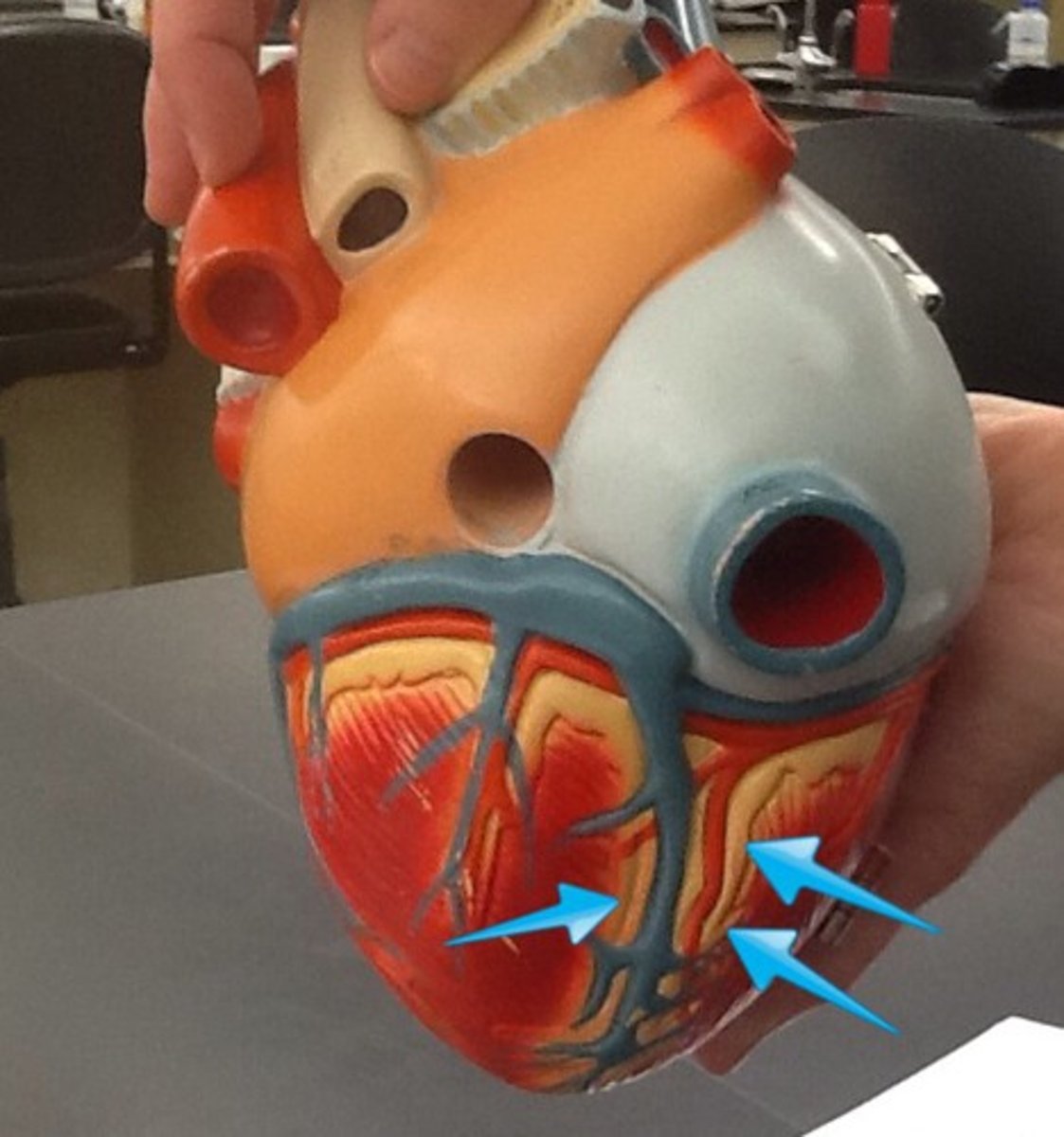
posterior descending artery
structure. sits @ posterior interventricular sulcus.
Left coronary artery
structure. On the coronary sulcus.
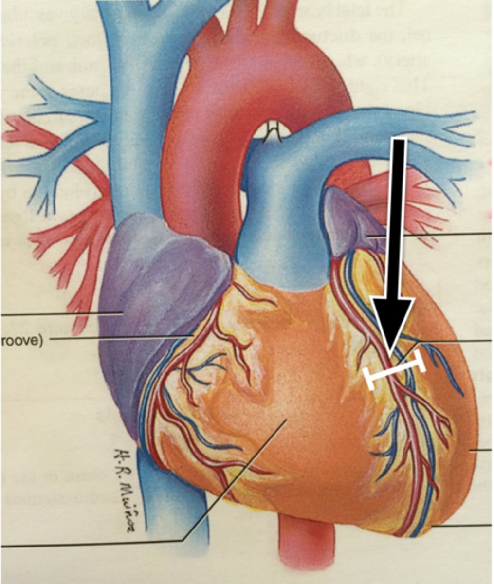
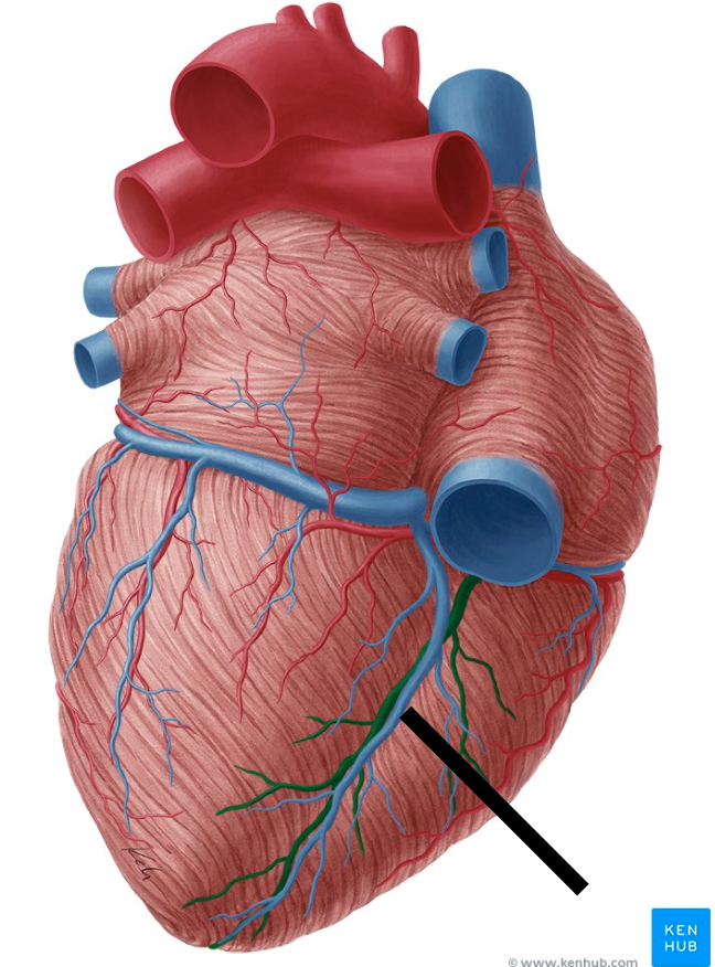
Left anterior descending artery
Structure. In the anterior interventricular sulcus.
circumflex artery
structure.
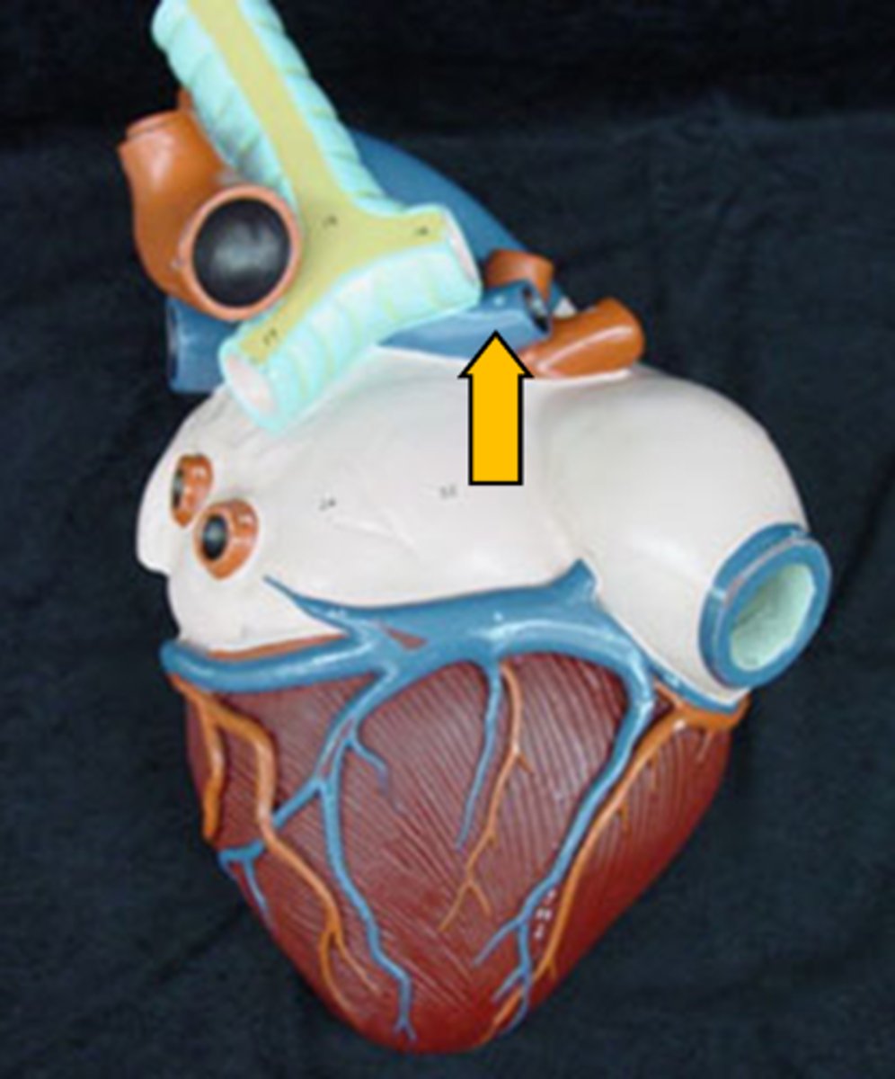
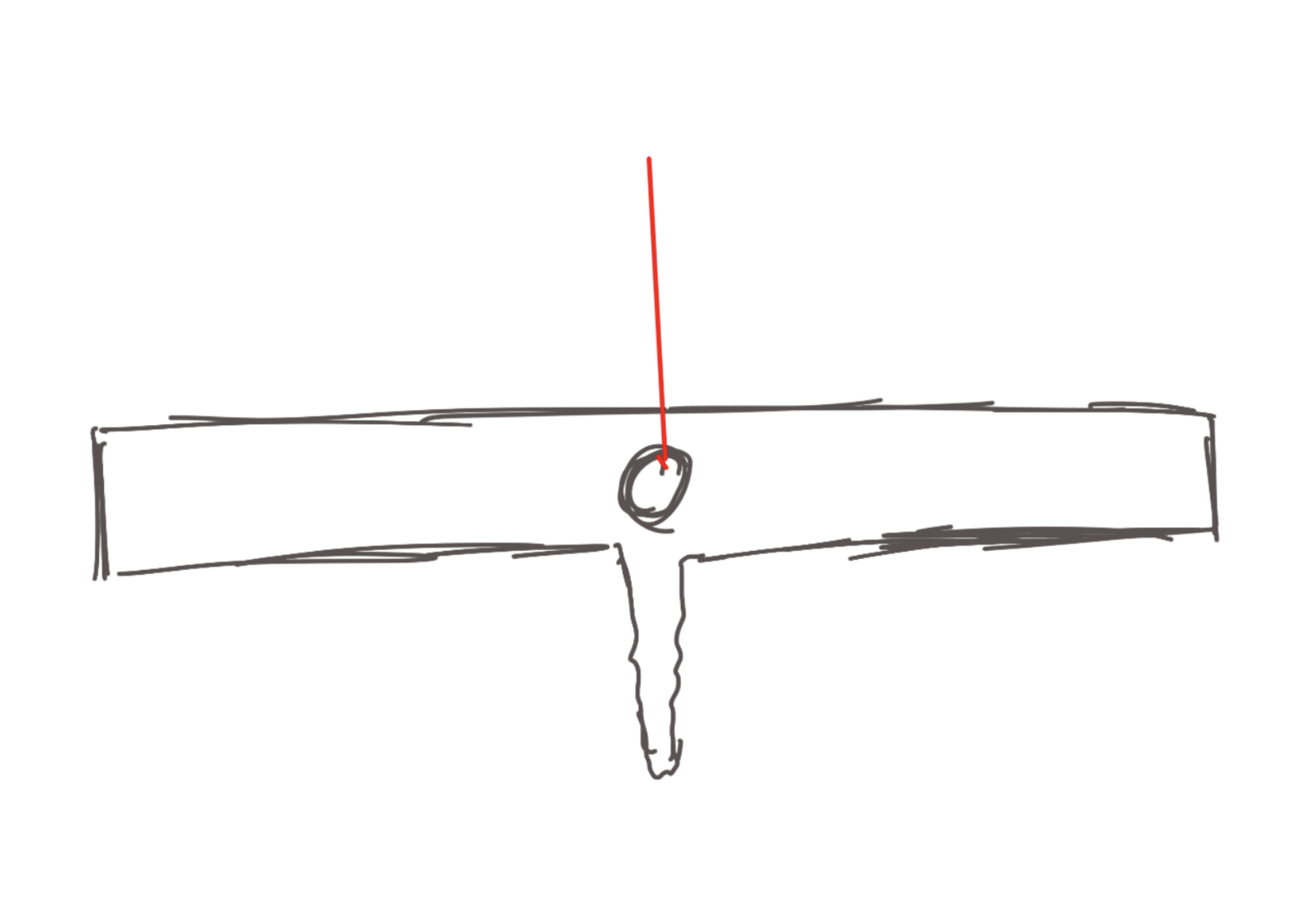
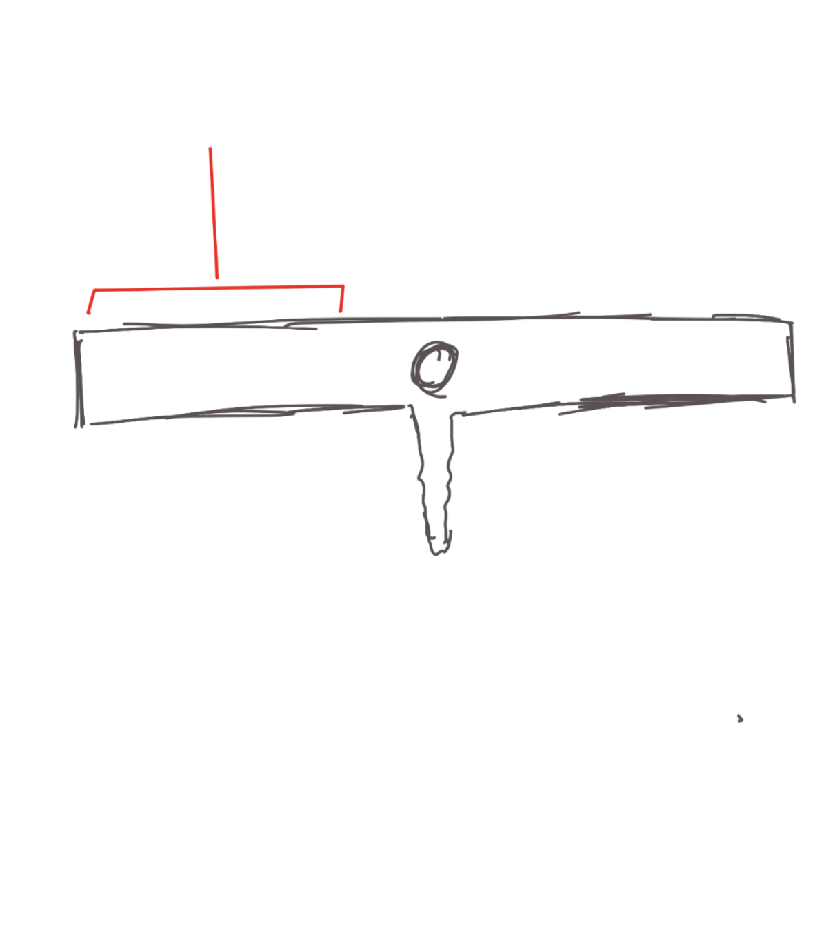
Cardiac veins
structure. T shaped
Coronary sinus
structure. Middle of the T-shape
Great cardiac vein
structure.
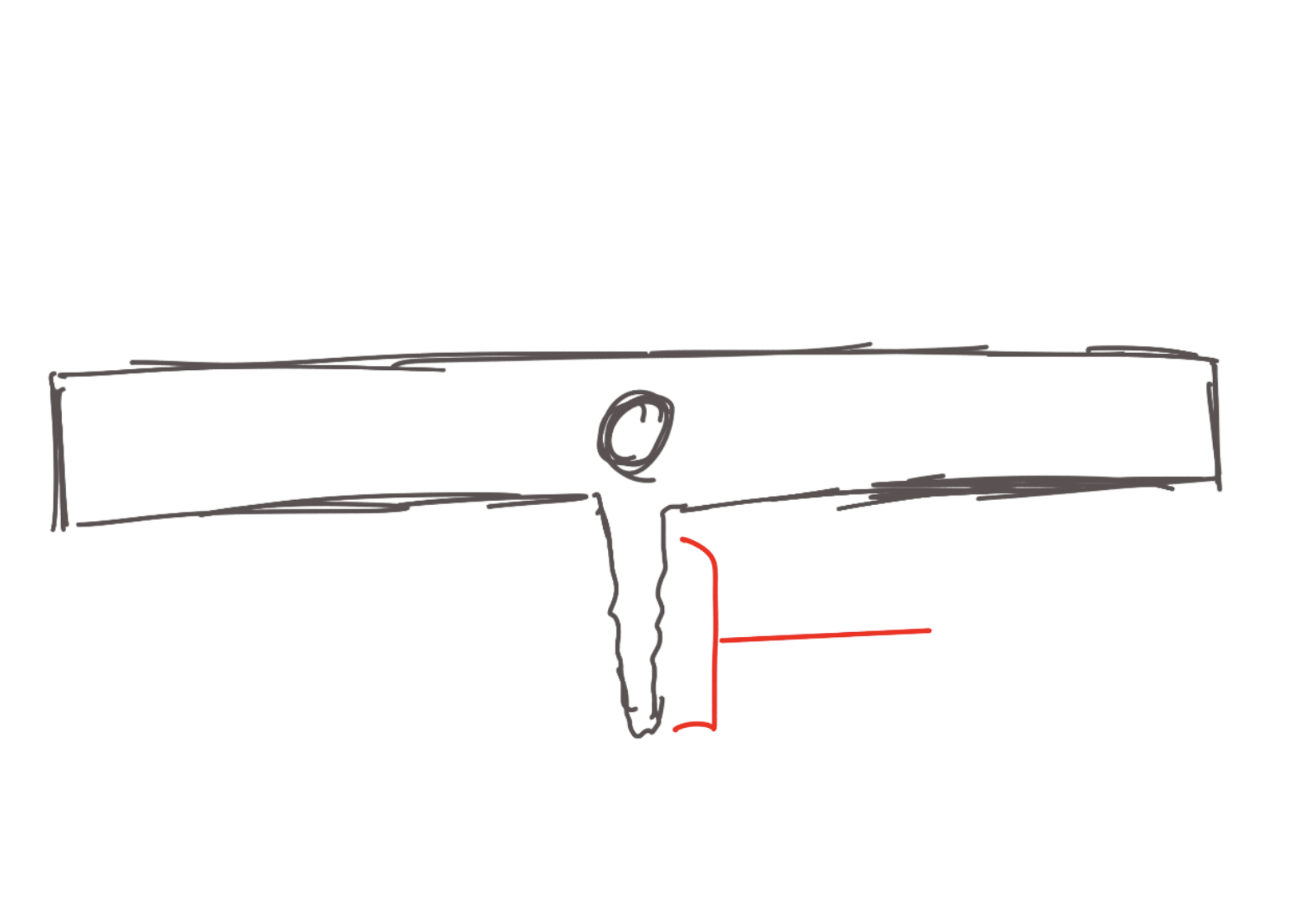
Middle cardiac vein
structure.
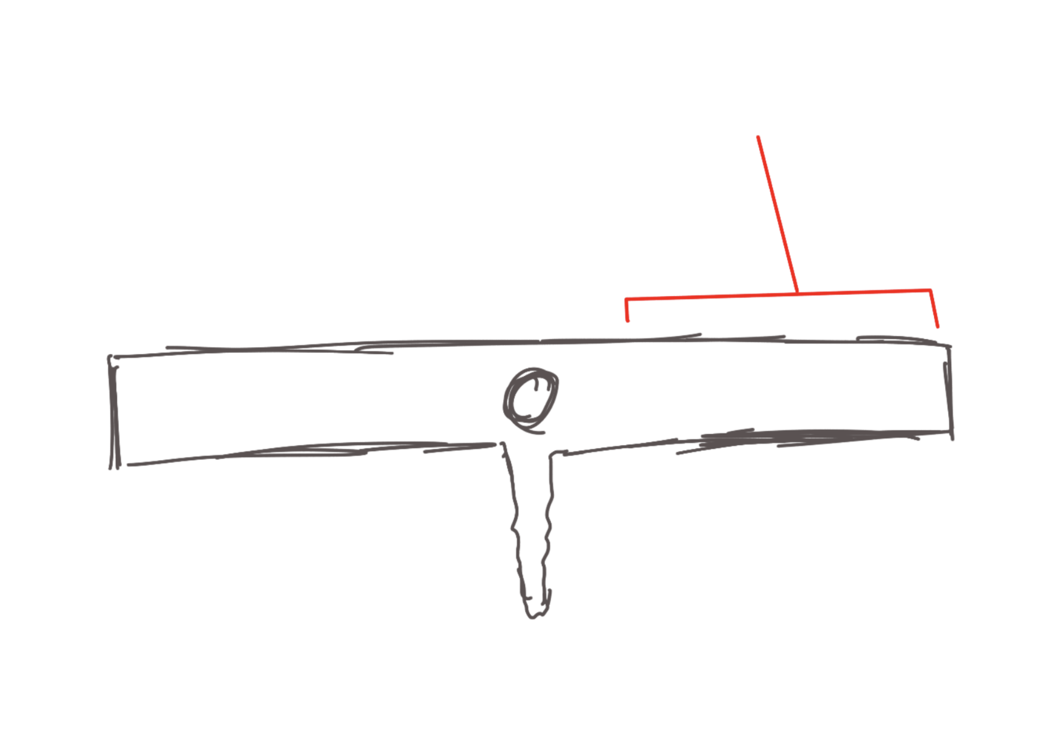
Small cardiac vein
structure.
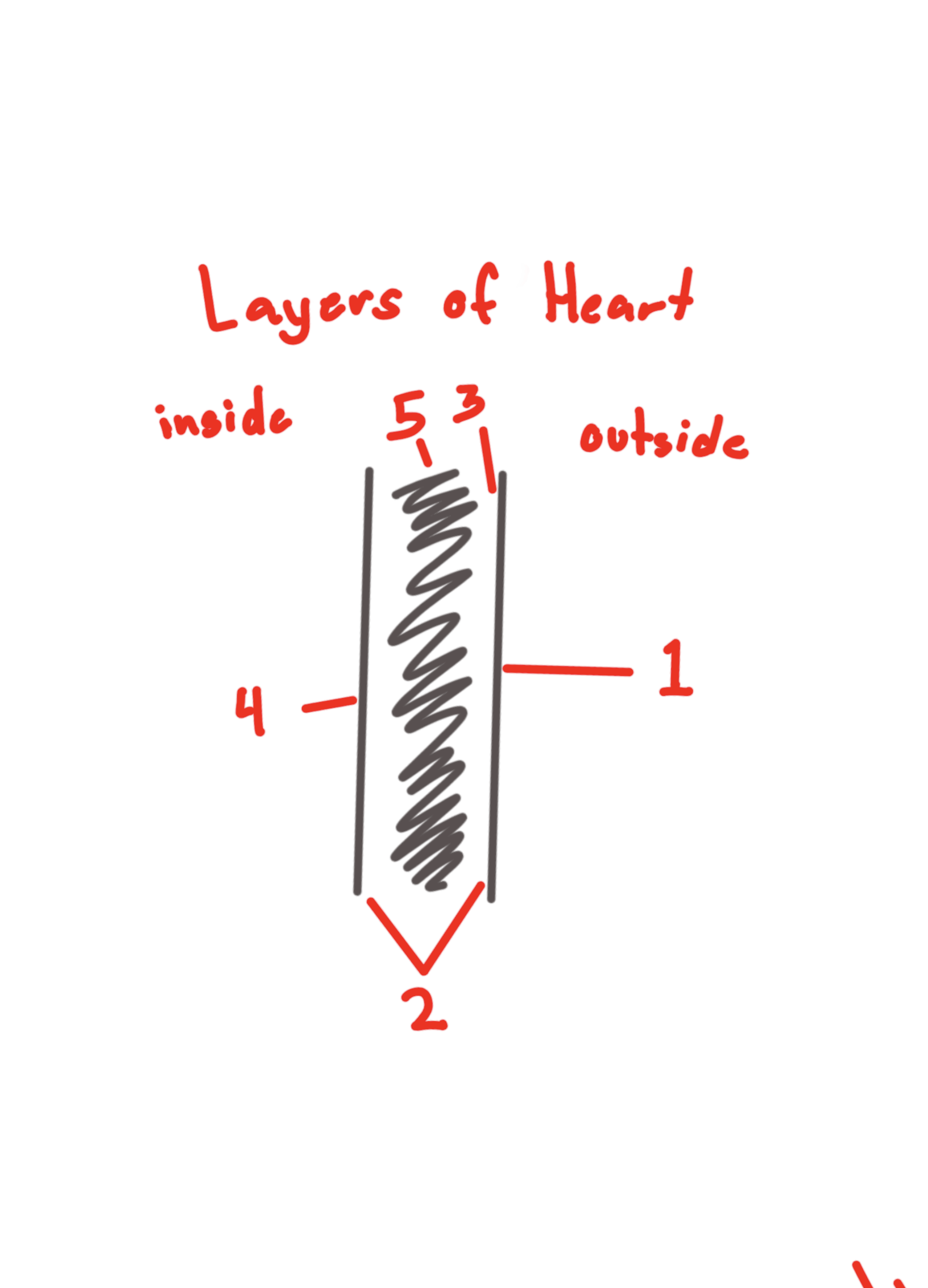
Fibrous pericardium
Covering. outer layer of the pericardium
#1

serous pericardium
Collective covering. In between the first and the third layer
#2

parietal layer of the serous pericardium
Feature. Behind the serous pericardium layer.
#3

visceral layer of the serous pericardium
Feature. the bare layer of the heart
#2

Pericardial cavity
(END OF S1) Space. between the 2 layers.
#5
Endocardium
inside circle. #1

Myocardium
thick muscular part. #2
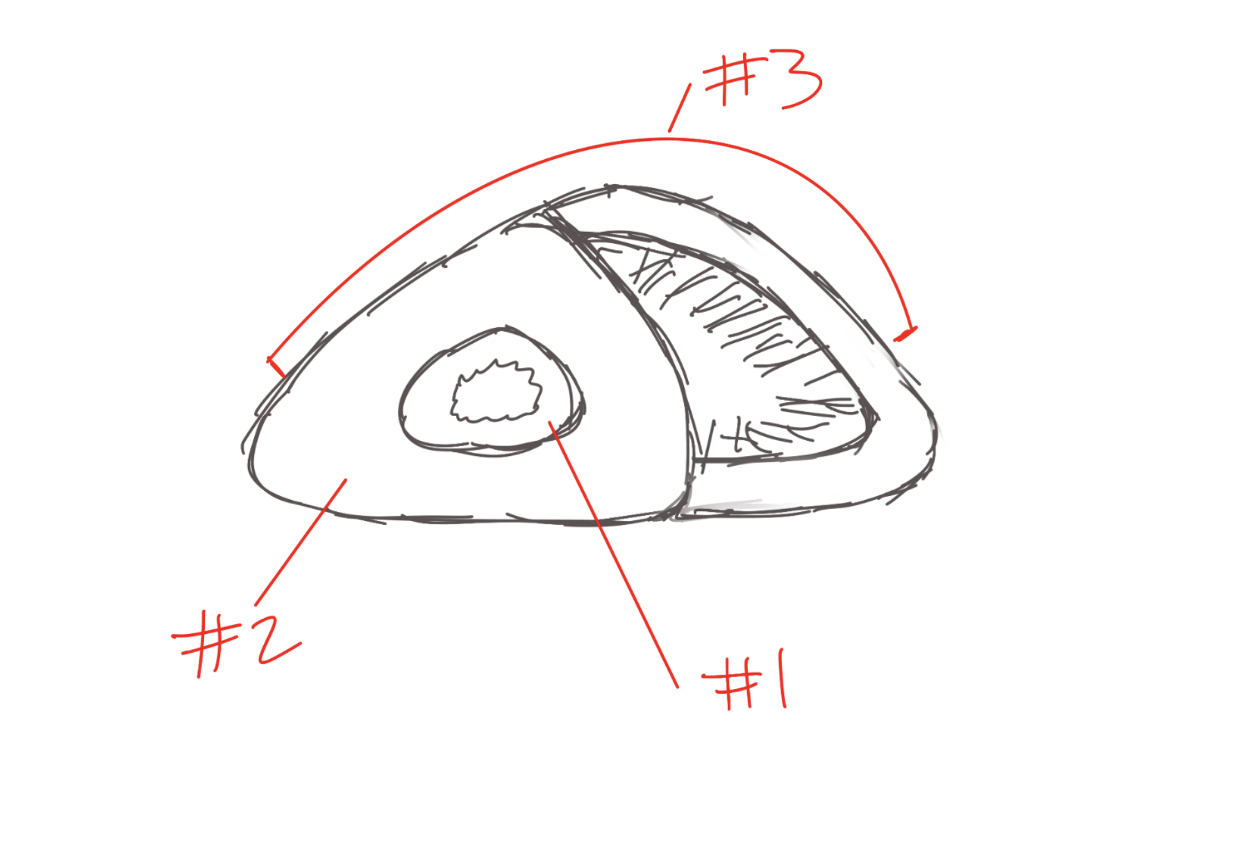
Epicardium
layer on the outside. #3

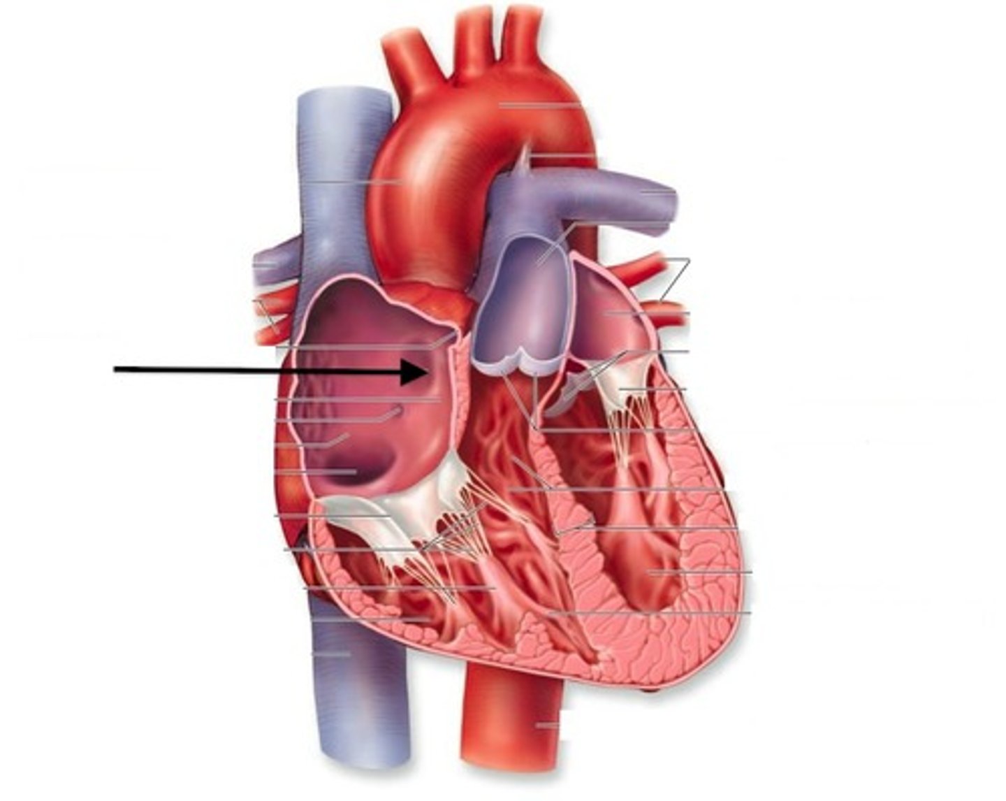
Interatrial septum
structure. DH They squeeze it.
Fossa ovalis
depression. DH BEAN
Right atrium
Charmber. DH, TA circles top right.
Pectinate muscle
structure. Inside flap on DH
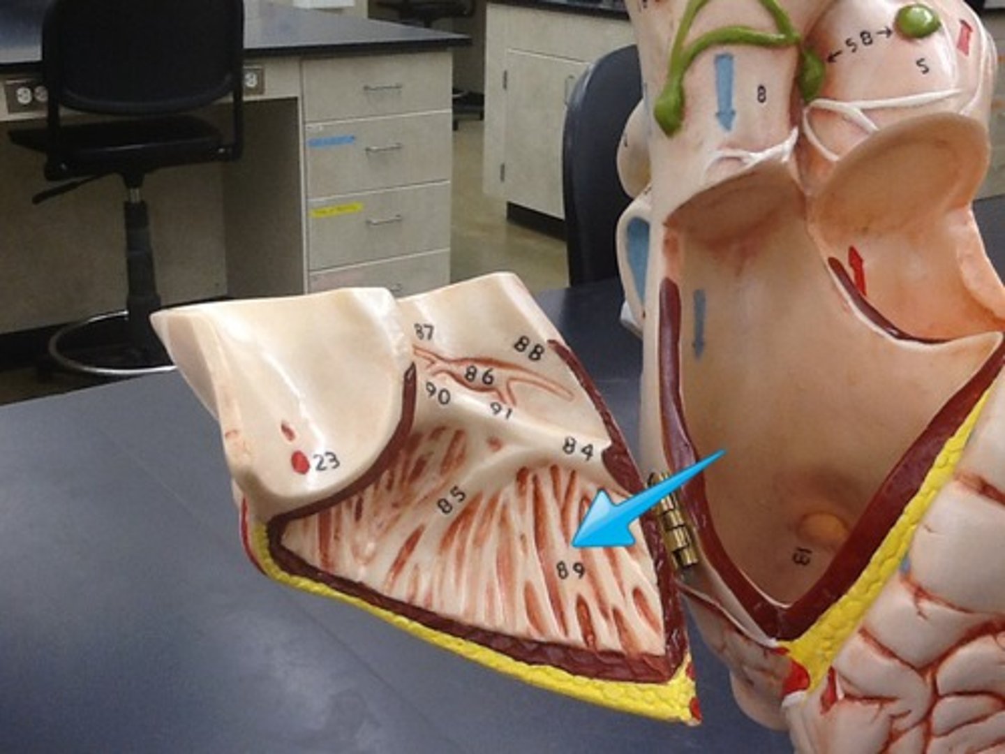
Sinus venarum
surface. Below pectinate muscle. #2 on this picture.
opening of the superior vena cava
Space. EFH
opening of the inferior vena cava
Space. Only one hole.
Opening of the coronary sinus
Space. Bellow fossa ovalis, the smallest hole?
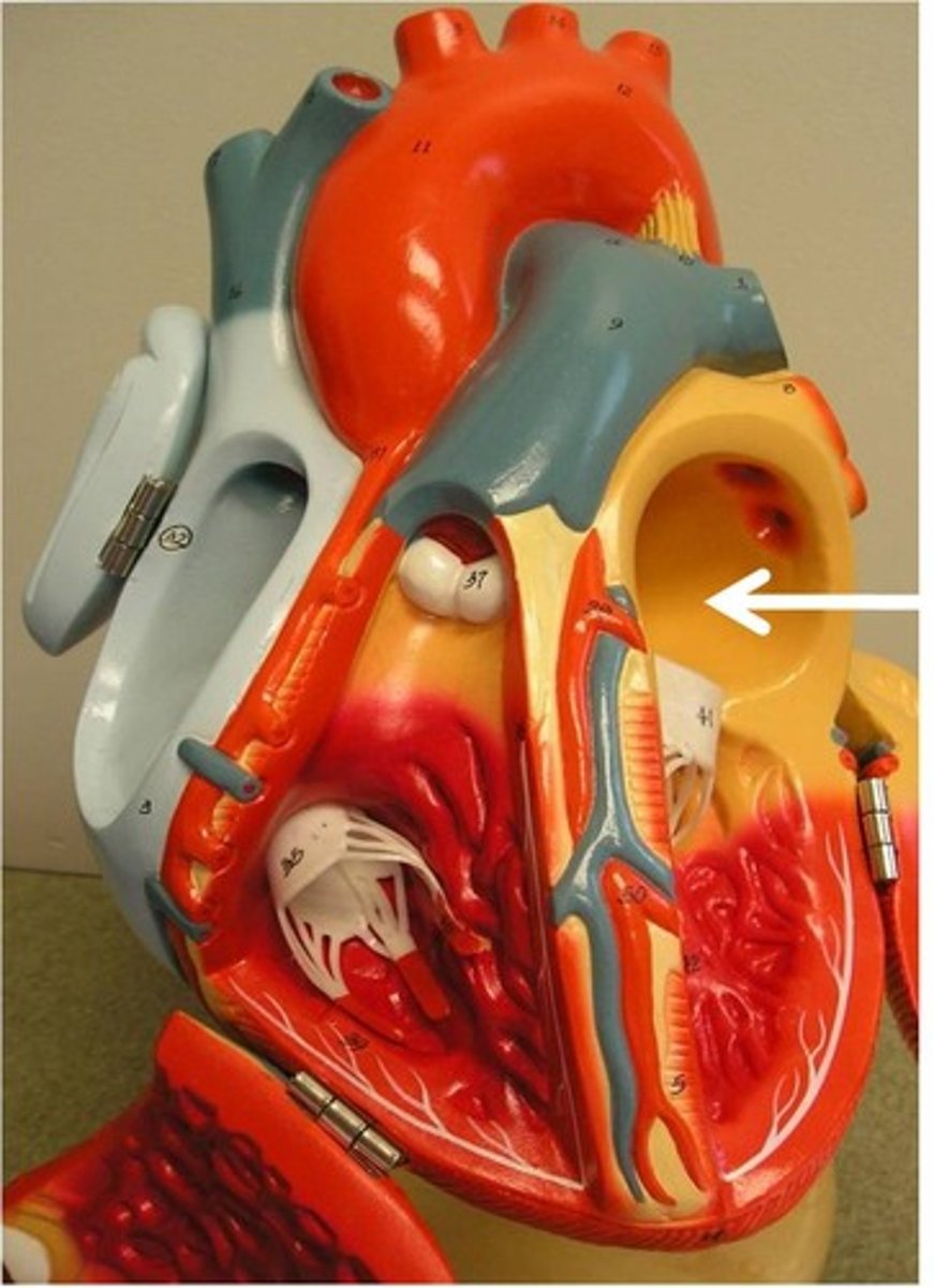
Left atrium
Chamber. EFH

Left auricle
Chamber. Ear on the DH.
Openings of the pulmonary veins
collective space. This but on the dark heart.
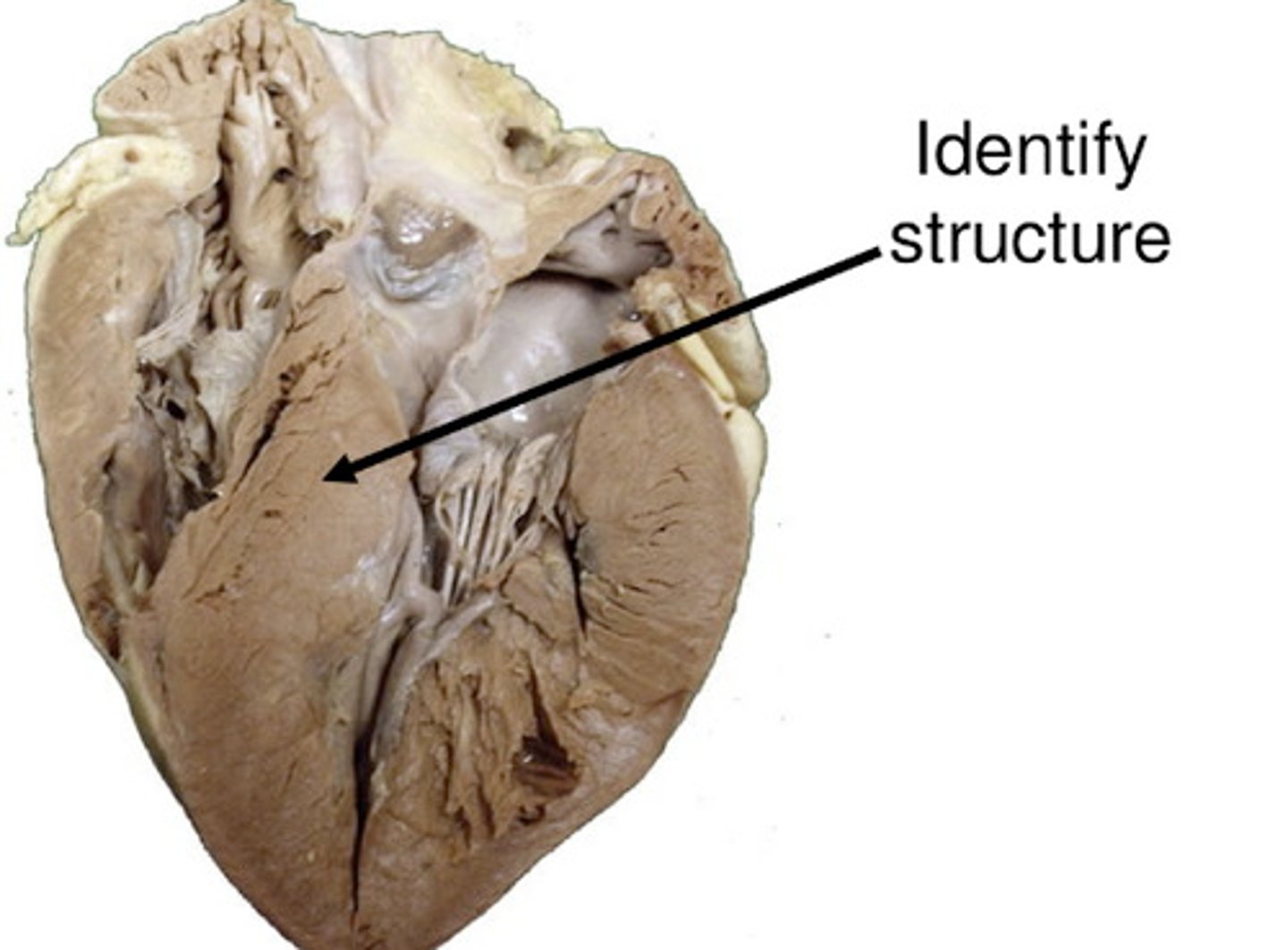
Interventricular septum
strucutre. DH

Muscular part of the interventricular septum
portion. DH
membranous part of the interventricular septum
portion. What is this portion in deep? DH
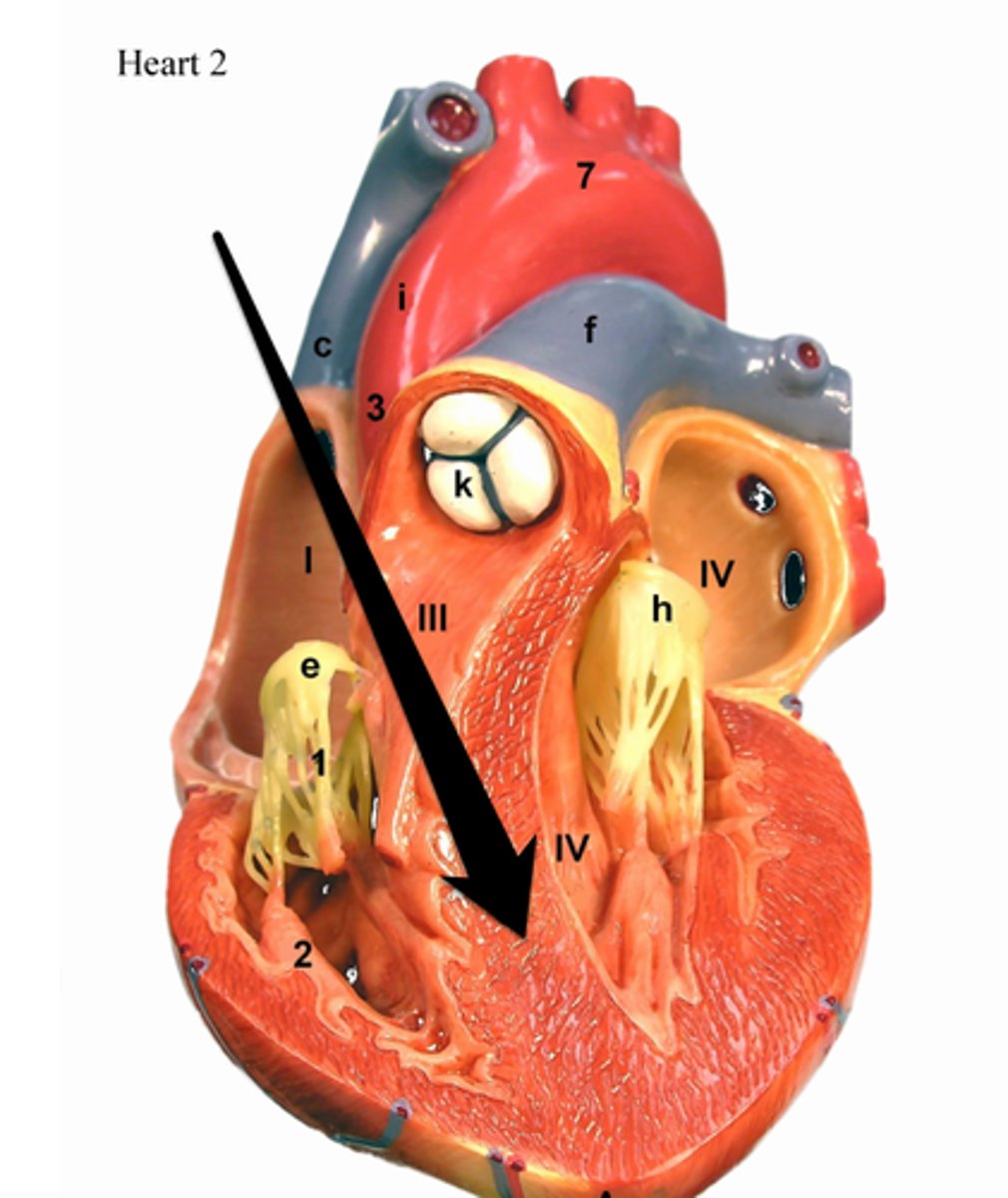
Right ventricle
Chamber
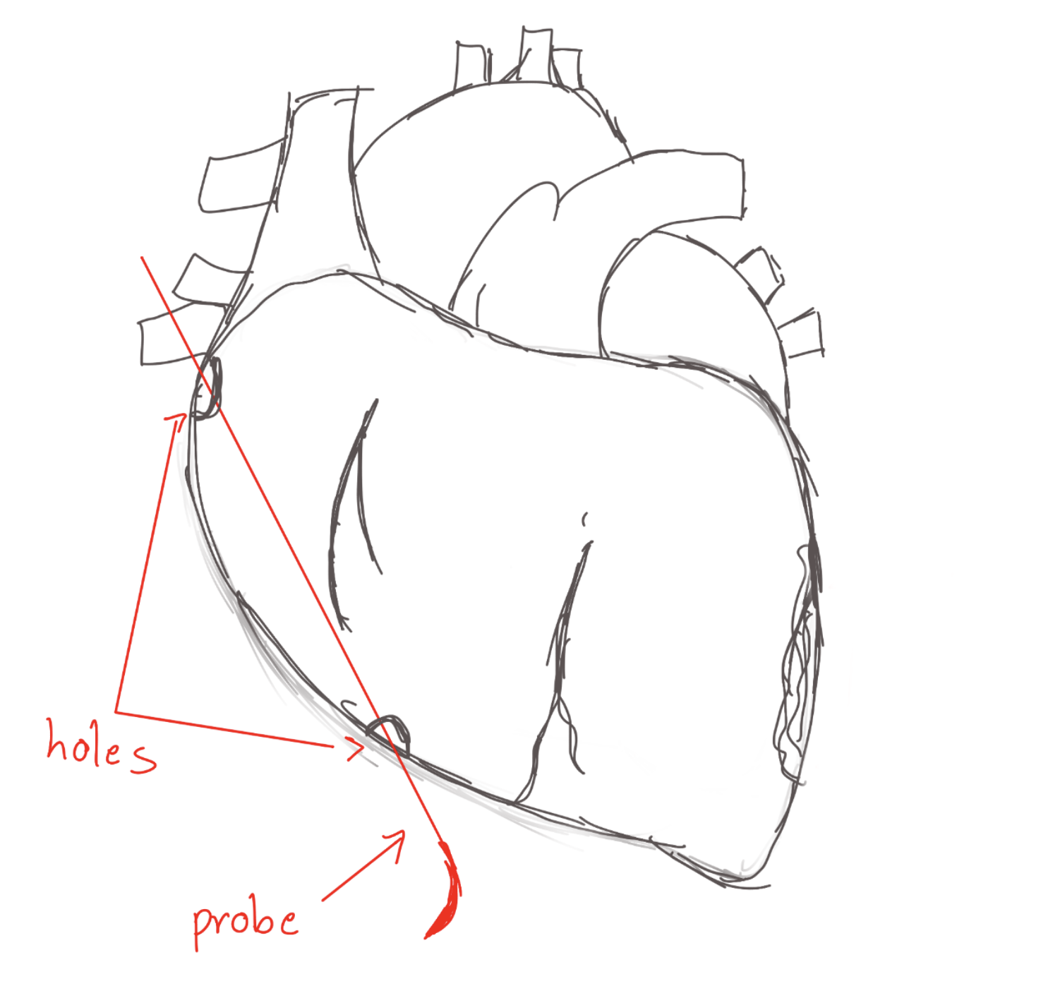
Right atrioventricular orifice
space. Basically the name. DH
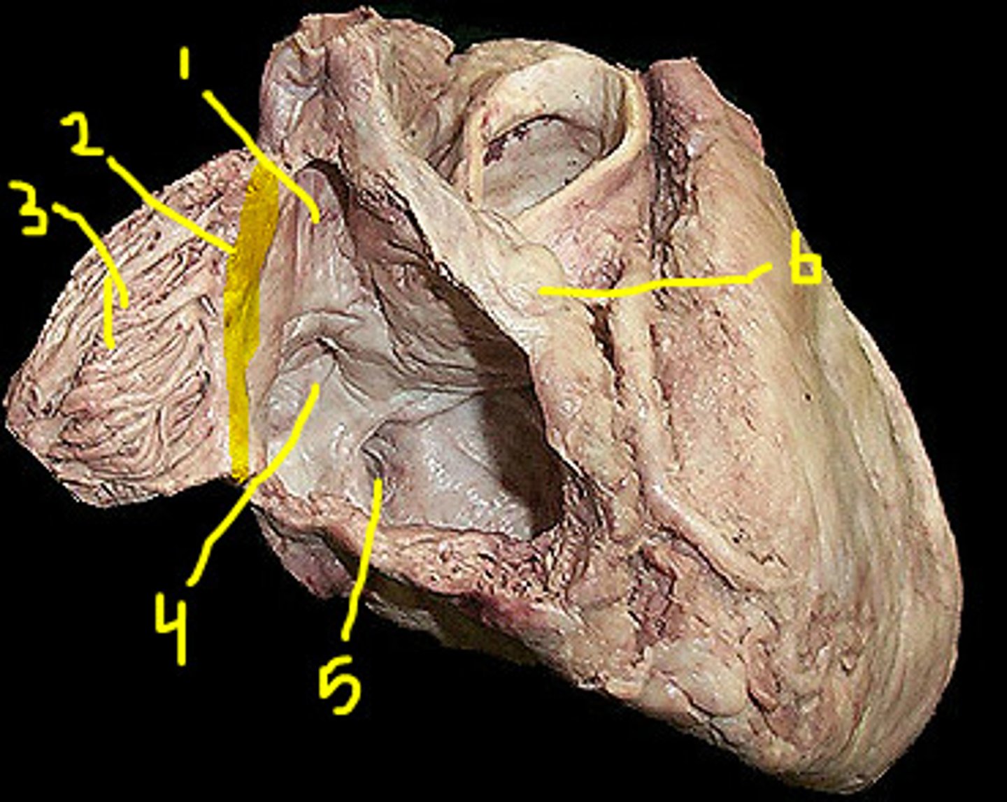
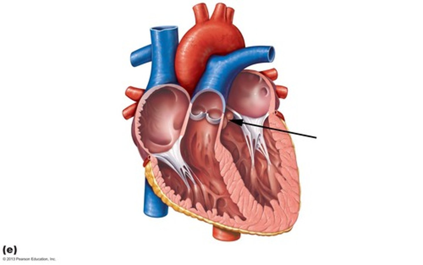
Tricuspid valve
structure. DH
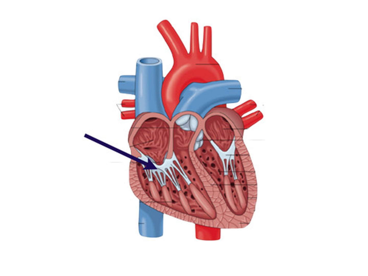
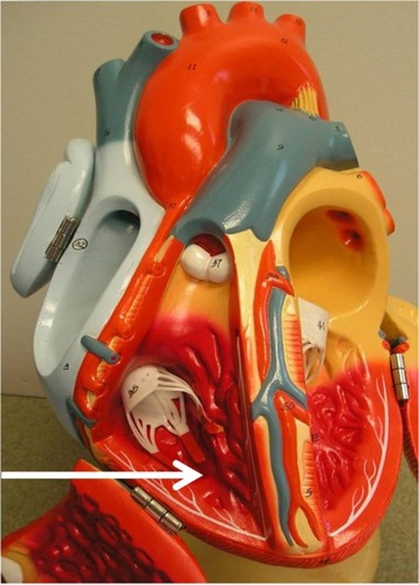
Papillary muscles (3)
structure. on both sides. DH.
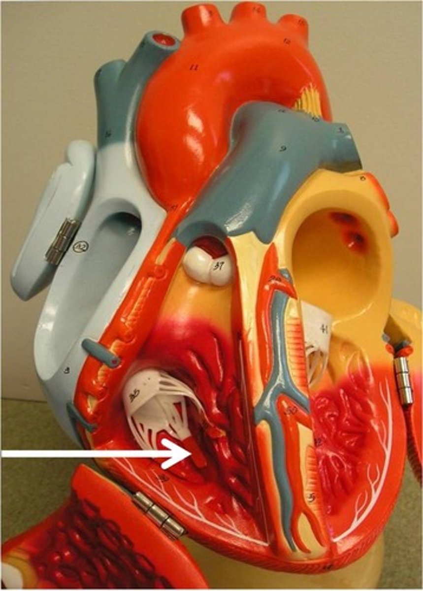
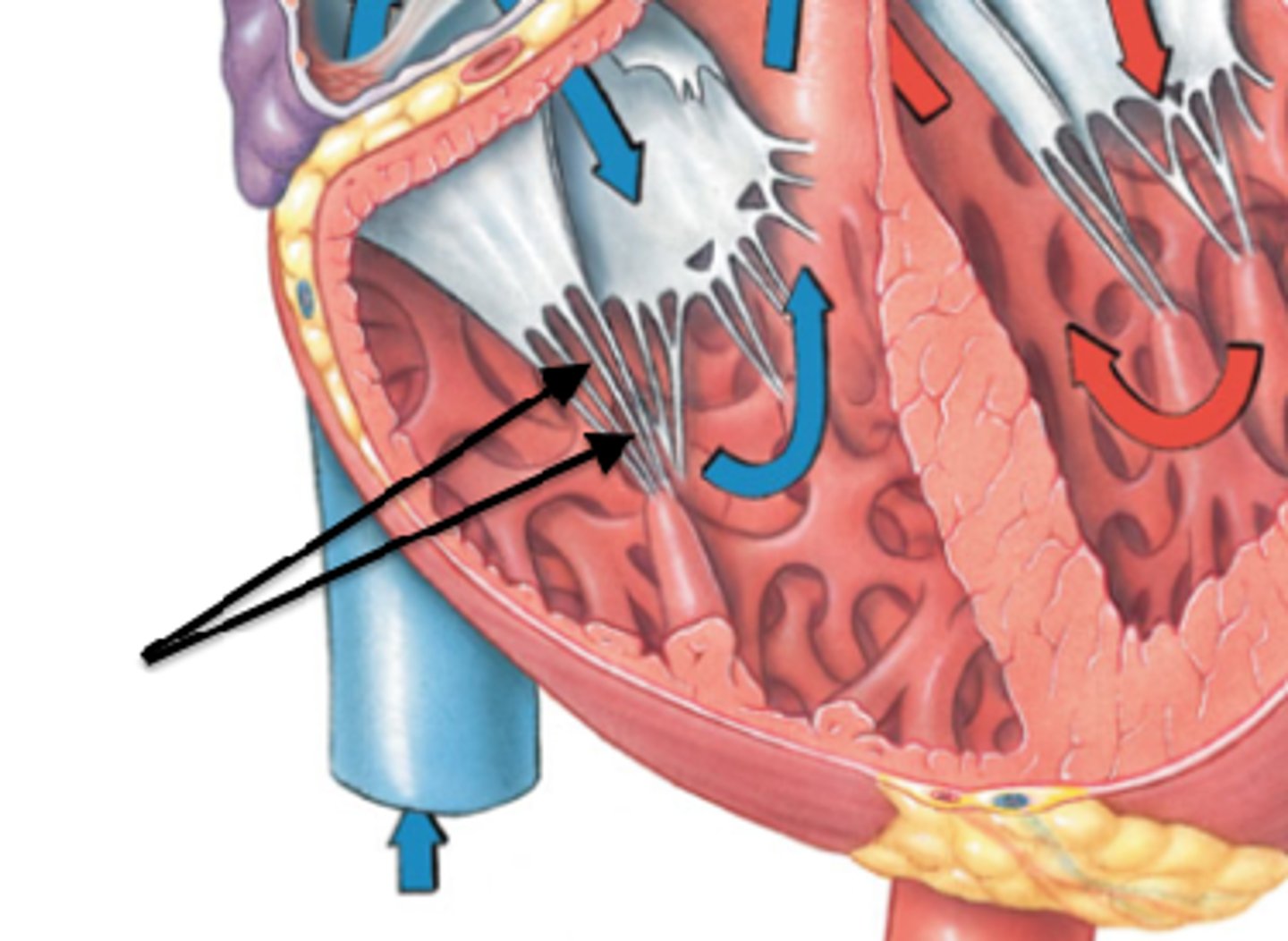
Tendinous cords
feature. Strings on top of the papillary muscle. DH.
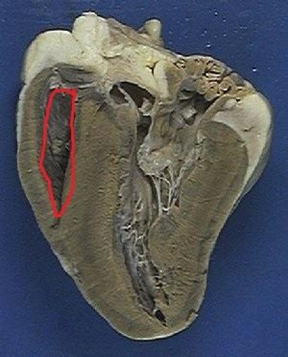
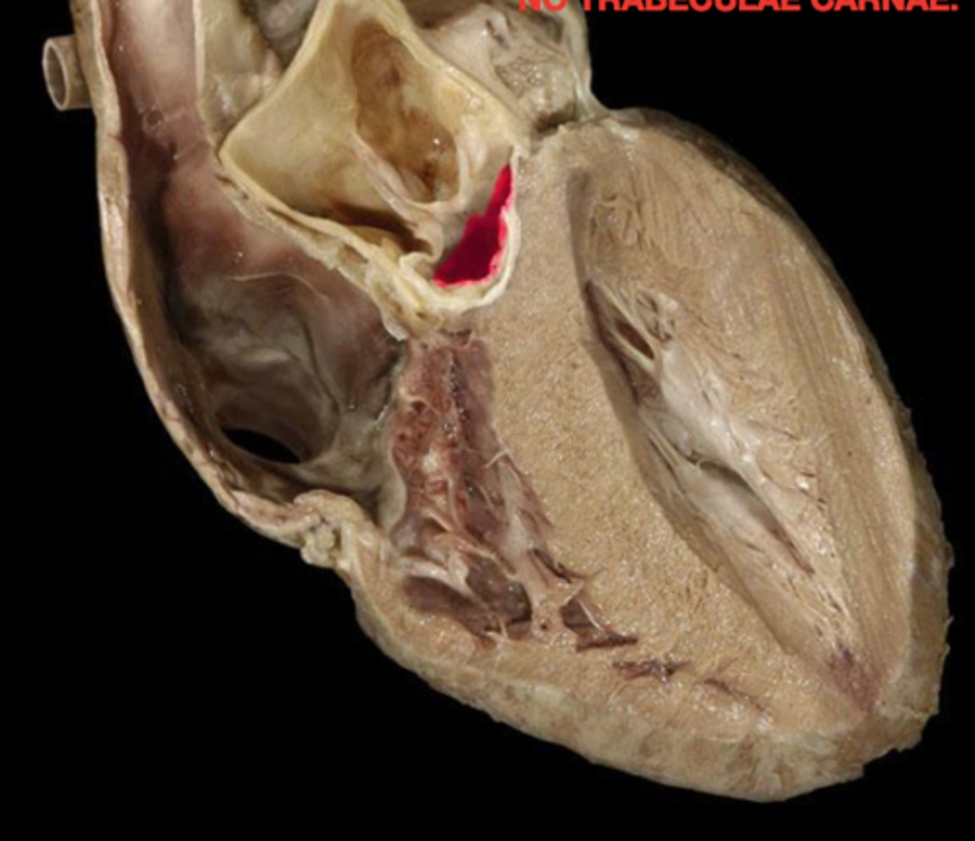
Trabeculae carneae muscle
Structure. Beef jerky, meat.
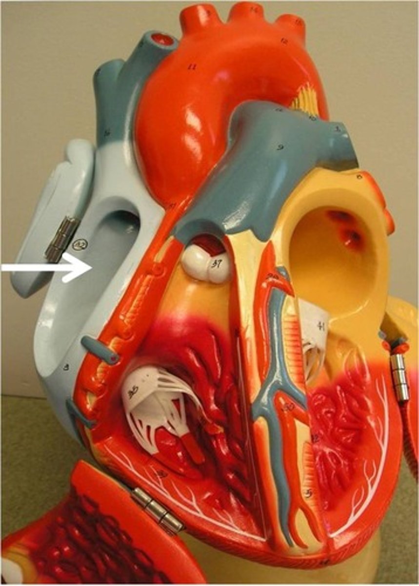
Conus arteriosus
Narrowing. In the little split in LH.
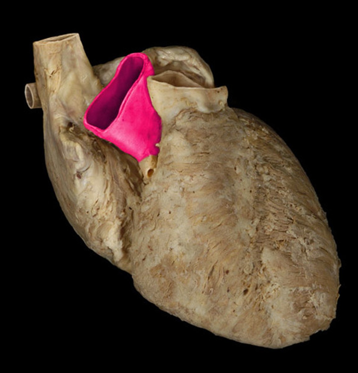
Opening of the pulmonary trunk
Space. Top of trunk in LH same area. TA circles the top.
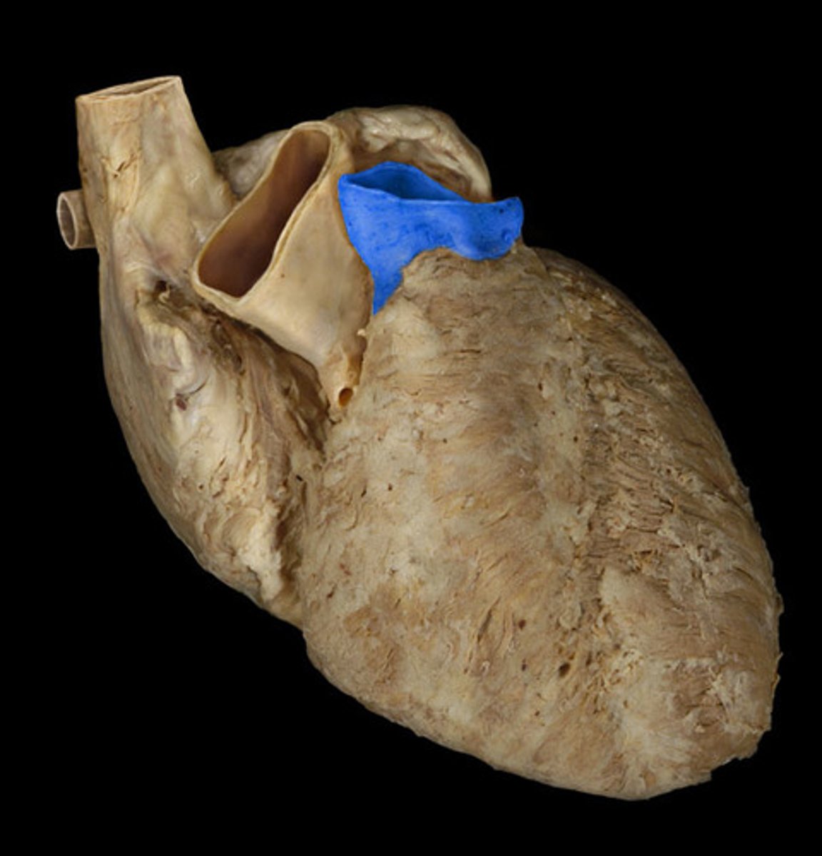
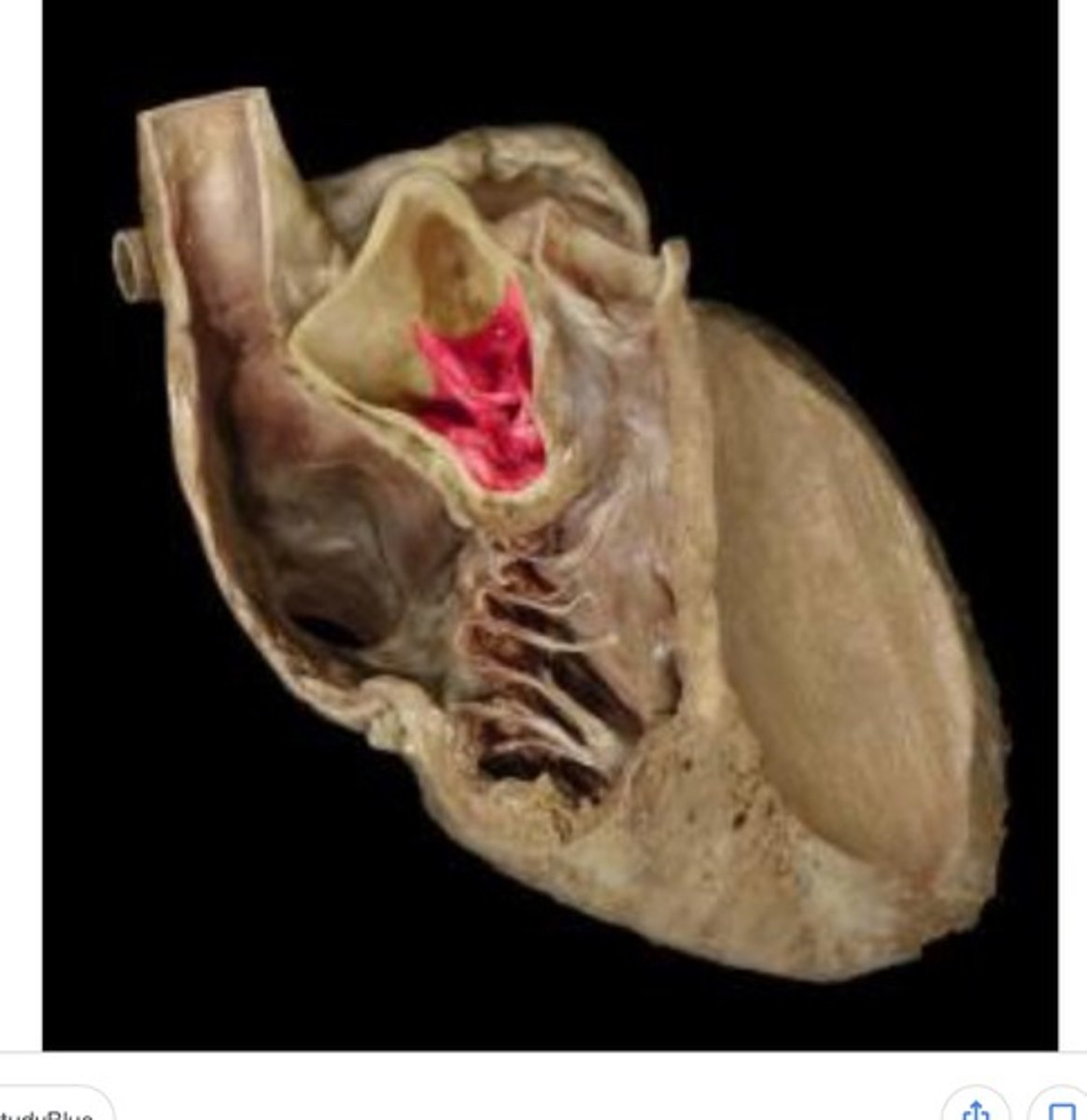
Pulmonary valve
Entire structure.

semilunar cusps of the pulmonary valve
structure. “what are these 3 components?”
Left ventricle
Chamber, DH. TA also pinches.
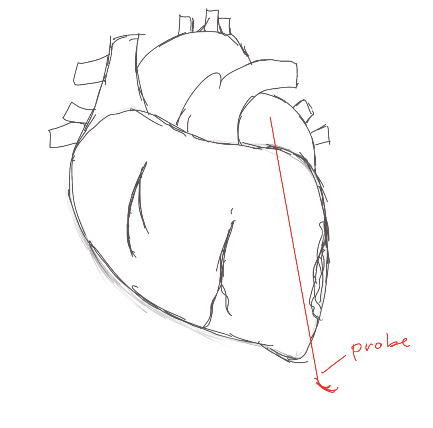
Left atrioventricular orifice
Space. Ta puts probes like the right one.
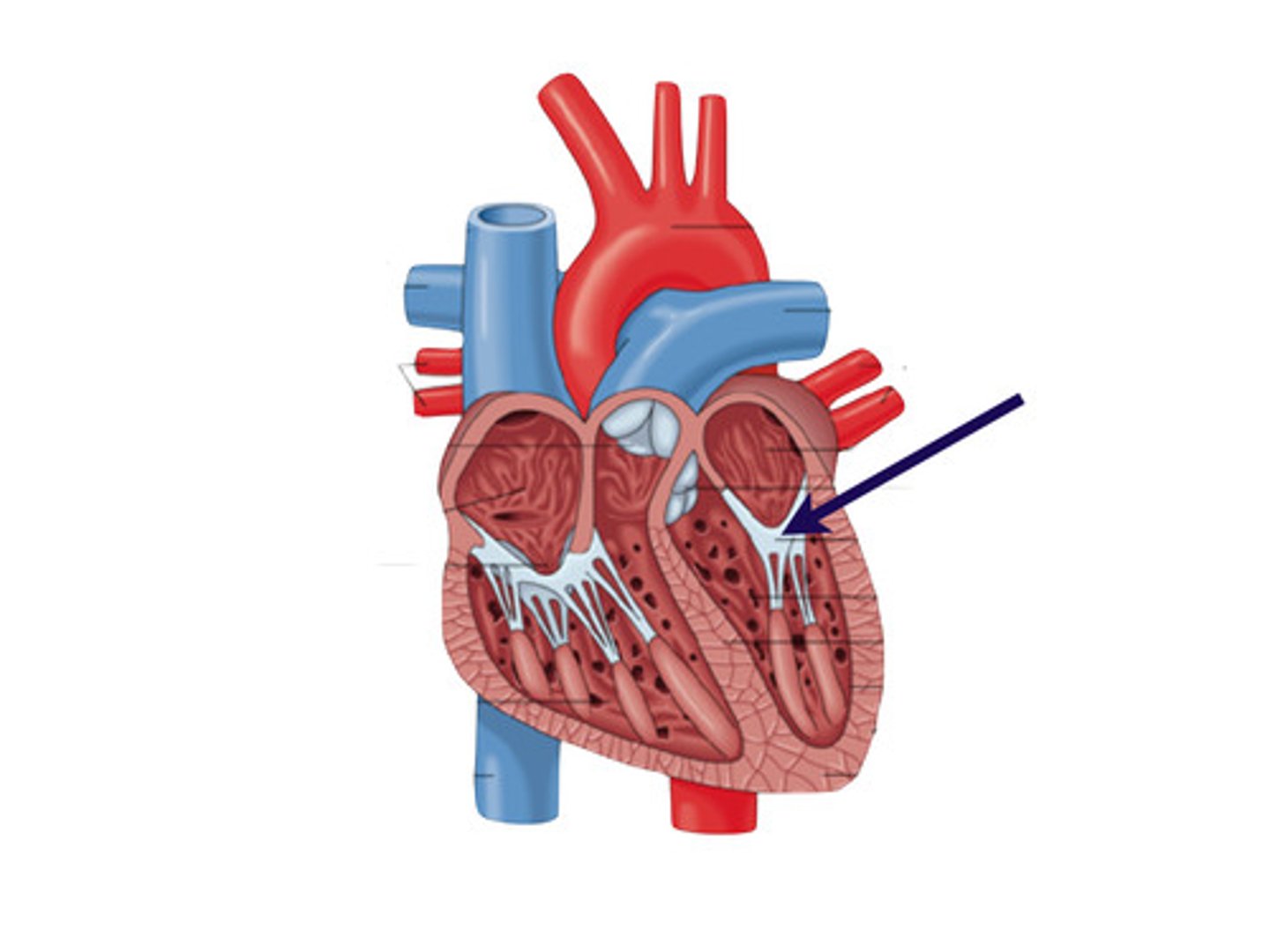
Mitral valve
Structure. EFS, inside left ventricle.
Aortic vestibule
Narrowing, In big split
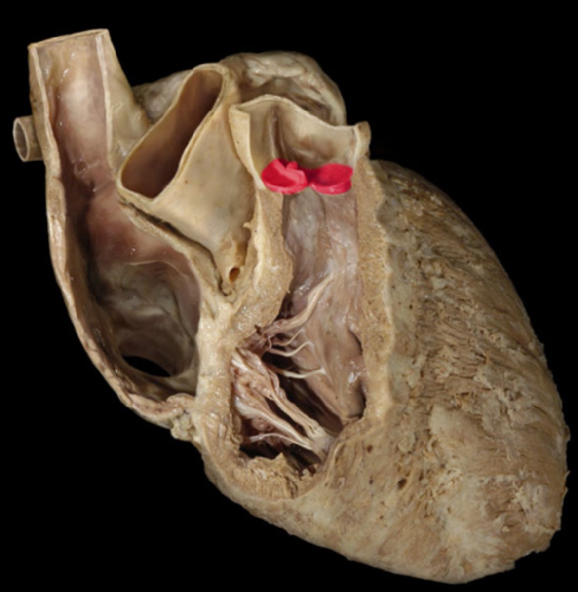
Opening of ascending aorta
space. Circle top, but closes the big split.
Aortic valve
Entire structure. LH.
semilunar cusps of the aortic valve
(END of S2) 3 components. The ones shown inside.
Pulmonary arteries
The thicker one (start of S3)
Pulmonary veins
thin ones
Ascending aorta
portion. Top of the heart, below artery.
Arch of aorta
curvature.
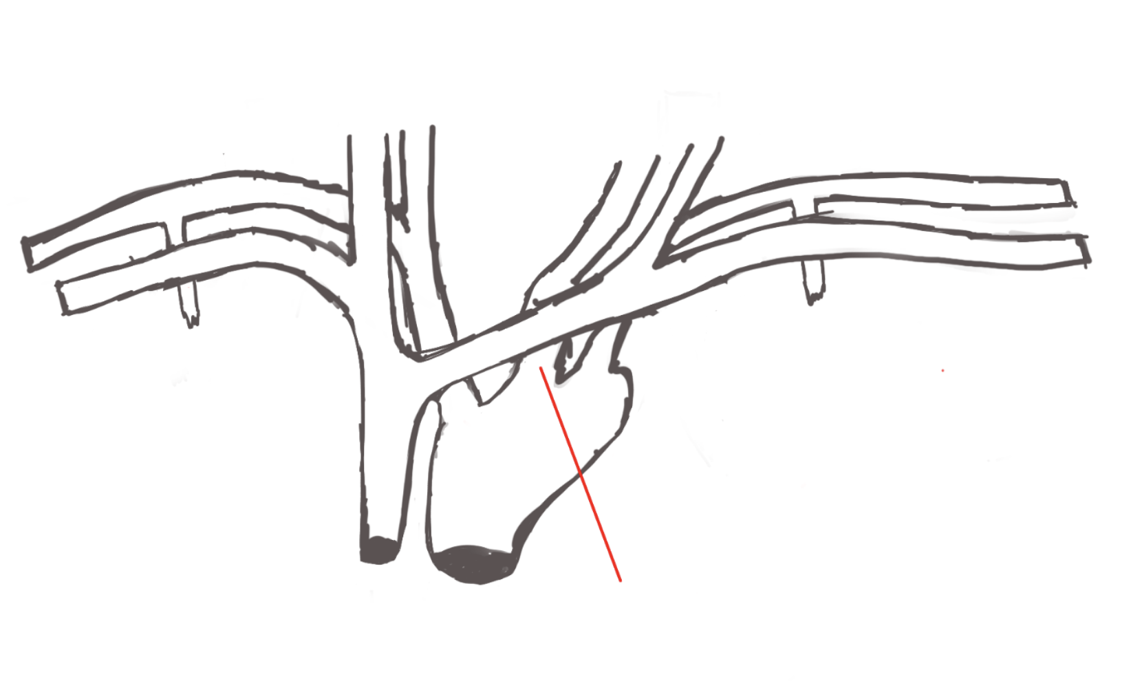
Brachiocephalic artery
Structure.
right common carotid artery
structure. Top of brachiocephalic artery
right subclavian artery
structure. Goes along clavical
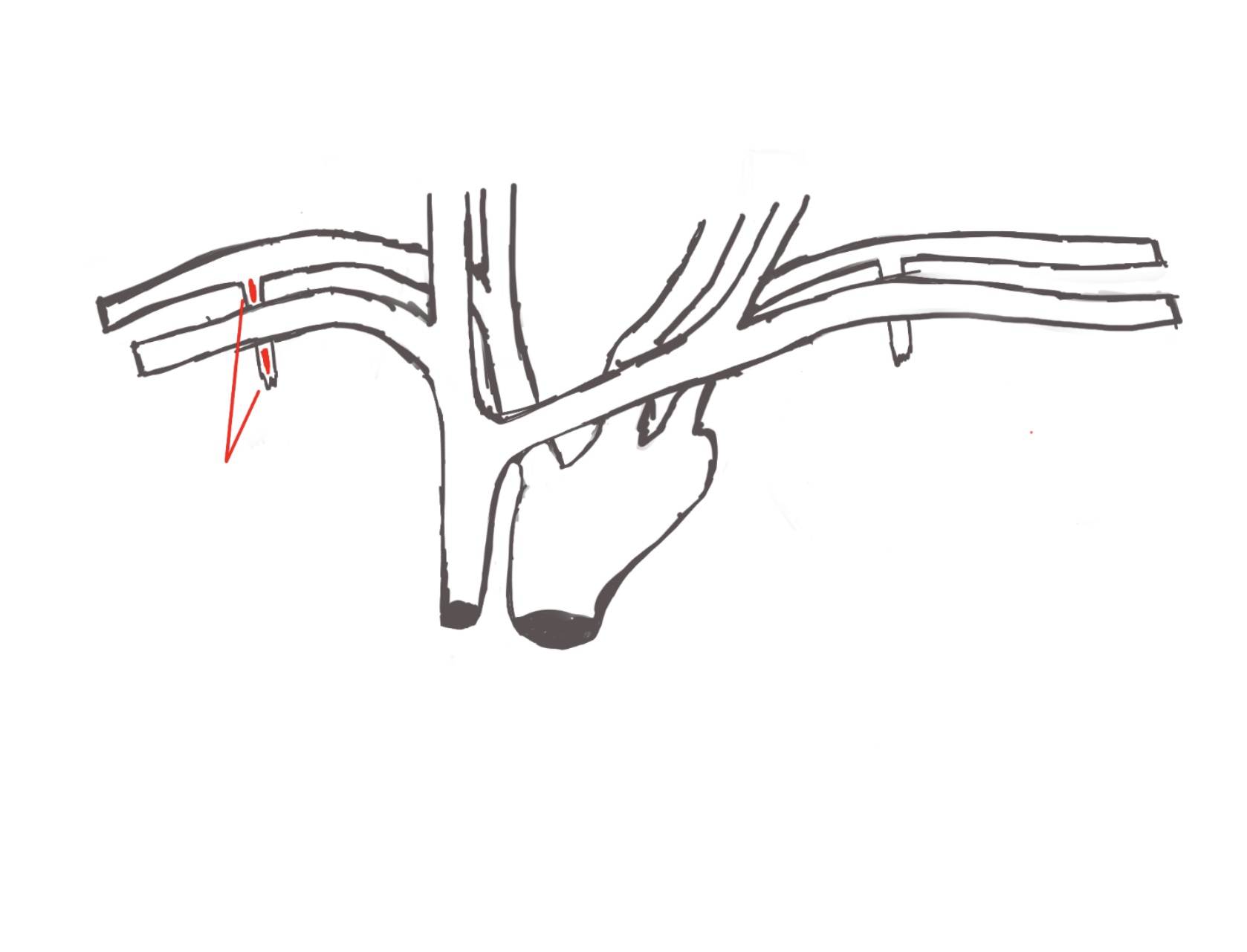
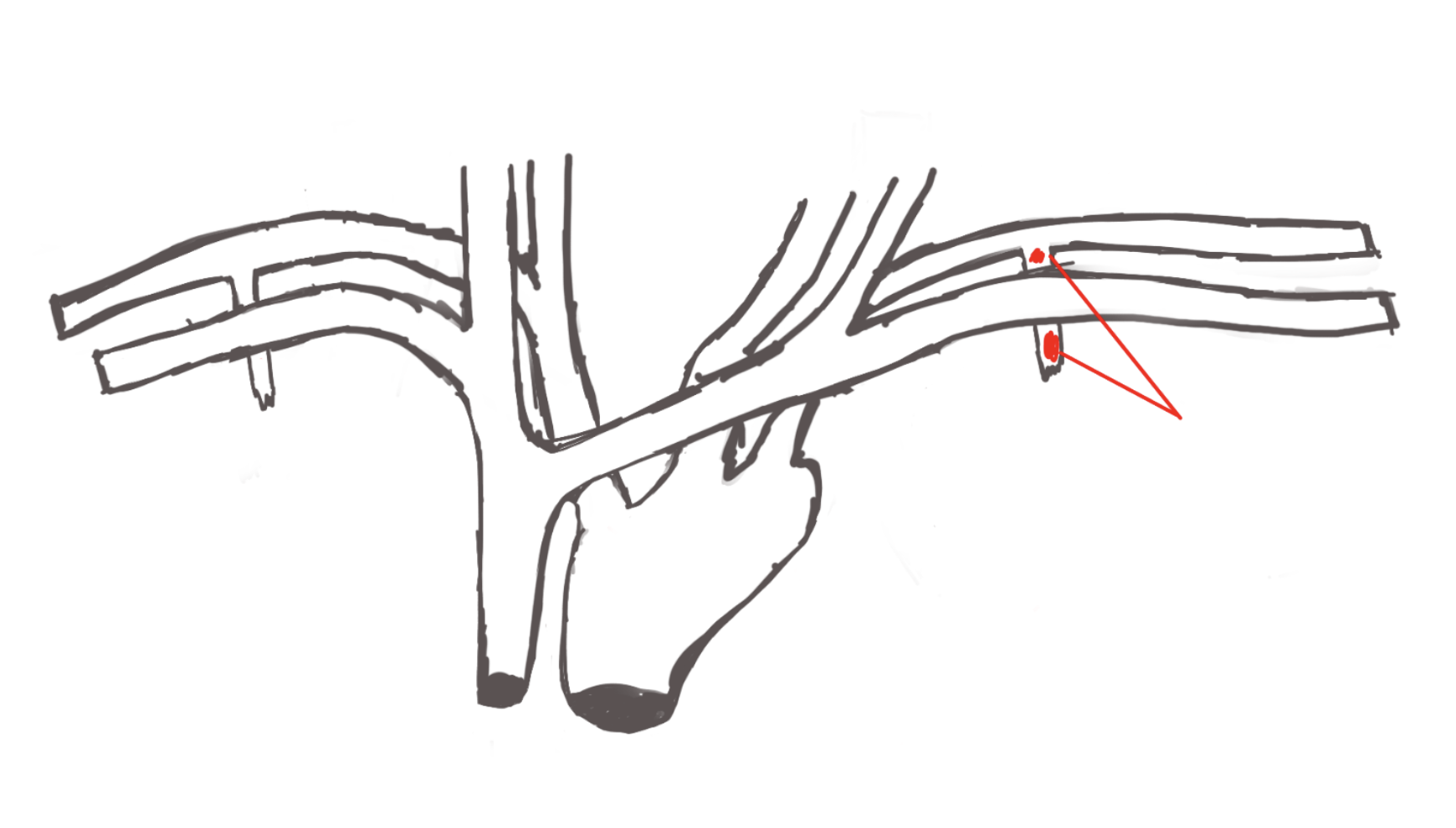
right internal thoracic artery
structure. goes to thoracic cut.
Left common carotid artery
structure.
Left subclavian artery
structure
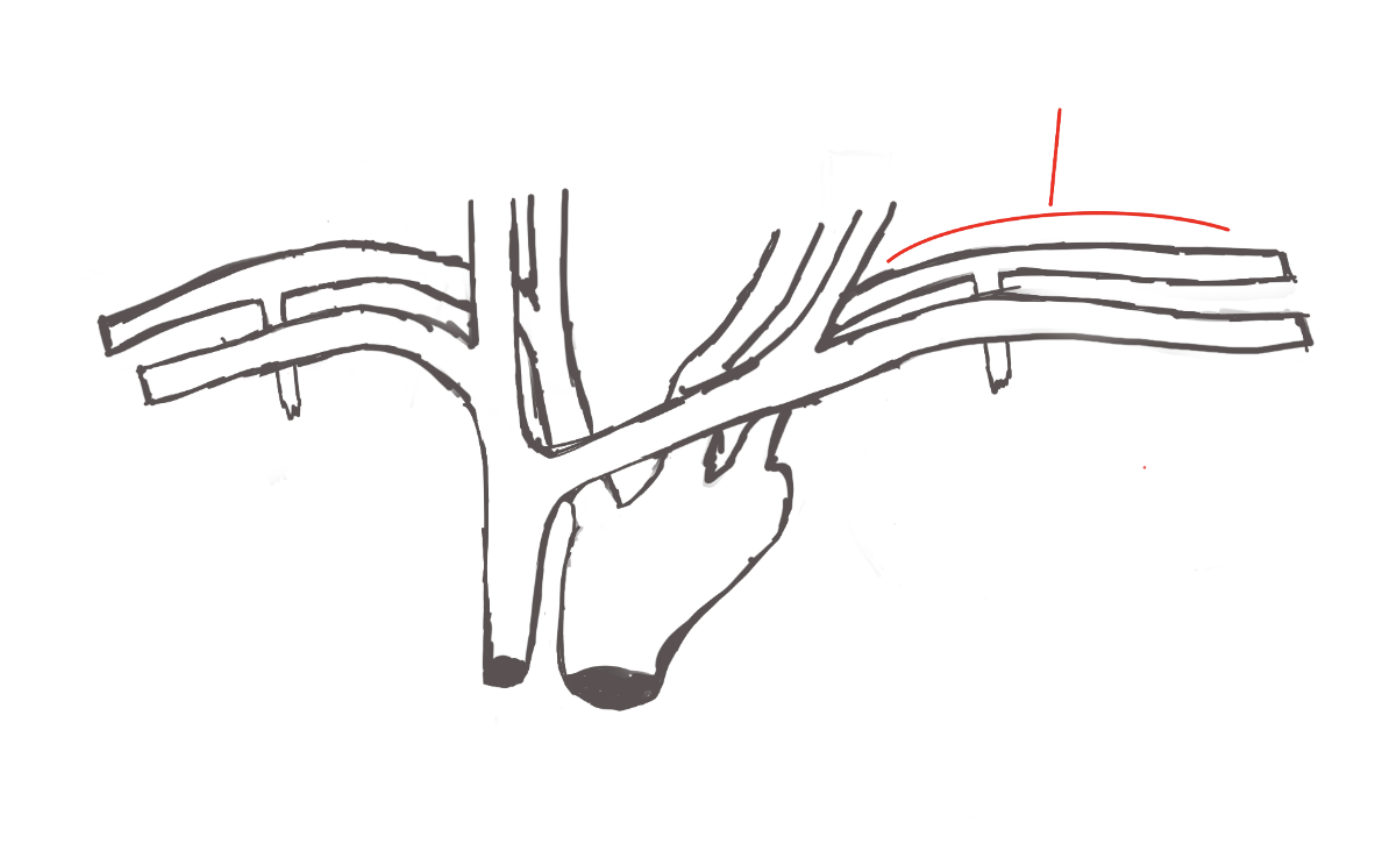
left internal thoracic artery
structure. left side also cut.
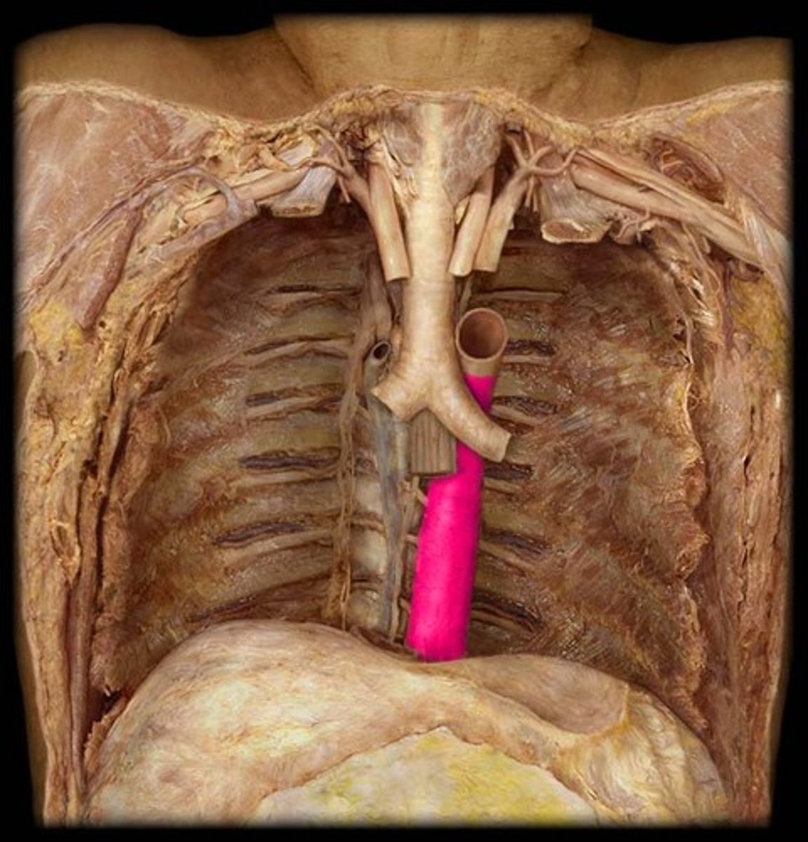
Thoracic aorta
portion. most left side thoracic area, takes the heart out.
Right posterior intercoastal arteries
The most posterior, artery.
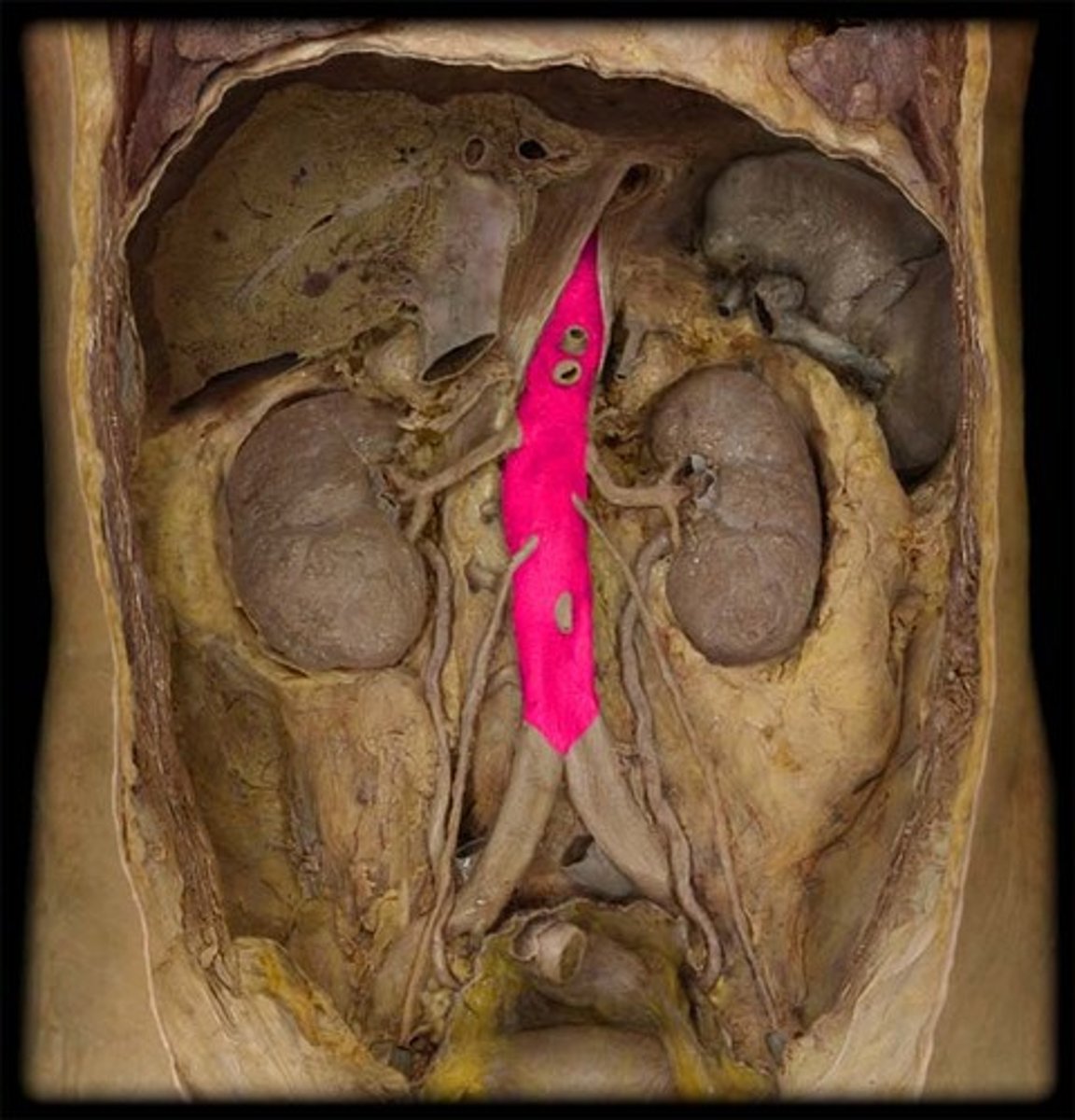
Abdominal aorta
portion. the tube!
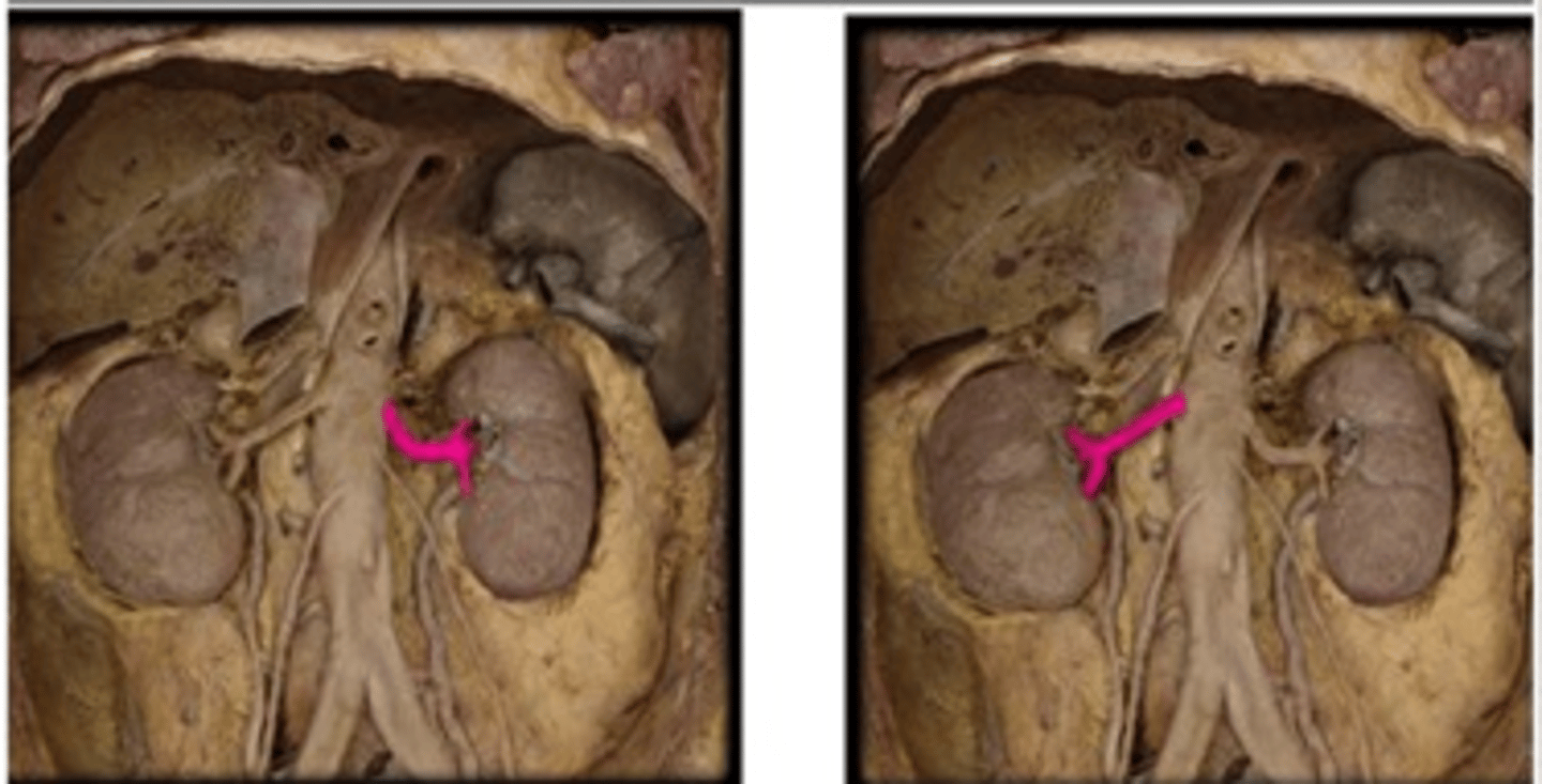
right renal arteries
the right is on top. we have to flip the kidney.
left renal arteries
below the kidney for left.
Celiac trunk
structure. on top of abdominal aorta