Diagnostic Imaging Final Review
1/108
There's no tags or description
Looks like no tags are added yet.
Name | Mastery | Learn | Test | Matching | Spaced | Call with Kai |
|---|
No analytics yet
Send a link to your students to track their progress
109 Terms
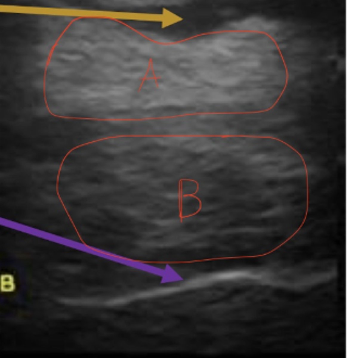
A is hyperechoic compared to B
How does A relate to B in echogenicity?
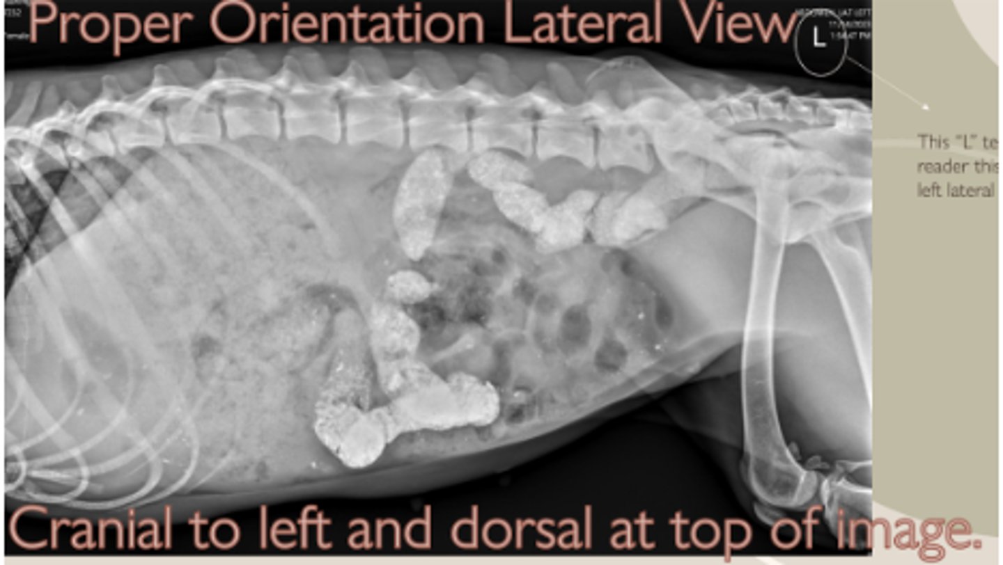
Cranial to the left
Which is oriented correctly?
Anechoic
Describe a fluid-filled structure in echogenicity.
Image with better detail
Which has the higher mAs?
Size, shape, number, location, margination, opacity
What are the 6 Roentgen signs?
CT scan
Which would be best for surgical bone repair?
Air, fat, water, bone, metal
Order of scale; black to white
to reduce scatter
Why do we use collimation?
Adjust the quantity of xrays produced
What does adjusting mAs do?
Positive contrast
What is the procedure for a ruptured bladder?
Free fluid
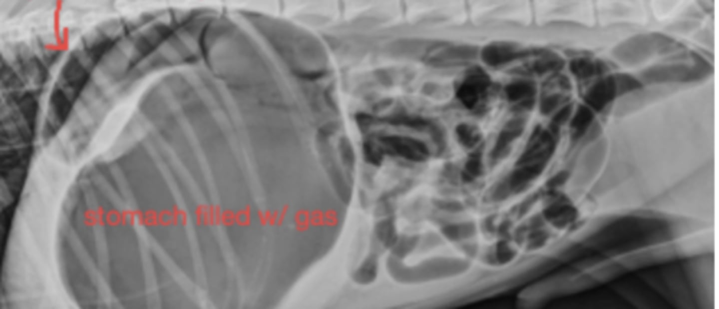
Radiograph; can't see much in abdomen - why?
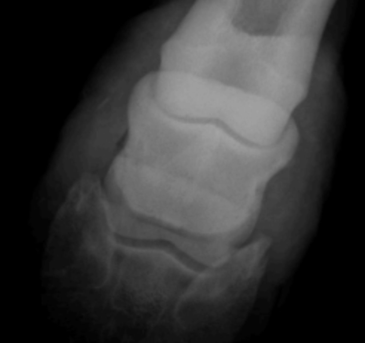
Laminitis
Horse lame in all four legs; image of hoof
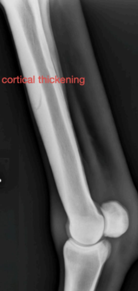
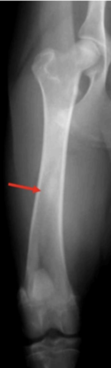
Metacarpal periositis
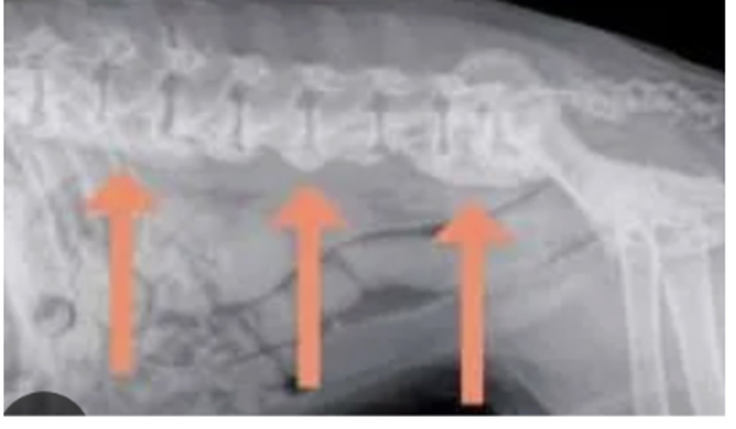
3 y/o horse with metacarpal pain; radiograph shows bone thickening- what caused this?
dorsopalmar oblique
Which shows the distal border of the navicular bone?
Lateromedial
What is the most important view for laminitis?
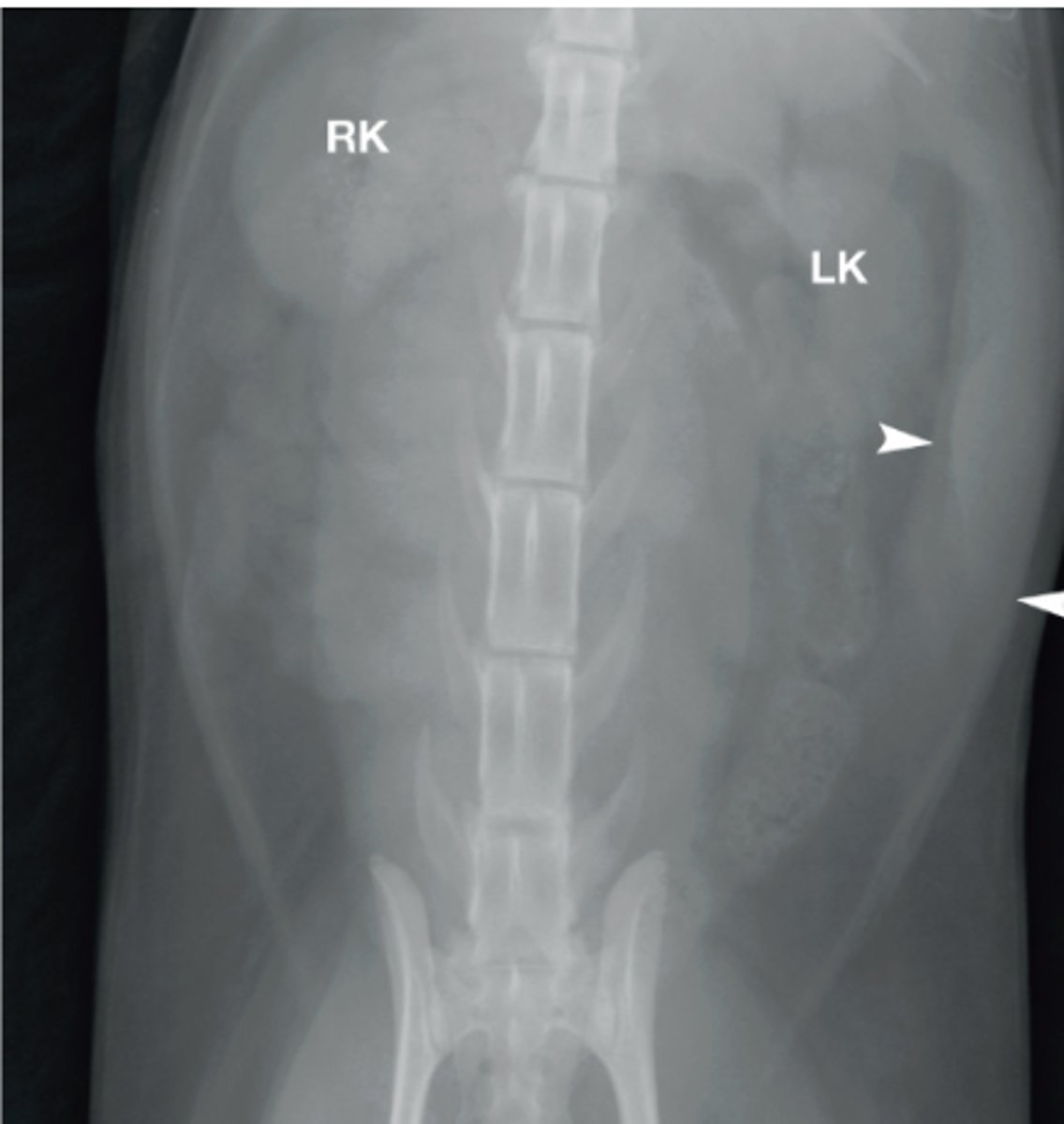
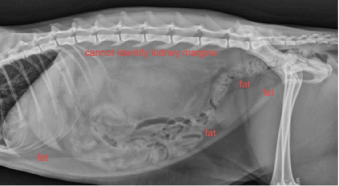
Renomegaly
Cat image with large kidneys and ureters- what is this called?
Odd behavior when under saddle
Lame on all four legs
Neck bent?
What is an indication for spinal radiographs?
Malalignment
What is wrong with this spine?
Linear foreign body
Dx of cat vomiting;
Sedation
2 yo chihuahua vomiting, sedated for radiographs, what is reason for splenomegaly?
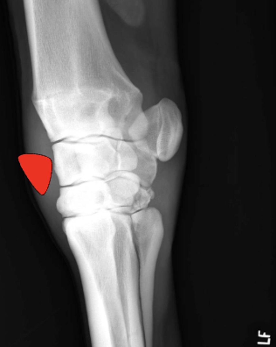
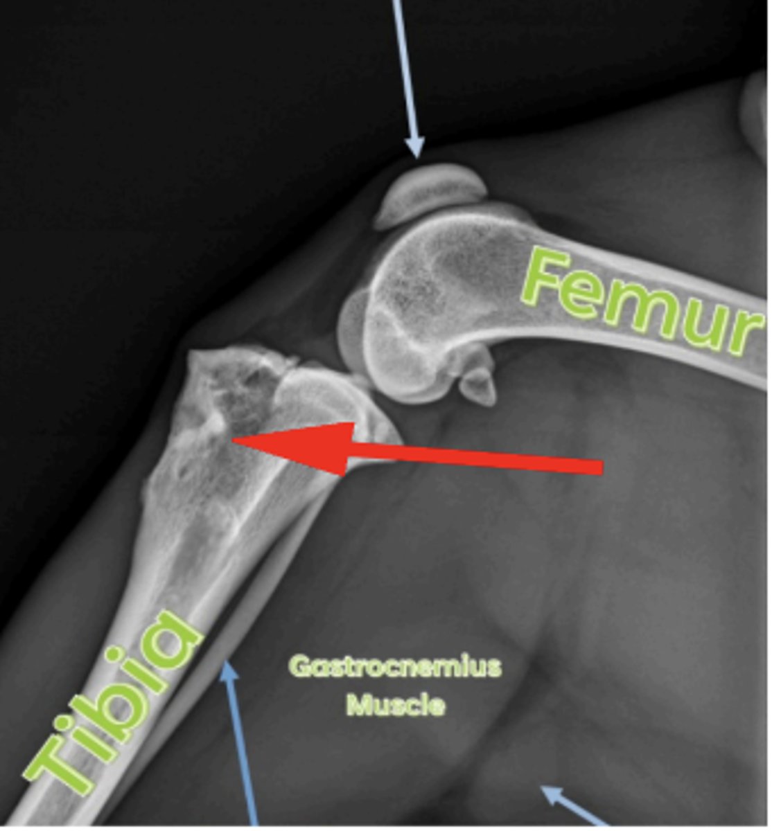
Medial femoral epicondyle
3 y/o horse lameness, effusion of stifle, where is the cyst?
left kidney
What structure is this?
Identify GI obstruction
Evaluate GI function
What are the uses of contrast?
T99
What radio drug is given for scintigraphy
Valgus
What is the term for lateral angulation?
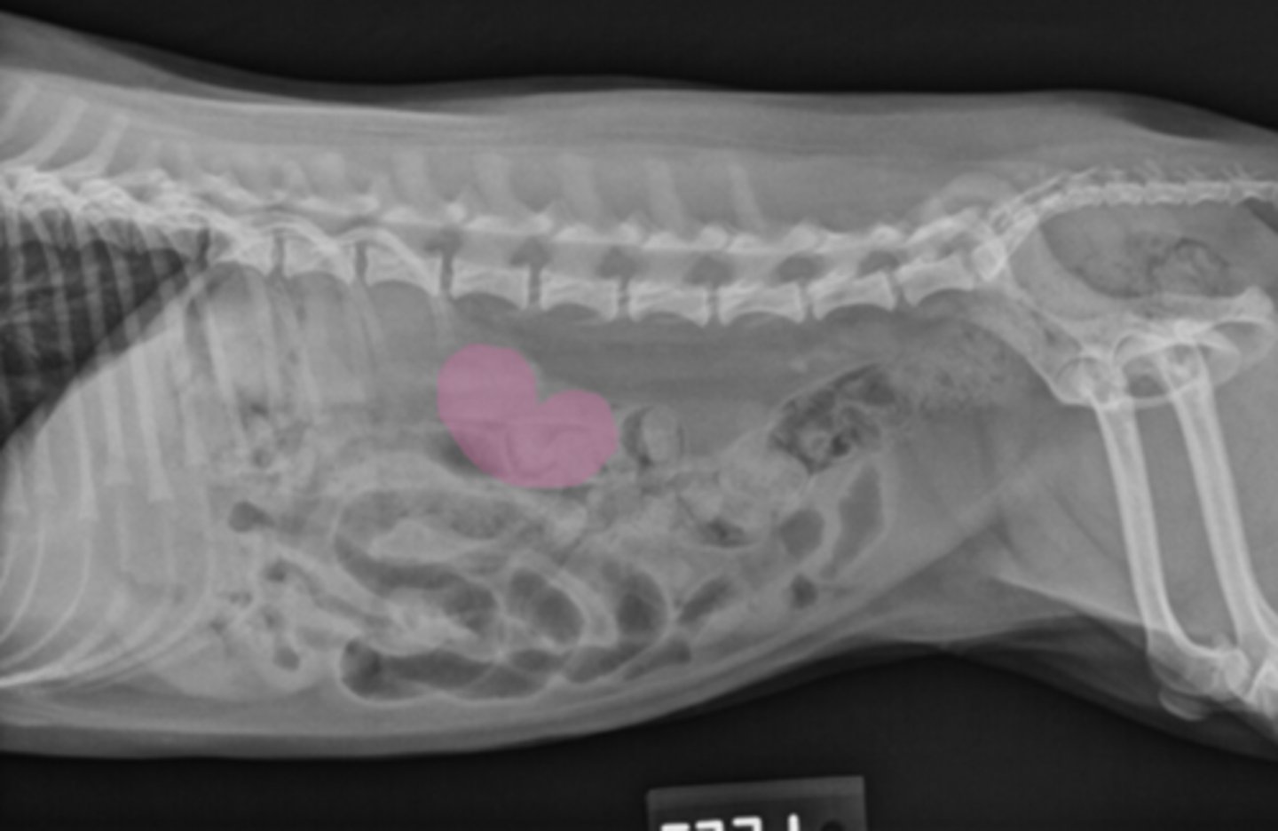
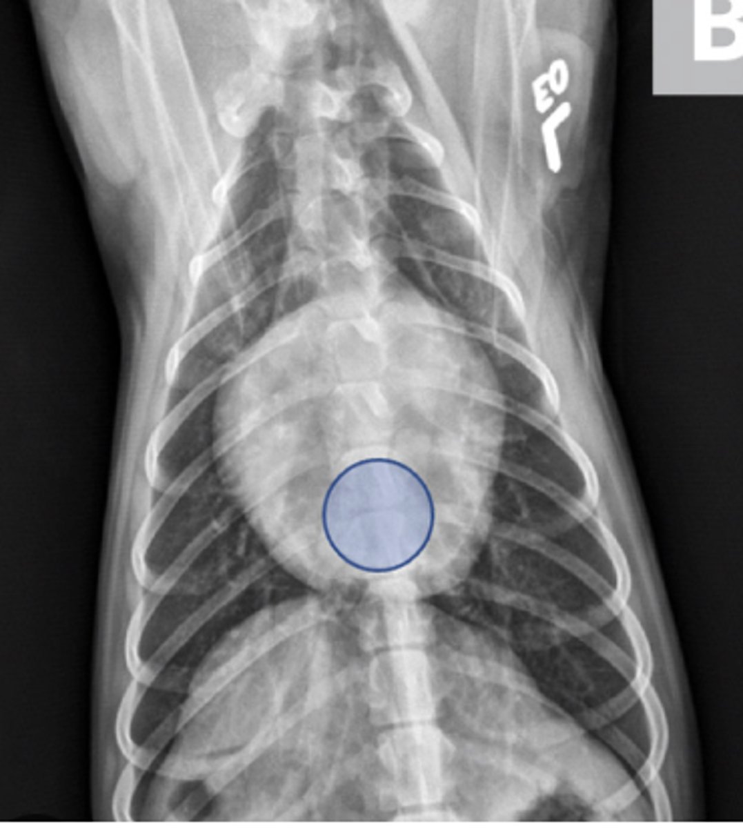
GDV
10 yo lab with abdominal distention, panting- what has caused this?
4 hours
What is the Max time for the stomach to empty?
Navicular syndrome
A Horse lame in four digits- what has caused this?

Fat
What are these opacities on the cat?
Dorsal and lateral
Where to put the marker for equine fetlock radiographs?
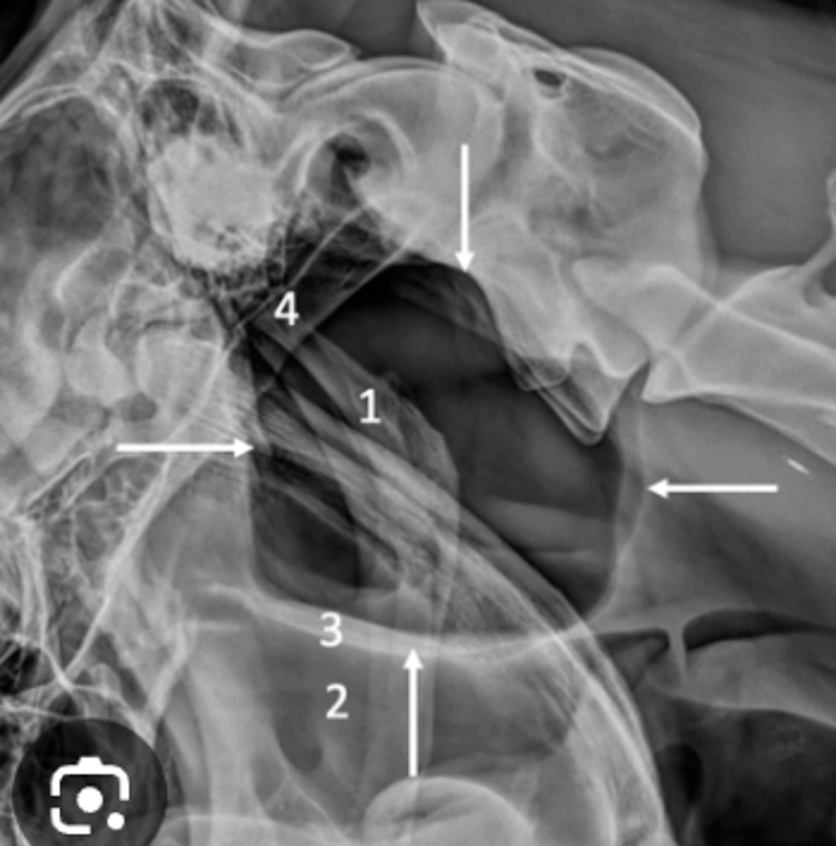
Guttural pouch
What structure is this in a horse?
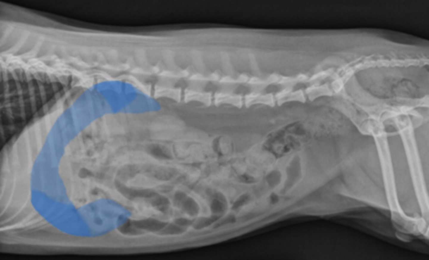
Caudal displacement of the stomach
Common with a large liver
Free gas
What is an indication for taking a horizontal beam radiograph?
adrenal gland
What organ is hard to see on radiographs?
Ureters
What is another organ that is hard to see on radiographs?
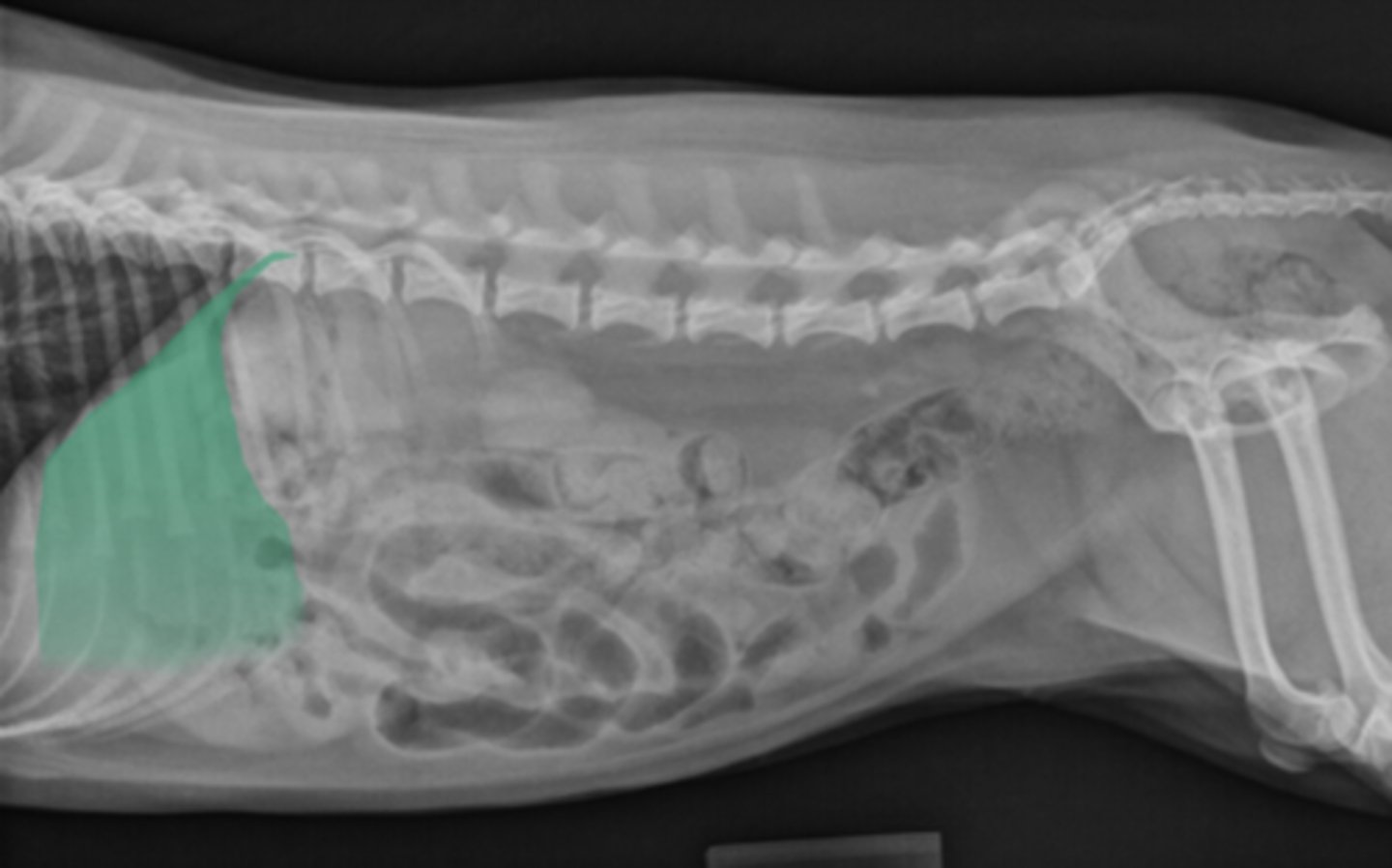
Liver
What organ is this?
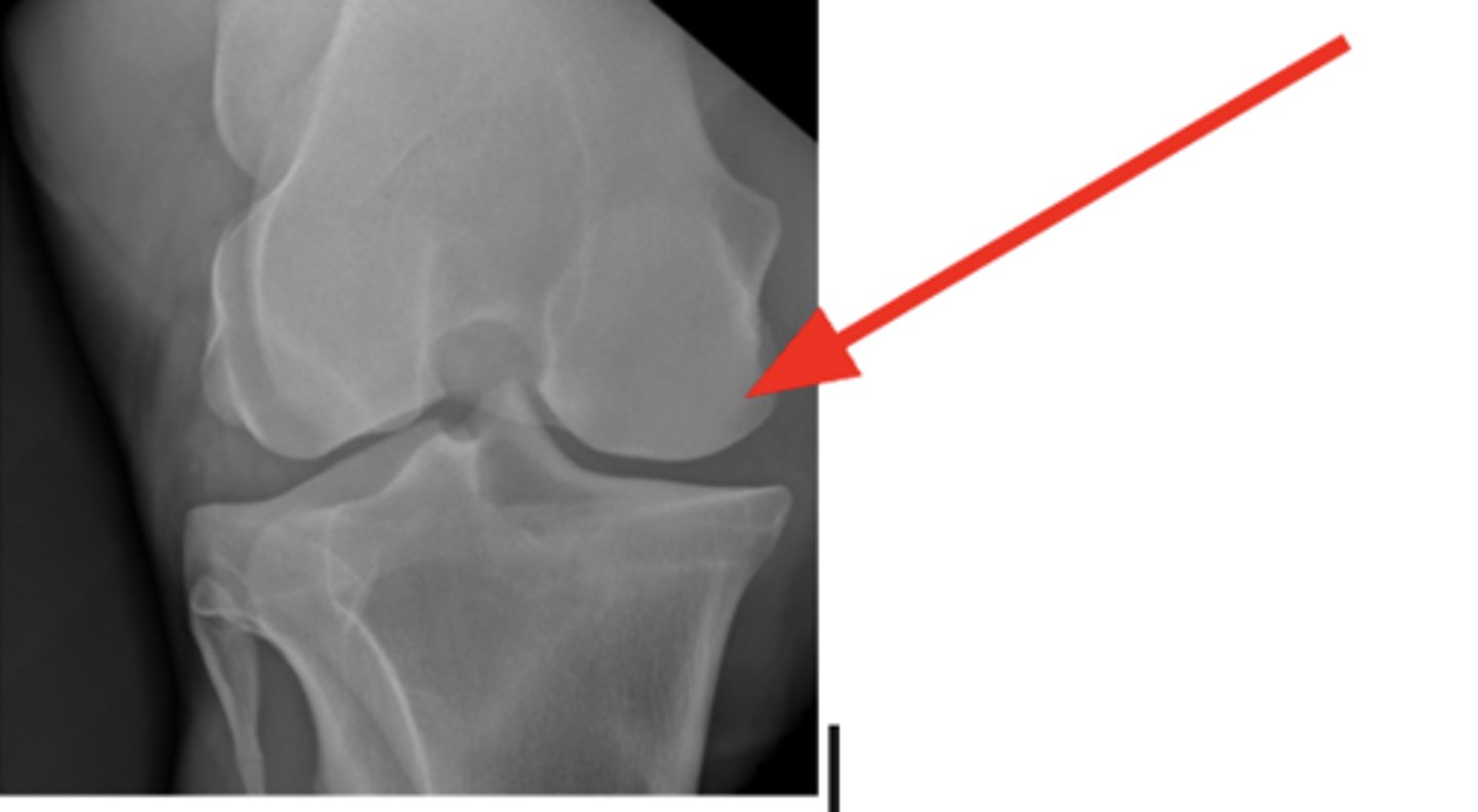
Dorsomedial
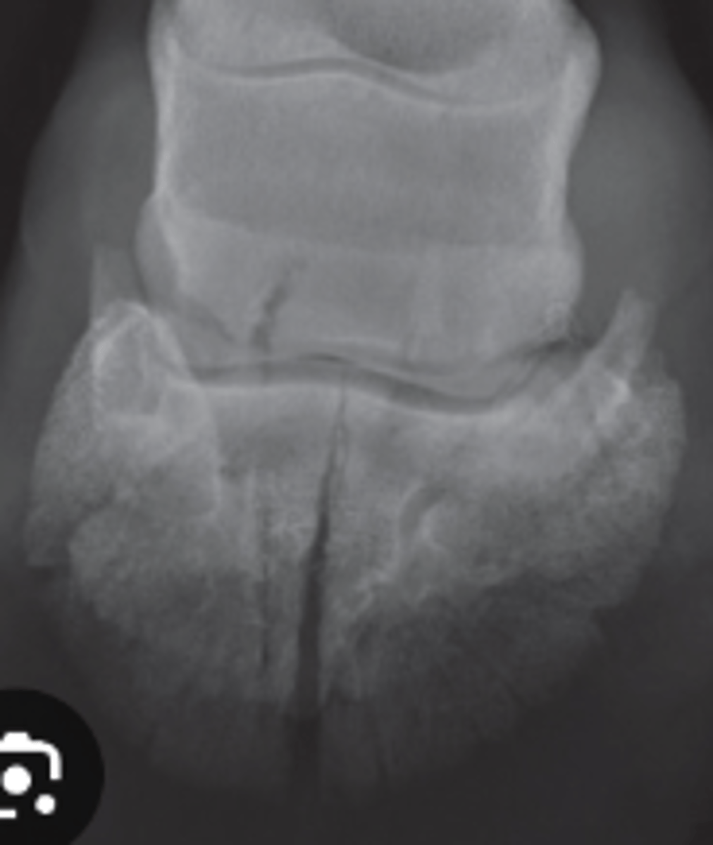
Where is this fragment located?
Type III
What fracture type is this?
Binds to hypoxyapetite
How is T99 taken up in the bone phase?
Costly to client
Not enough contrast material
What are the pitfalls of GI contrast?
Spleen
What organ is this?
OCD in distal tibia/intermediate ridge
2 yo horse w talocrural joint effusion; left hock lesion
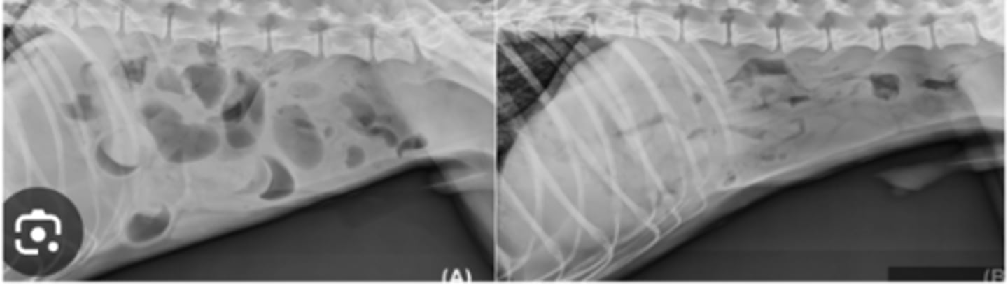
Functional ileus
Dog vomiting and after radiographs a second radiograph reveals gas has decreased- what kind of ileus is this?
Accumulates at bone turnover sites
uses: 18F-sodium fluoride (18F-NaF) and 18Ffluorodeoxyglucose (18F-FDG)
What is the mechanism of PET scan?
Hepatomegaly
What is an enlarged liver called?
Gastroenteritis
X-rays were taken and then taken again hours later, gas was passed. What is this?
CT scan
What Imaging is used for repairing fractures?
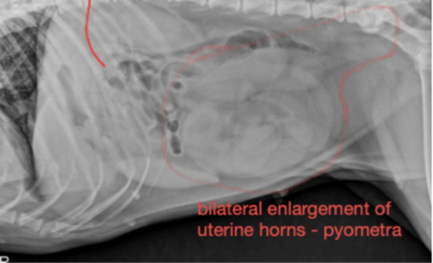
Pyometra
Female dog with vaginal discharge, vomiting, lethargy-what has caused this?
Megaesophagus
What is the most common radiographic abnormality associated with persistent right aortic arch in a dog?
Cardiomegaly from left atrial enlargement
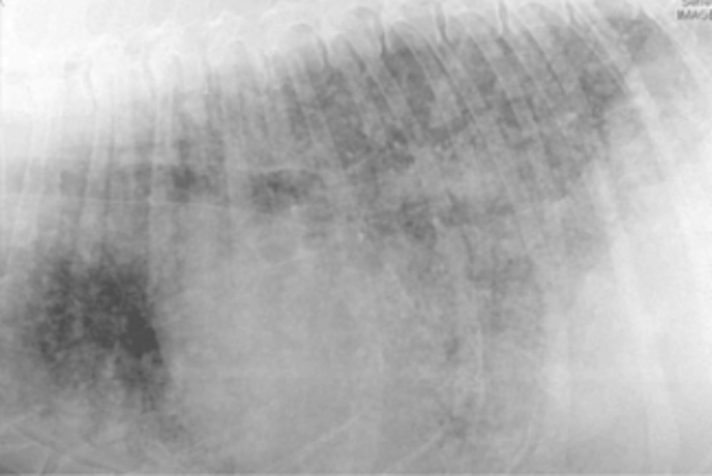
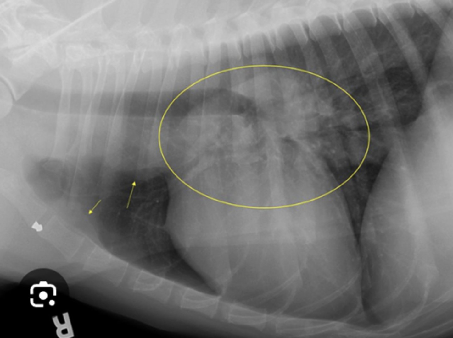
Image of 6yo terrier with cardiac murmur and raspy lung- what do you see?
hepatomegaly/splenomegaly
Which of the following is a characteristic on radiographs of right heart failure?
bronchial
What lung pattern is this?
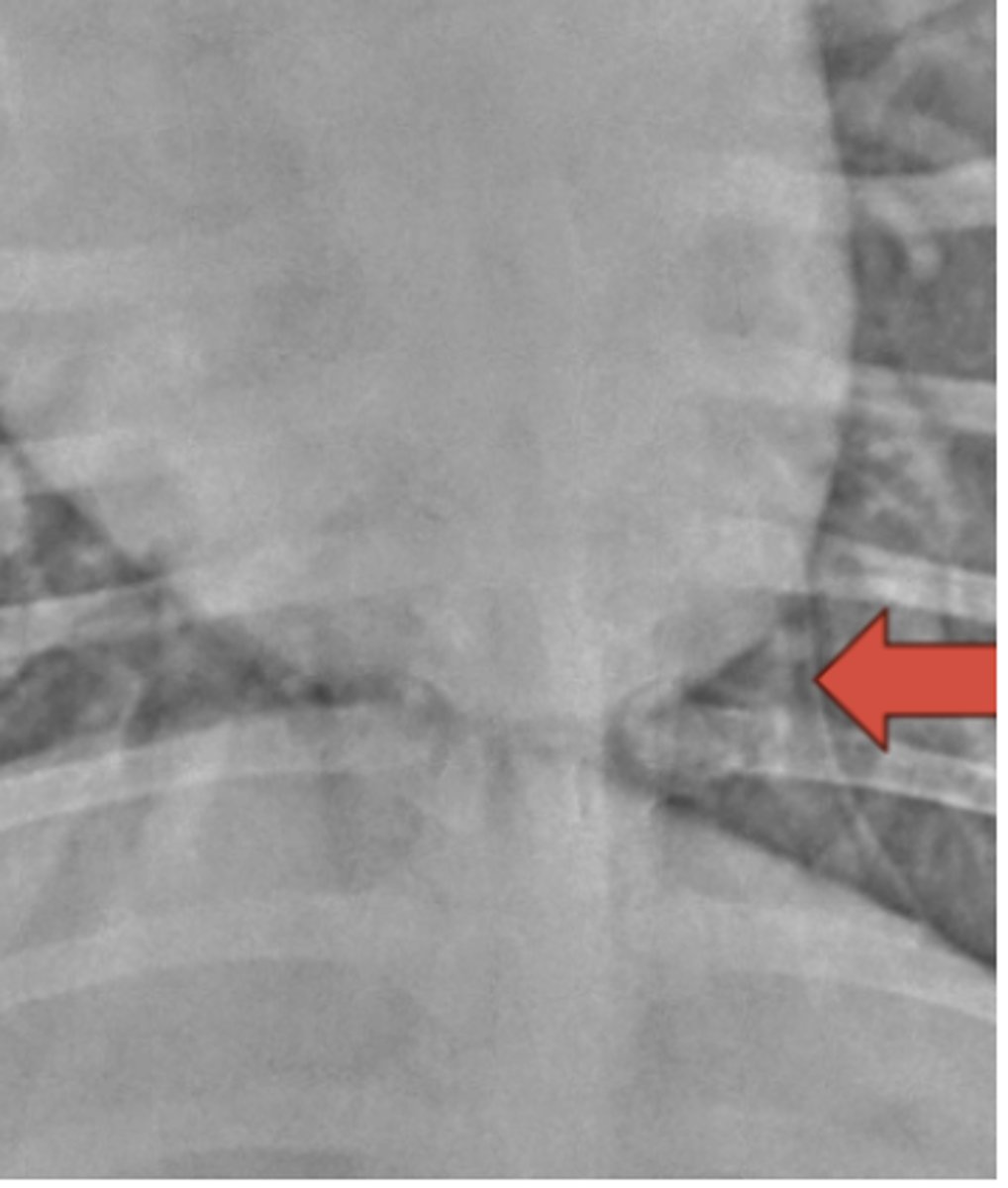
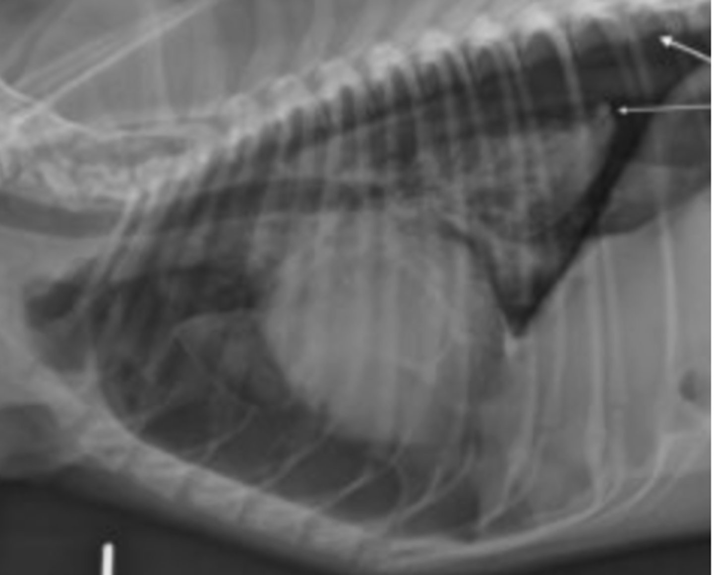
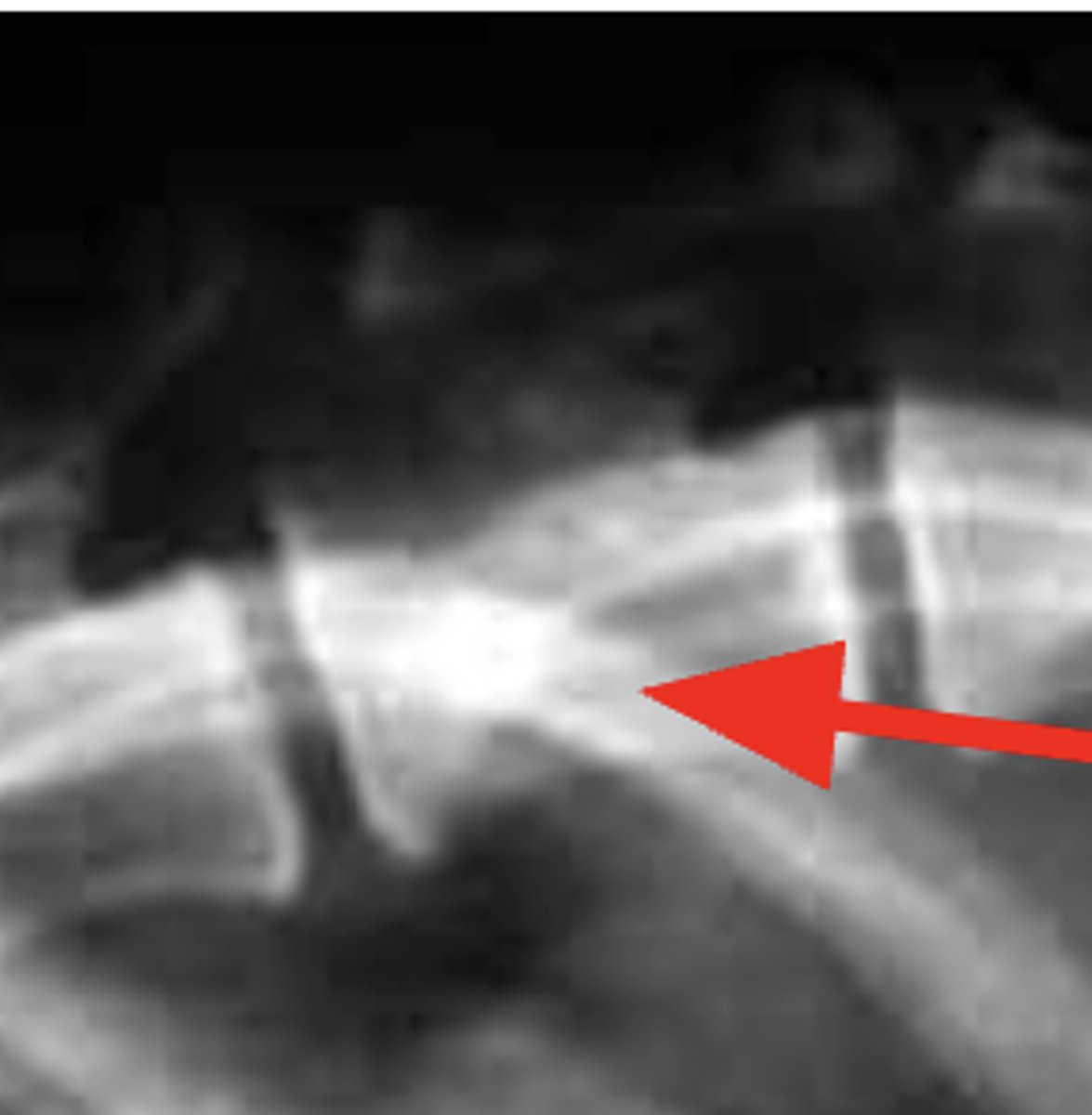
Mediastinal reflection
What structure is indicated by the arrow?
Collimate to the thorax only, pull the thoracic limbs cranially, line up the rib heads
What changes would you make to this radiograph of a feline?

Structure interstitial
What lung pattern is this?
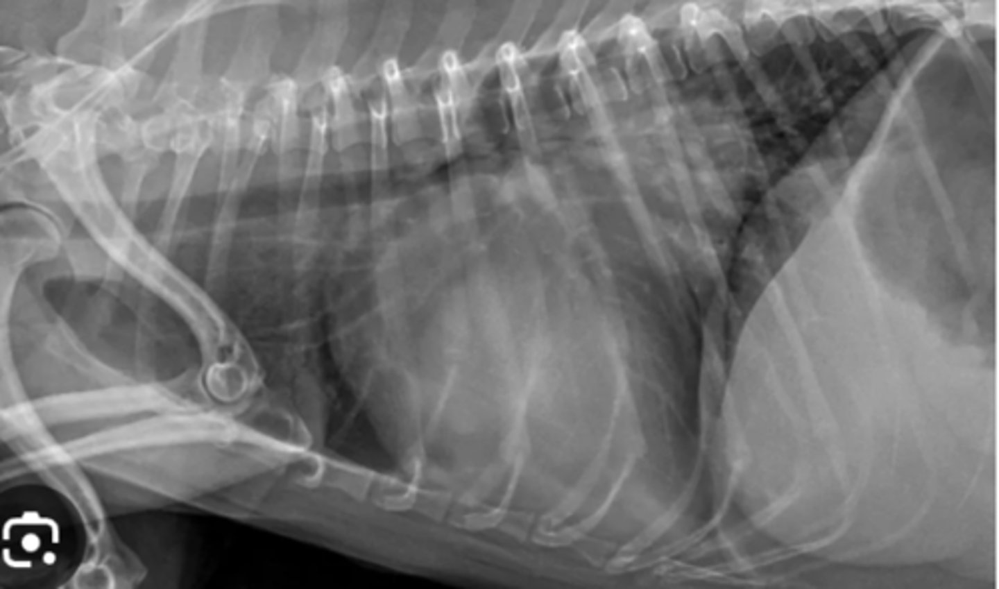
Pneumothorax
Dog with dyspnea and radiograph shows heart lifted dorsally- what has caused this?
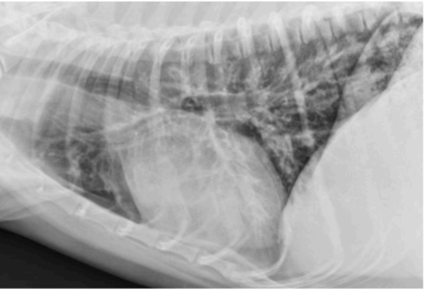
Use this image with this question "Rocky', a 4-year-old Gordon Setter, is presented with a rapid onset of dyspnea. Lung sounds are decreased. The owner is not much help but says "Rocky' was fine yesterday. Only one radiographic view is available, because the patient is distressed. What is your diagnosis based on this lateral radiograph?
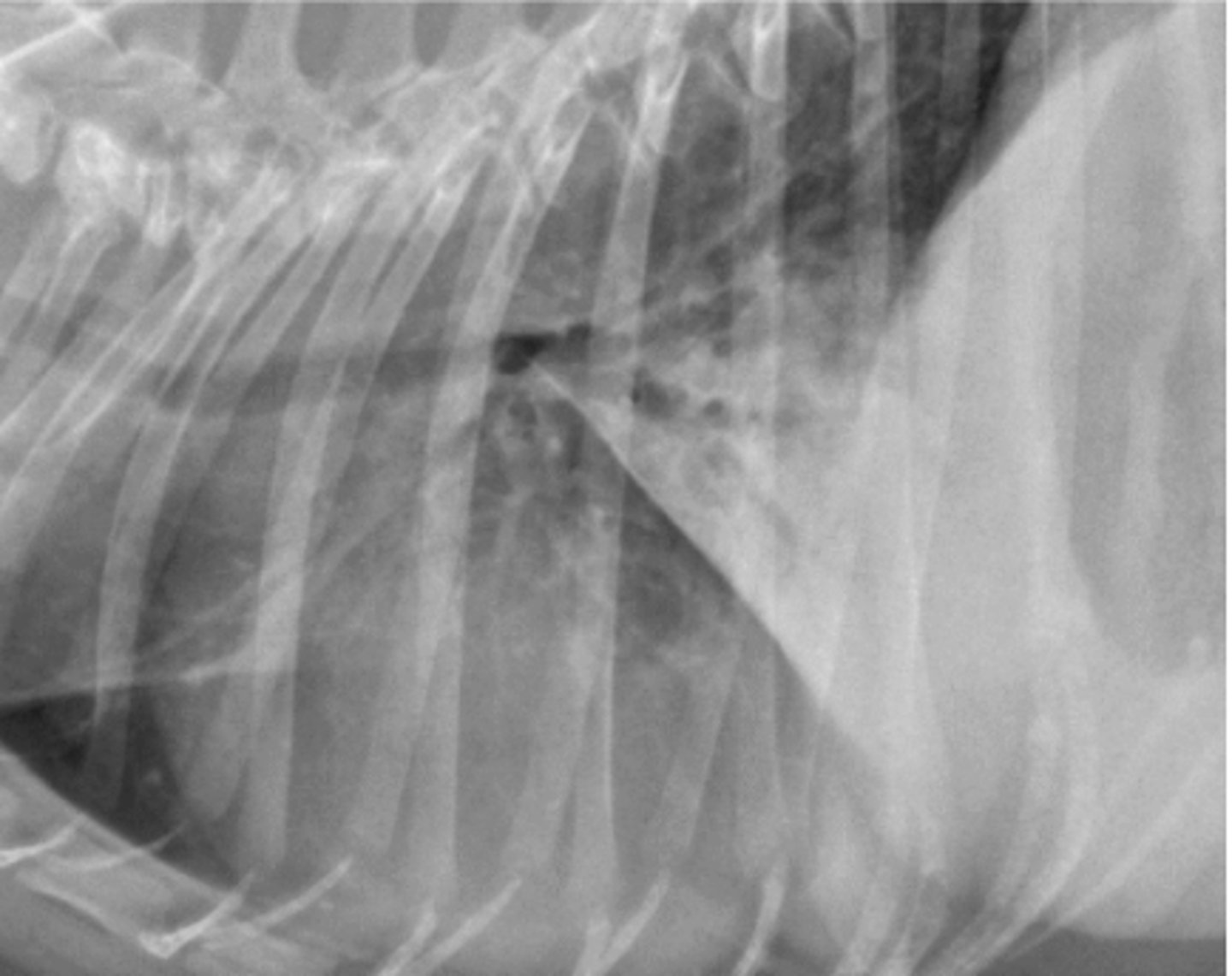
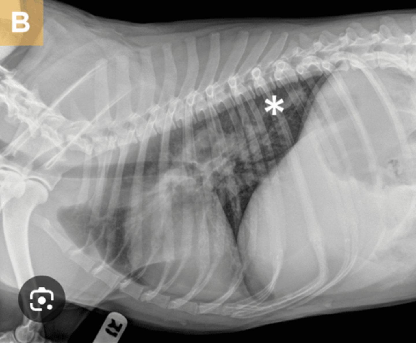
Pleural effusion
Image where it's hard to see heart; what is the principle finding?

Greater curvature of caudo-dorsal heart margin
Splitting of mainstem bronchi
In regards to left atrial enlargement, which of the following can be seen on a radiograph?
Generalized cardiomegaly
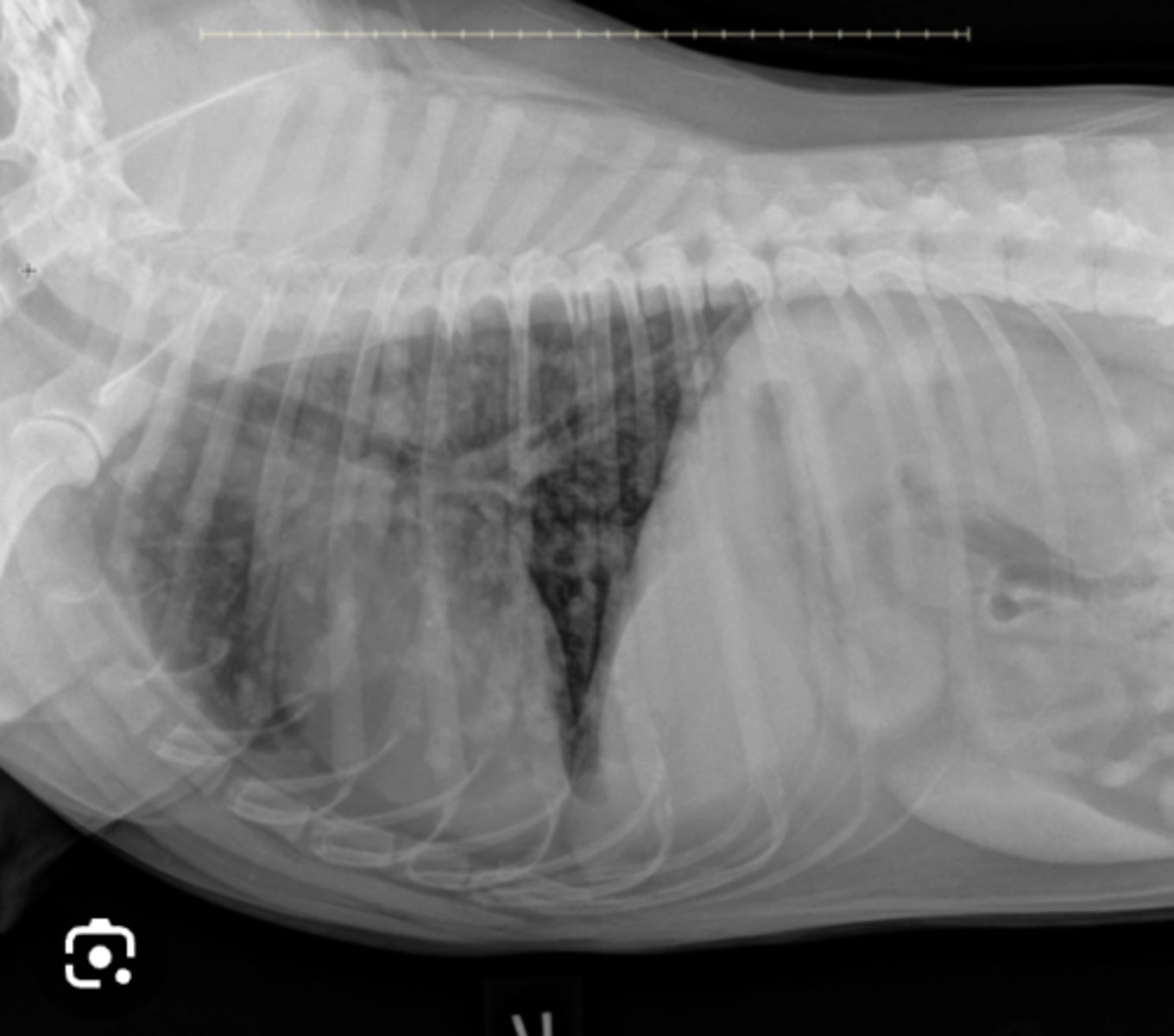
Use this image with this question. Using the provided feline images, how would you describe the cardiac silhouette?
Megaesophagus
Patient with vomiting and regurgitation shows this radiograph- what do you see?
Bronchial
What Lung pattern of young cat with respiratory signs is this?
<2.5
Which is the appropriate size of a feline cardiac silhouette?
Tracheobronchial lymph node
What lymph node is this?
Lobar signs
What is a characteristic of an alveolar pattern?
Tortuous pulmonary arteries
What radiographic finding of this dog with Heartworm disease?
Metastatic disease
Use this image for this question. A 12-year-old canine neutered male patient presents for inappetence, labored breathing, and weight loss. Based on the lung pattern in this thoracic radiograph, and the clinical history, what is your radiographic interpretation?
Increased horizontal location of the heart
What is a normal variation of a geriatric cat?
seen on the image
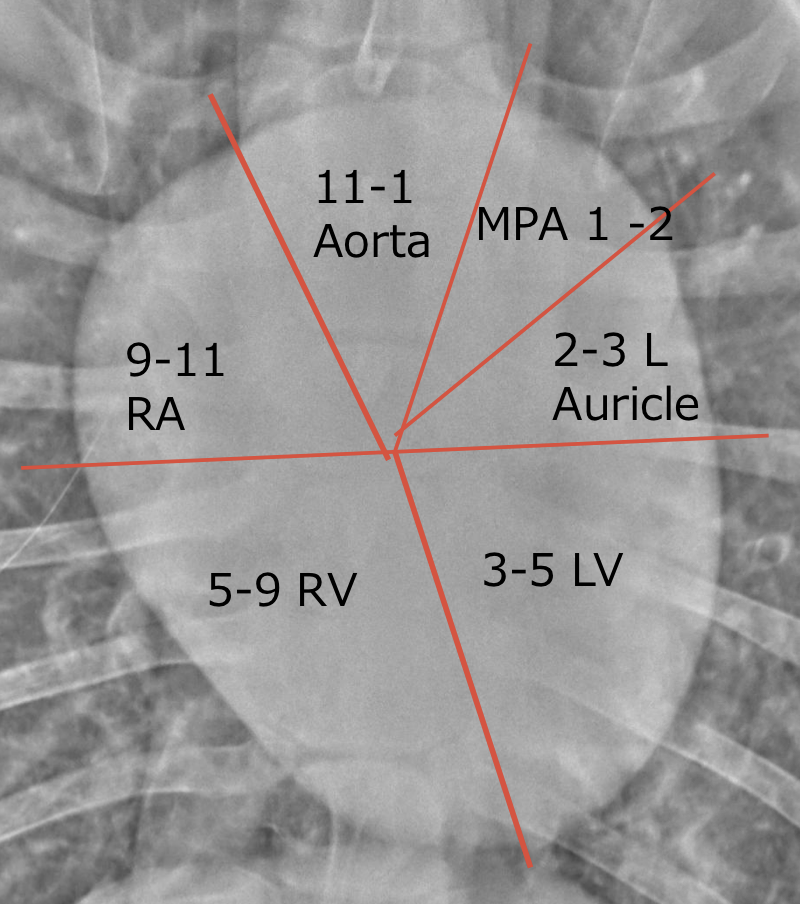
Where is the left ventricle
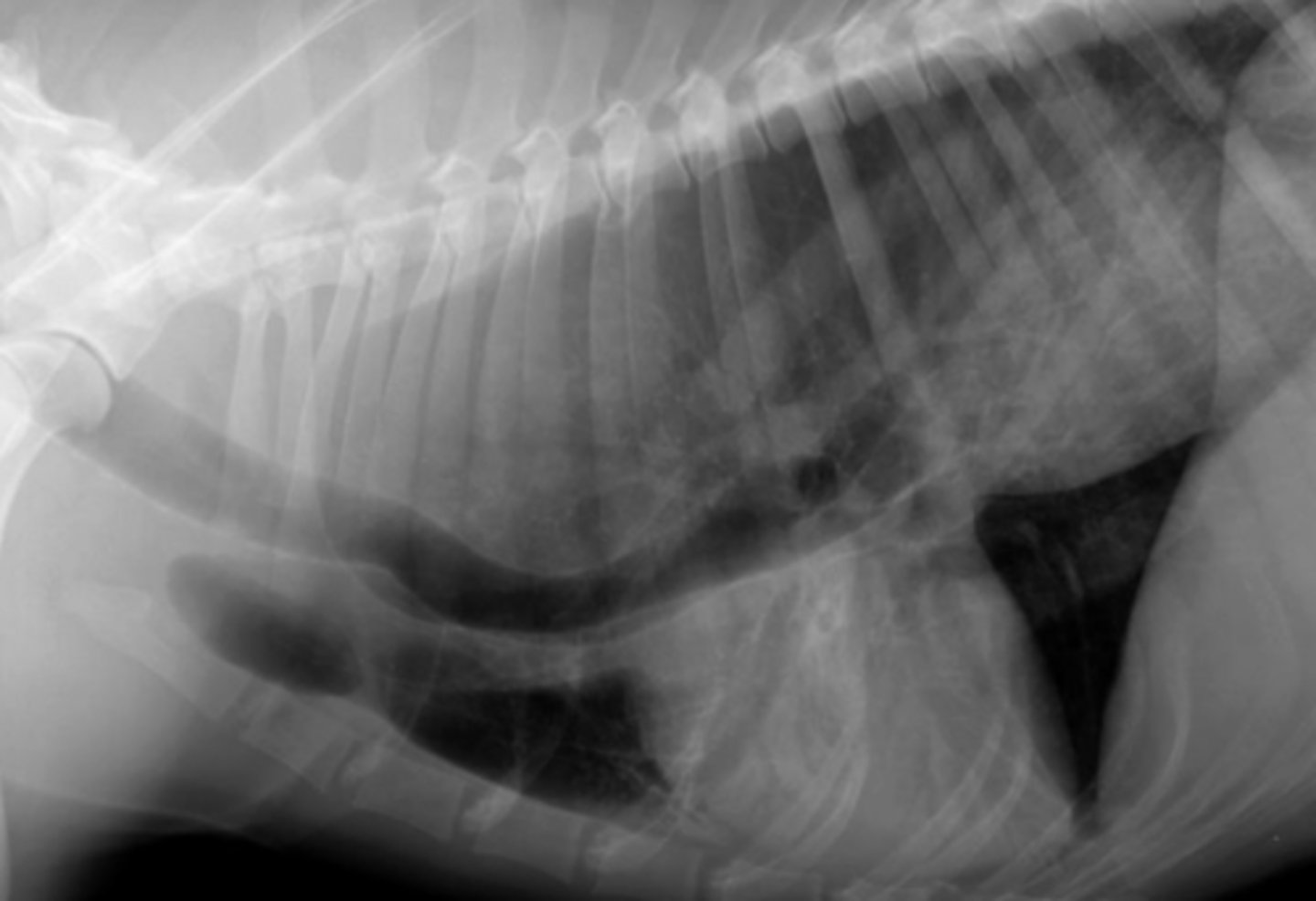
megaesophagus
What can cause ventral displacement of the trachea?
3
How many radiograph views for the thorax?
Pulmonary artery enlargement
What radiographic changes are seen with heartworm disease?
Dilated cardiomyopathy
What is a Common cause of a globoid heart?
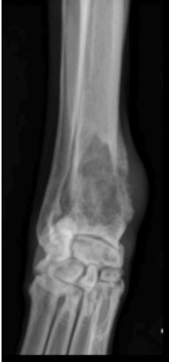
Panosteitis
10 y/o German Shepherd comes in with lameness on the right hind leg; what is the radiographic finding?
Hypertrophic osteodystrophy
6 month old Lab with distal limb swelling and the metaphysis is warm to the touch.- What is wrong with it?

Tibial tuberosity
What anatomical location is indicated by the arrow?
Alveolar
Which lung pattern prevents visualization of blood vessels?
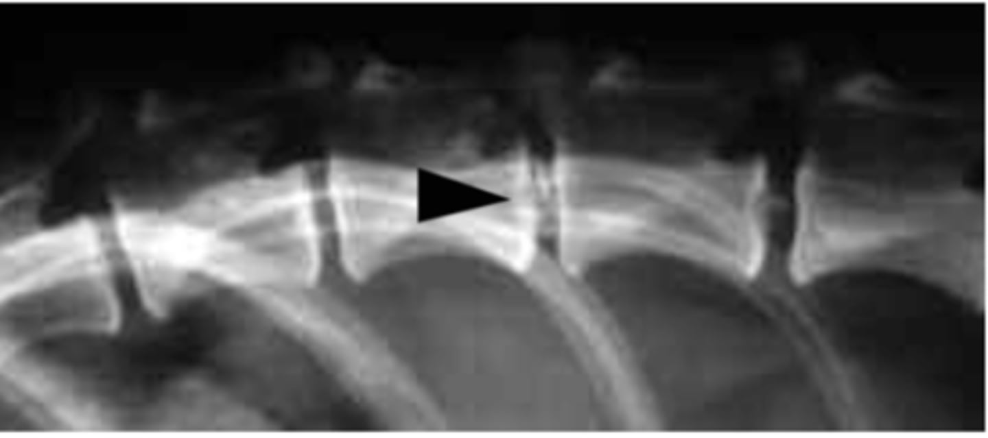
Mineralization of intervertebral discs
What is going on with this radiograph?
Comminuted
What fracture type has bone in multiple pieces where you can't find and definite origin of fracture?
Spondylosis deformans
What abnormality is shown in this radiograph of the spine?
They should be aligned in a straight line
In a VD radiograph, how should you align the spinous processes?
Vertebral body
What is the anatomical structure indicated?
Long zone of transition
What is characteristic for an aggressive bone lesion?
Complete
Closed
Comminuted
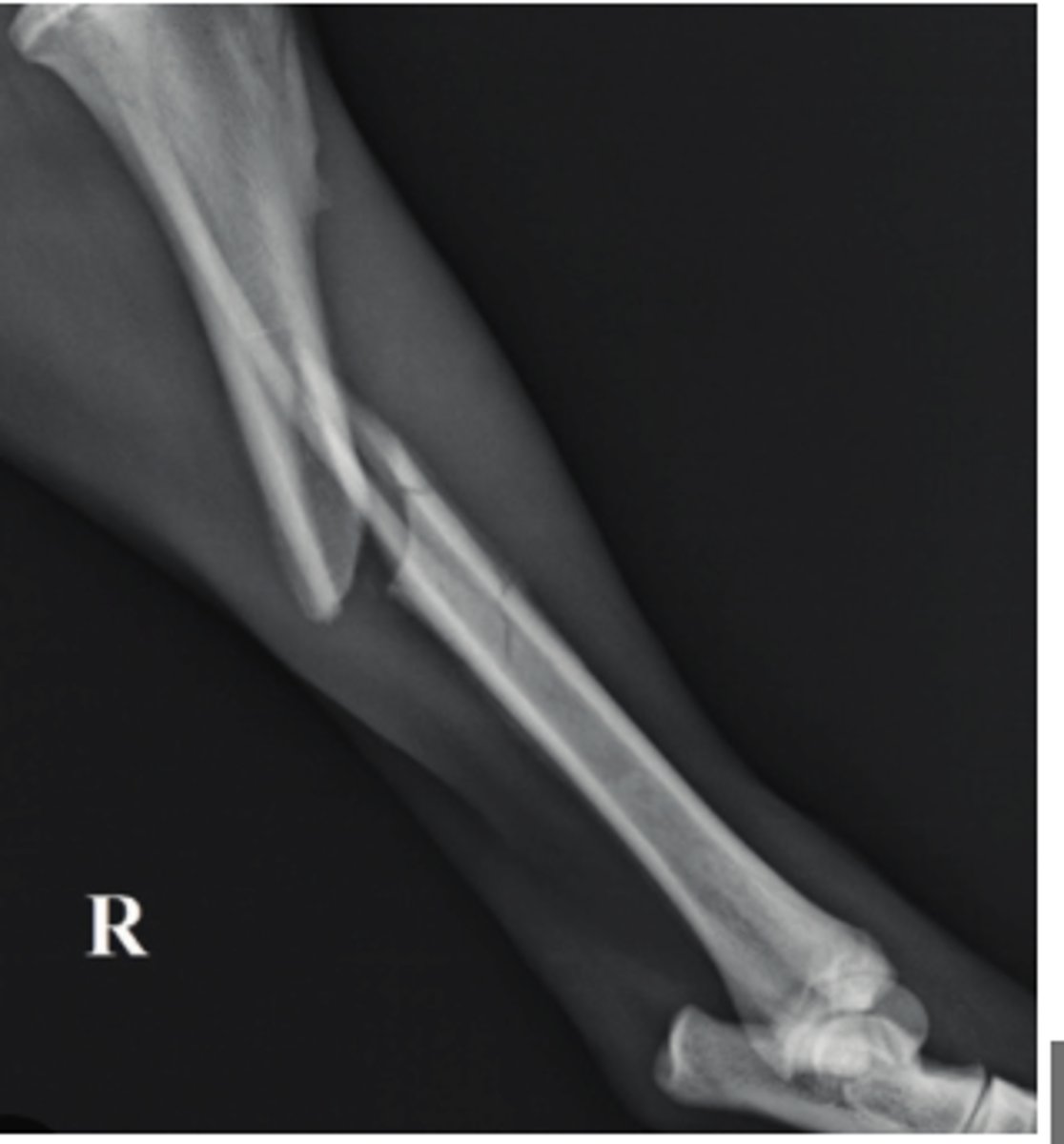
What describes this bone fracture
For viewing lesions of the spinal column you may have missed on radiograph
Indication for myelography?
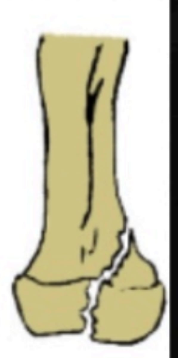
Type IV
Which Salter-Harris fracture is pictured?
General enlargement with left atrial enlargement
8 y/o FS german shorthair pointer presents with exercise intolerance after hunting; what do these radiographs show about the cardiac silhouette?
5 mm
What is the minimum size for a nodule to show up on radiographs?
Primary bone tumor
6 y/o dog lame on the limb shown- what kind of tumor is this?
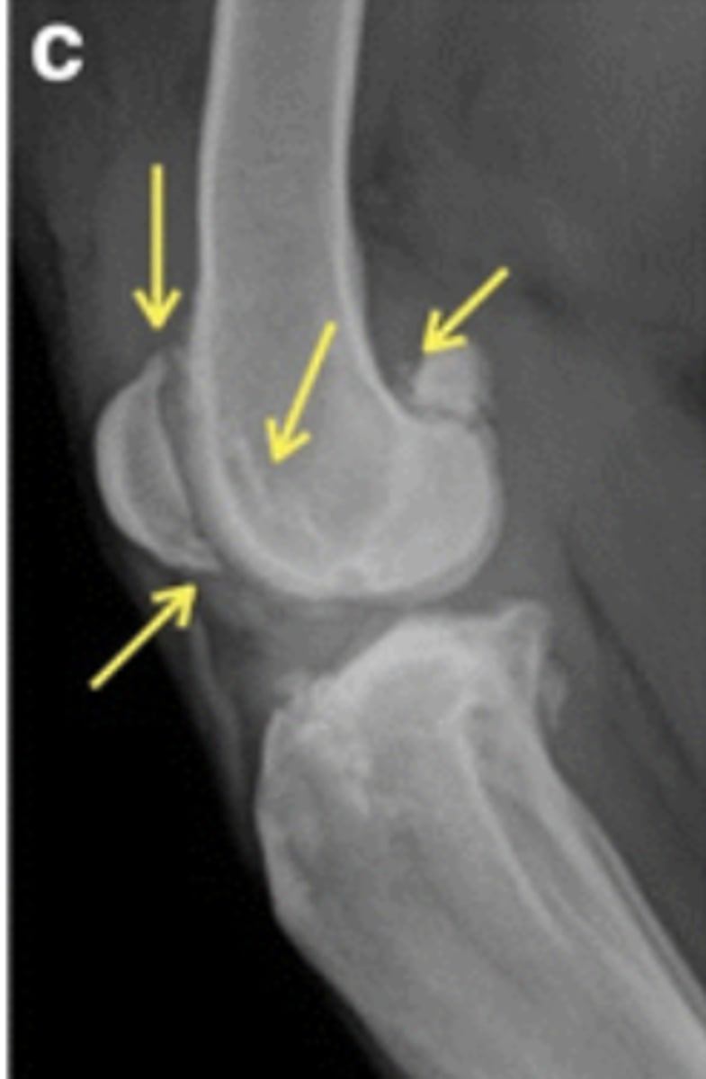
Mineralized cartilaginous flap
What is a Common finding of OCD?
Caudal portion of humeral head
Location of OCD in the shoulder joint
Mastiff
What breed is most common to have a retained cartilaginous core?
Incongruity
What radiograph condition a result of premature physeal closure?
Ill-defined margins
Irregular periosteal proliferation
Cortical lysis
Define an aggressive bone lesion?
narrowed intervertebral space
What is seen in IVDD?
Osteophytes
A Wheaton terrier with chronic lameness shows this:
To evaluate ligamentous injury
What is the primary reason for stress radiographs of a joint?
asymmetric head & unilateral epistaxis
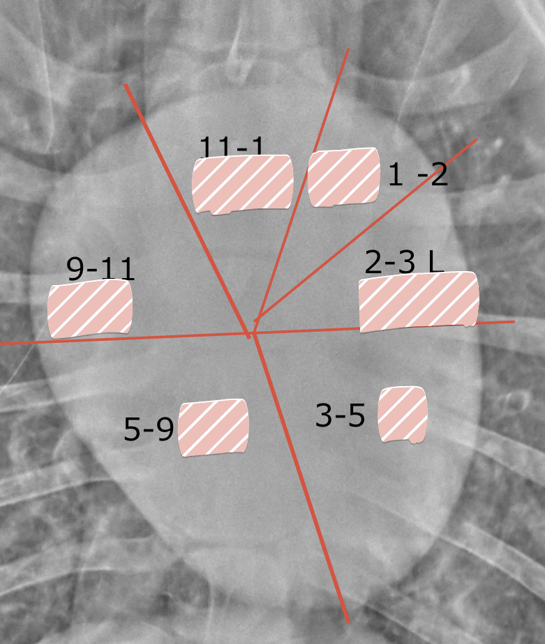
What 2 would be indications of the skull radiographs?
Femoral condyles
With lateral radiographs of stifle joint, what should be aligned?
Core lesion
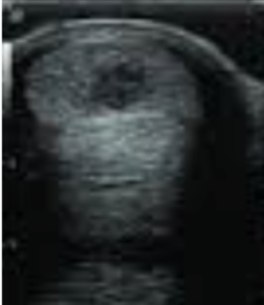
7 year old thoroughbred presents with acute lameness, swelling of metacarpal?; ultrasound shows this- what is this?
Mucosa to lumen
How do you measure the small intestine for dilation?