CNBY 645 Exam 3: Paper 1
1/39
There's no tags or description
Looks like no tags are added yet.
Name | Mastery | Learn | Test | Matching | Spaced |
|---|
No study sessions yet.
40 Terms
Tumor microtubes
Long cellular processes extended by glioma cells and riven by pathways involved in neurodevelopment and neuroplasticity
Crucial for invasion and proliferation
Connect single tumor cells to a functional communicating network
Unknown involvement in the neuron-glioma interactions
Paracrine mechanisms
Cell-to-cell communication in which a signal is released by a cell and acts on a neighboring cell
Autocrine mechanisms
cell-to-cell communication in which a signal is released by a cell and acts on the same cell that produced it
Neuroglinin-3 neuronal protein (NLGN3)
A neuronal cell adhesion molecule (CAM) that is a key regulator of synaptic function, found at both excitatory and inhibitory synpases
Crucial for synapse development and forms connections between neurons
Can be secreted by active neurons to promote tumor cell proliferation
NGS
neurogliomal synapses
Signal travels from neuron synapse to glioma cell
Electron microscopy (immuno-electron microscopy)
A technique that uses antibodies labeled with electron-dense markers to visualize the precise location of molecules within a cell or tissue using a transmission electron microscope
What type of tumor studying? Cells, tissues, xenografts?
Studied glioblastoma cells and used xenografts of patient-derived human GB cell lines into mice
dSTORM
Direct stochastic optical reconstruction microscopy
A super-resolution imaging technique
Achieves ~20nm resolution, far beyond standard light microscopy
Works by turning fluorescent molecules “on and off” randomly and reconstructing their precise location
In the paper, dSTORM was used to visualize synaptic proteins at glioma tumor microtubules (TMs) and confirm the presence of bona fide postsynaptic structures
HOMER1, HOMER2, and HOMER3
Postsynaptic density scaffold proteins found at excitatory (glutamatergic) synapses
Role:
Organize and cluster receptors (like AMPARs and mGluRs)
Anchor them to postsynaptic density
In the paper: HOMER1/2/3 served as markers of postsynaptic specialization on glioma TMs
Detection of HOMER proteins in glioma TMs confirmed that glioma cells form postsynaptic-like sites at neuron-glioma synapses
VGLUT1 (vesicular glutamate transporter 1)
A presynaptic protein that loads glutamate into synaptic vesicles
Marker of glutamatergic presynaptic boutons (where glutamate is released)
In the paper: VGLUT1 puncta were found were found aligned with glioma postsynaptic AMPARs (GluR1 subunits), proving that the input gliomas receive is glutamatergic
AMPAR
A glutamate receptor that mediates fast excitatory neurotransmisiion
Plays a crucial role in learning and memory by enabling synaptic plasticity
Glutamate
An excitatory transmitter in the CNS
5-ALA
5-aminolevulinic acid
Is a fluorescent dye that causes tumor cells to glow and helps distinguish between cancerous and healthy tissue
NMDAR
A glutamate receptor that opens a channel allowing calcium influx, triggering a cascade of events that result in long-term changes in synaptic strength
Slice culture
A technique that uses a thin slice of tissue to analyze for research purposes
Co-culture
A technique in which two or more different cell types are grown together in the same culture to study their interactions
Patch clamp
Uses a glass micropipette filled with electrolyte solution to record the electrical currents and voltage across a small patch of cell membrane or across a cell
Reveals insights into neuronal activity, cellular function, and the effects of drugs
sEPSCs
spontaneous excitatory post-synaptic currents
Small, rapid changes in a neuron’s membrane potential that occur spontaneously and increase the likelihood of the neuron firing an action potential
Arises from random, localized release of neurotransmitters
Often mediated by non-NMDA receptors, which cause an influx of positive ions and depolarize post-synaptic cell
Helps explain fundamental synaptic transmission, the role of different receptors and how neurons respond to normal activity
CNQX
Cyanquixaline
A competitive AMPAR antagonist
TTX
Tetrodotoxin
A powerful neurotoxin (often found in pufferfish) that blocks voltage-gated sodium channels in nerve and muscle cells
NASPM
1-napthylacetyl spermine
Acts as a selective antagonist that inhibits calcium-permeable AMPPA receptors
Spontaneous slow inward currents (SICs)
Excitatory events in neurons caused by the release of glutamate from astrocytes that activates extrasynaptic NMDARs
Have slow kinetics (rise/decay time) and large amplitude
Thought to synchronize small groups of neurons and regulate synaptic plasticity
Blocked by NMDA receptor antagonists and illustrate a NMDA receptor dependency
Glioma calcium currents
These are changes in electrical charge inside glioma cells caused by calcium flowing through ion channels, particularly calcium-permeable AMPA receptors (AMPARs)
In glioma, calcium currents:
Signal through tumor cell networks (via tumor microtubes)
Promote cell invasion and proliferation
Are triggered by neuronal activity at neurogliomal synapses (NGS)
Multiphoton Laser Scanning Microscopy (MPLSM)
A powerful live imaging technique that uses two-photon or multiphoton excitation
Allows researchers to look deep into the living brain of mice with minimal damage
In this study: used to record calcium activity in glioma cells in real time through the cranial window
Cranial window
A surgical procedure where part of the skull is removed and replaced with a transparent cover
Enables repeated in vivo imaging of the brain (using MPLSM)
In this study: it was used to visualize glioma cells in live mice, track their calcium signals, and measure invasion velocity
Channelrhodopsin-2 (ChR2)
A light gated ion channel from algae used in optogenetics
When expressed in neurons, it allows researchers to control neuronal activity with blue light
In this paper: stimulating ChR2-expressing neurons with blue light increased neuronal firing —> drove synchronized calcium signals in glioma cells
Epileptiform activity- gabazine
gabazine is a drug that blocks GABA-A receptors, which normally mediate inhibitory (calming) signals in the brain
Blocking them induces hyperactive, seizure-like activity (epileptiform activity)
In this paper: Gabazine was used to mimic epileptic conditions in vivo —> this increased glioma calcium currents and enhanced tumor network activity
Anesthesia
Isoflurane impacts on calcium networks: isoflurane is commonly used anesthetic
In glioma-bearing mice:
Isoflurane suppressed calcium transients and reduced coordinated glioma network activity
Also slowed glioma cell invasion in vivo
Shows how brain activity levels directly impact glioma cell behavior
Invasion velocity- repetitive in vivo imaging
Researchers tracked how fast glioma cells moved (their invasion velocity) using time-lapse in vivo imaging through the cranial window
Glioma cells with functional AMPARs and neuronal input invaded faster
Anesthesia (isoflurane) or genetic AMPAR blockade (GluA2-DN) reduced invasion velocity
GluA2-DN-GFP
Dominant-negative (DN) mutant version of GluA2 (tagged with GFP for visualization) was expressed in glioma cells
This blocks AMPAR signaling in those cells
Result: Glioma cells with GluA2-DN-GFP showed slower invasion and reduced proliferation compared to control glioma cells
Optogenetic construct
A genetic tool that makes glioma or neuron cells light-sensitive (e.g. by expressing ChR2)
When illuminated, neurons fire —> this increases glutamatergic synaptic activity onto glioma cells
Used to prove that neuronal activity directly stimulates glioma growth and invasion
Perampanel
A clinically approved antiepileptic drug that selectively blocks AMPA receptors
In the study:
Chronic perampanel treatment in mice slowed flioma growth
Reduced glioma cell density increases over time
Suggests repurposing perampanel as glioma therapy
Main goal of study
Hopes to find that neurons and glioma cells form direct, bone fide chemical synapses called neurogliomal synapses (NGS)
Figure 1
Goal: Show that glioma cells physically form bona fide synapses with neurons
Methods: Immuno-EM images
Key takeaway: NGS are consistently formed in incurable human gliomas and mouse models thereof but not in less malignant primary brain tumors, suggesting a specific contribution to the malignant features of astrocytic gliomas, including GB
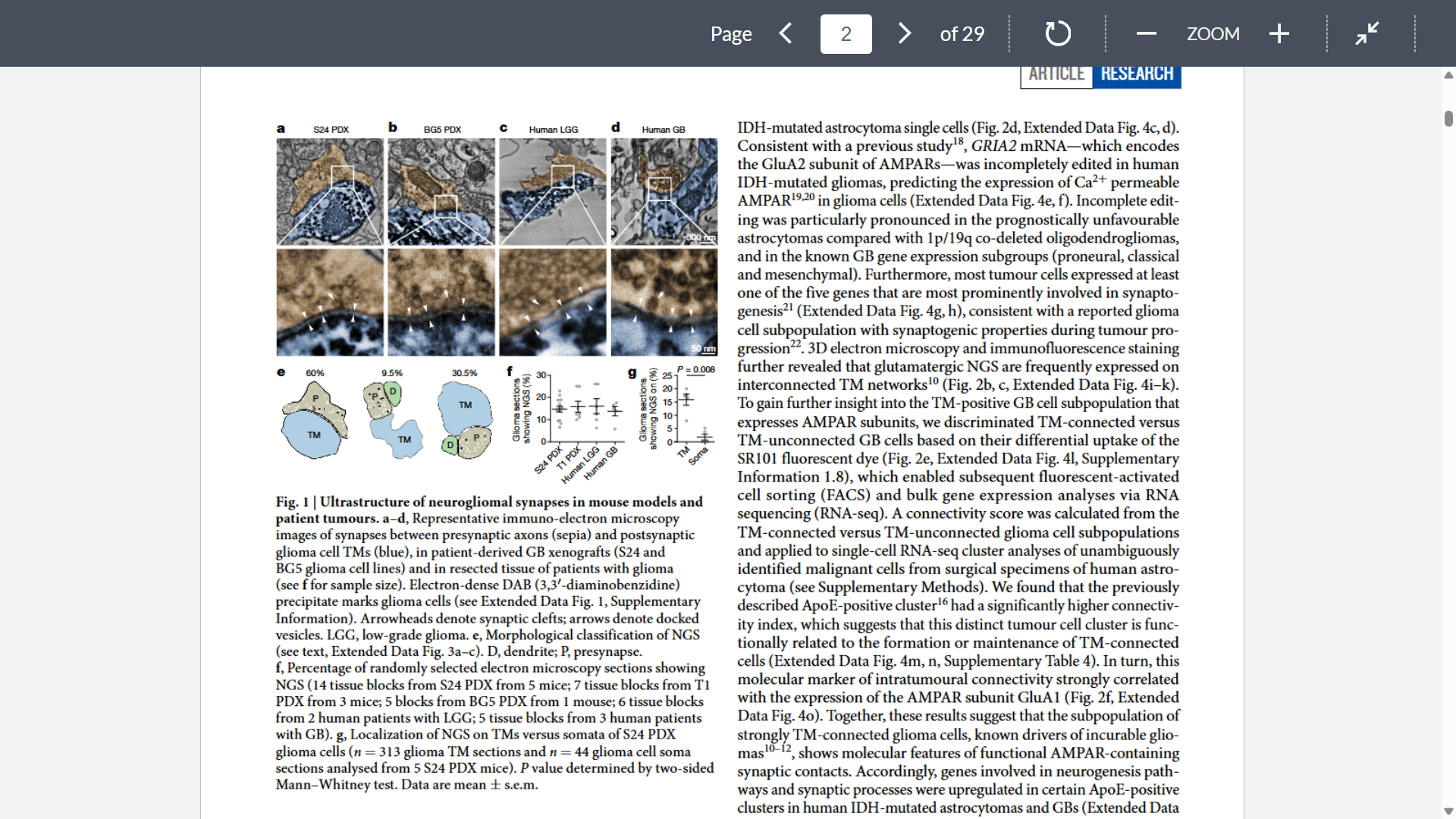
Figure 2
Goal: Show glioma cells express synaptic proteins and AMPA receptors
Methods: dSTORM micoscopy, confocal imaging, sRNAseq, EM
Key numbers: Across xenograft models and resected human GB material >80% of AMPAR signals on glioma TMs colocalized with presynaptic signals
Key takeaway: Glioma cells molecularly organize like neurons at NGS with AMPAR-rich synaptic contacts, especially in TM-connected subpopulations
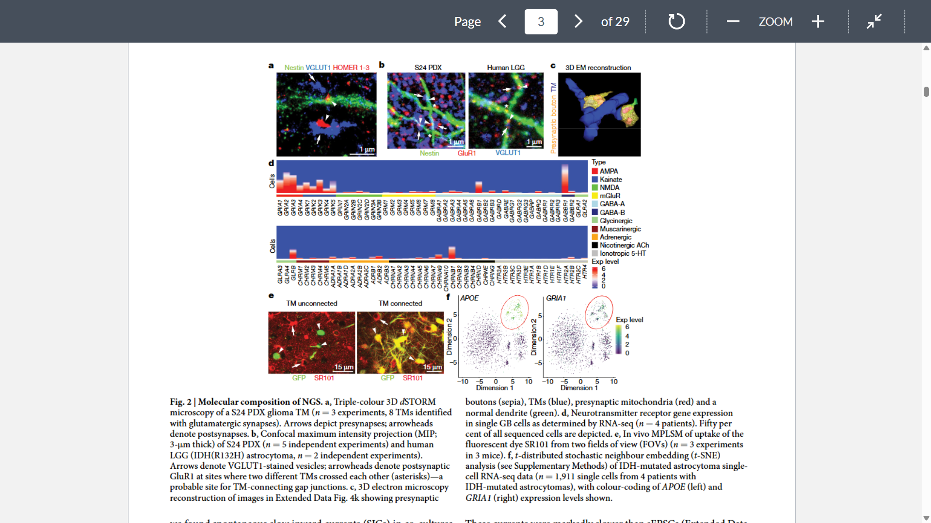
Figure 3
Goal: prove that NGS are functional synapses
Methods: patch clamp, sEPSCs
Key takeaway: all of this data combined with knowledge from previous studies indicates that there is a relevant electrophysiologically heterogeneous functional input of NGS on GB cells
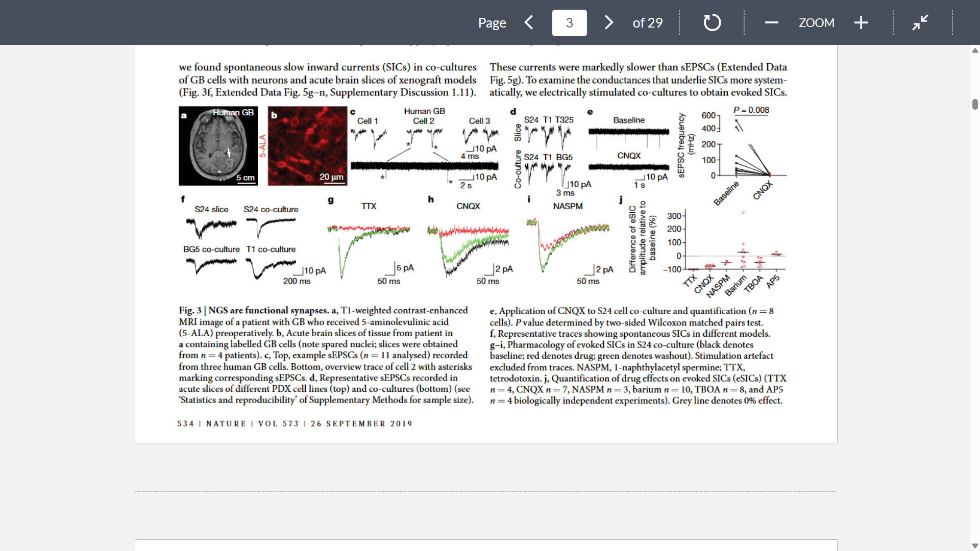
Figure 4
Goal: determine what are the effects in the glioma cells of the NGS and currents
Key takeaway: Calcium can enter GB cells via AMPARs and possible additional conductances and that neuronal activity can cause time-locked and clinically relevant calcium signals in GB cell networks
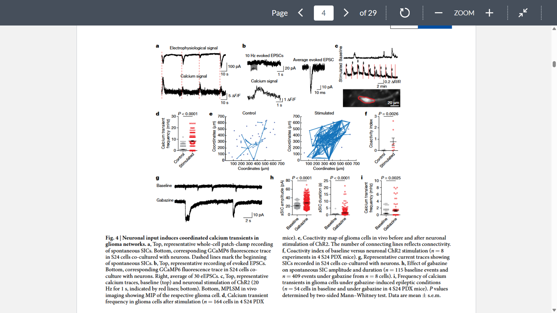
Figure 5
Goal: analyze the consequences of NGS and its effect on tumor growth
Shows that isoflurane treatment visibly slows invasion of tumor microtube-bearing GB cells

Conclusion
Reframes gliomas as active participants in neural circuits
Gliomas hijack CNS synaptic mechanisms by forming function NGS, providing excitatory AMPAR-mediated input to glioma cells; this drives calcium signaling within tumor microbe-connected networks and drives invasion/proliferation
Suggesting seizures in glioma patients may not just be symptom of tumor growth but also actively fuel tumor progression by increasing neuronal activity
Therapeutic: drugs that block synaptic input (like perampanel) may be repurposed for glioma treatment