Anatomy exam 3 - general study
1/31
There's no tags or description
Looks like no tags are added yet.
Name | Mastery | Learn | Test | Matching | Spaced | Call with Kai |
|---|
No analytics yet
Send a link to your students to track their progress
32 Terms

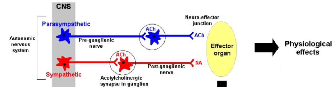
preganglionic neuron
A neuron of the Autonomic nervous system. The cell body is in an autonomic nucleus in the brain or spinal cord (CNS). The axon goes to an autonomic ganglion (PNS) and synapses with a ganglionic neuron.
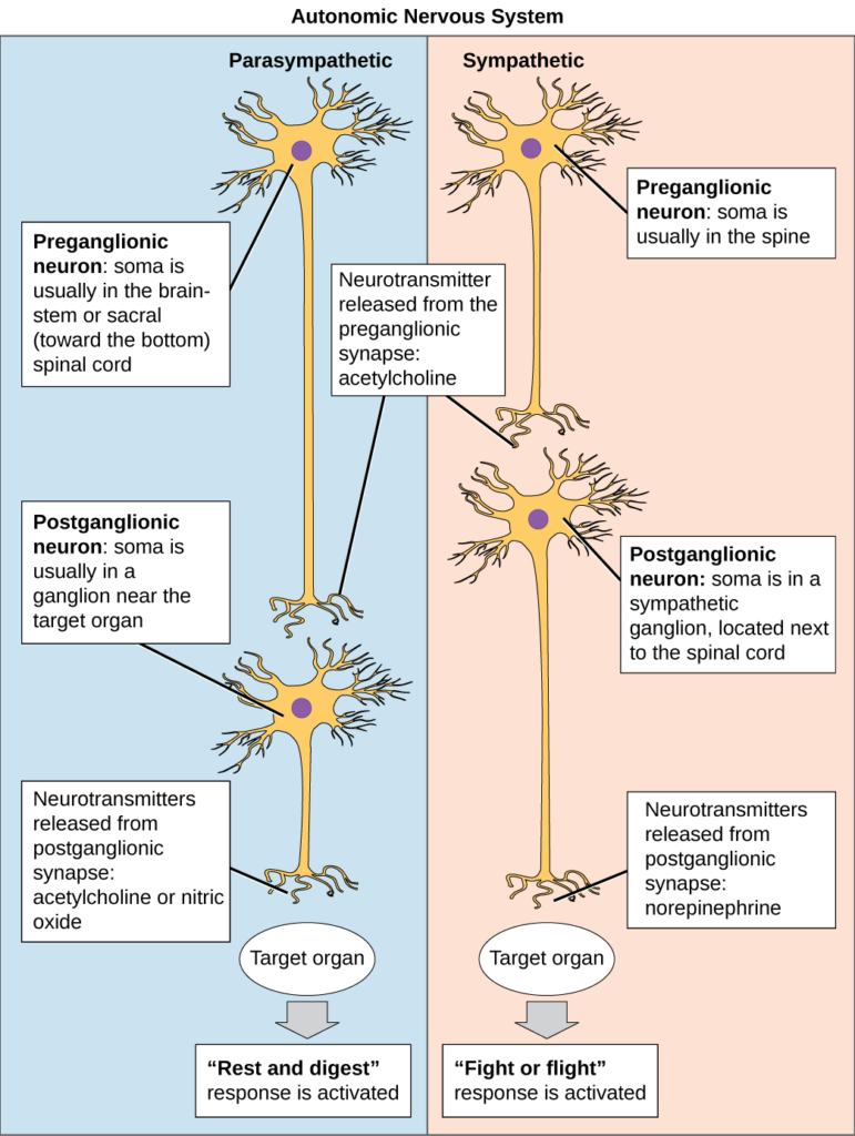
ganglionic neuron aka postganglionic neuron
A neuron of the Autonomic nervous system. The cell body is in an autonomic ganglion (PNS). The axon goes to an effector.

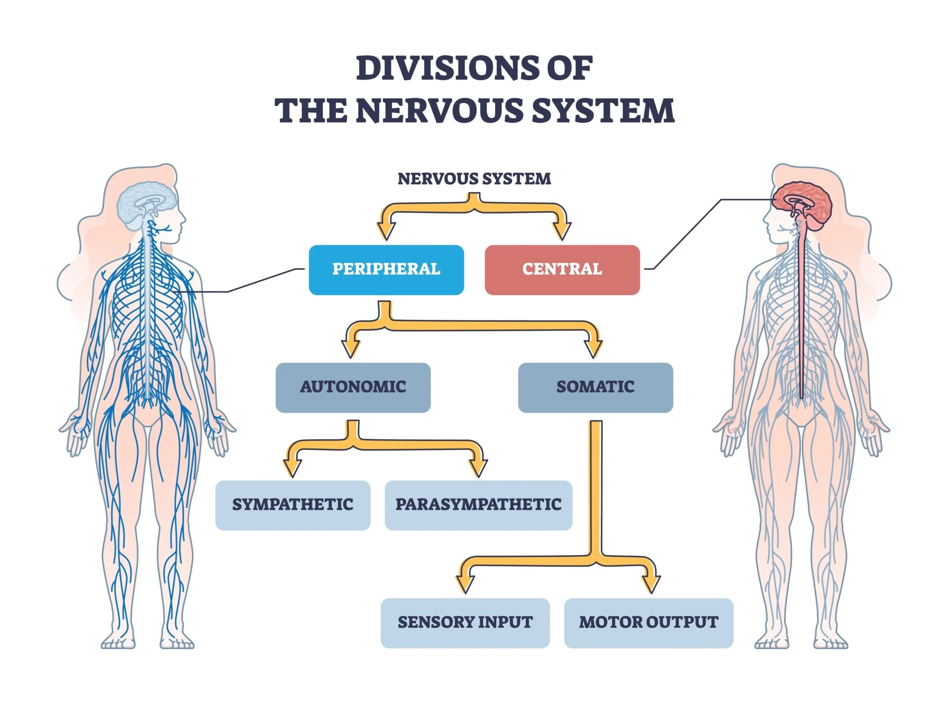
Autonomic nervous system
Part of the peripheral nervous system (PNS, the nervous system outside of the brain / spinal cord) that regulates involuntary physiologic processes, including heart rate, blood pressure, respiration, digestion, and sexual arousal.
Has preganglionic and postganglionic, aka ganglionic, neurons.
Effectors: cardiac muscle, smooth muscle of blood vessels and organs, glands, and adipocytes.
Divisions: sympathetic, parasympathetic, and enteric nervous systems.
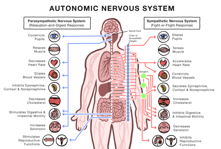
Sympathetic nervous system
Division of the autonomic nervous system. The fight or flight section. It’s ganglia are close to the CNS and far from effectors (effector = thing effected it seems). It’s nerves are in the thoracic and lumbar regions.
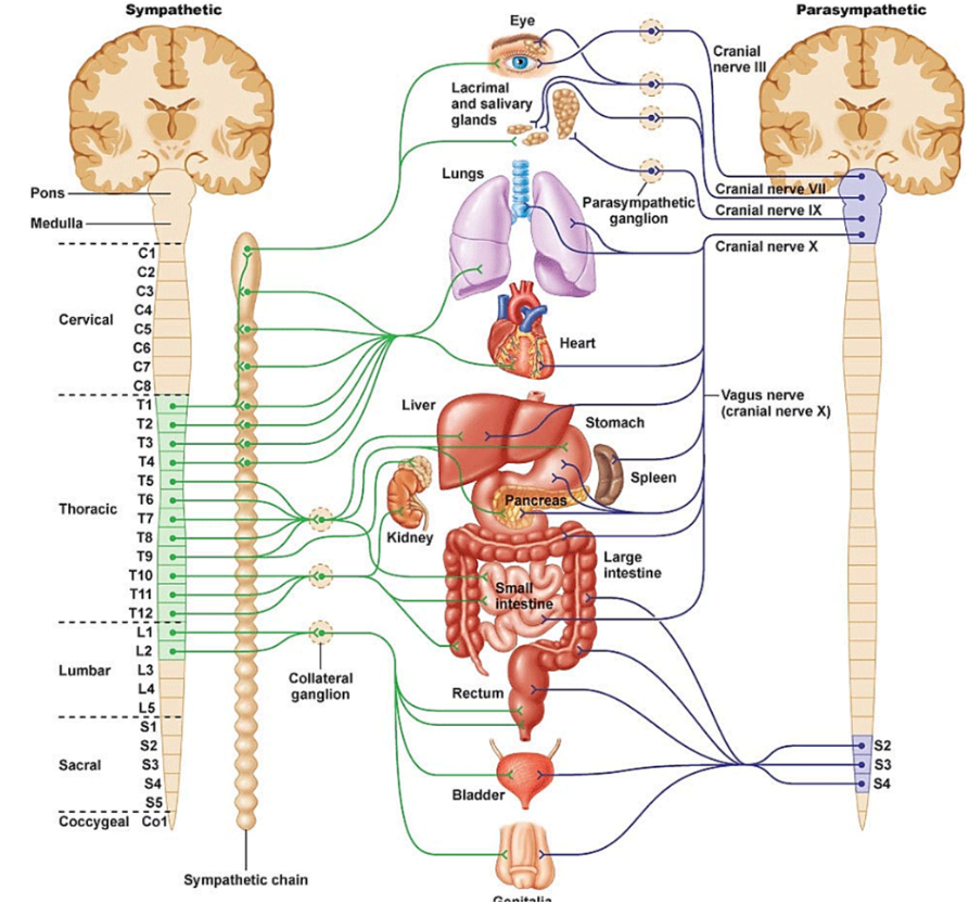

Parasympathetic nervous system
Division of the autonomic nervous system. The rest and digest section. It’s ganglia are far from the CNS and close to effectors. It’s nerves are in the cranial and sacral regions.

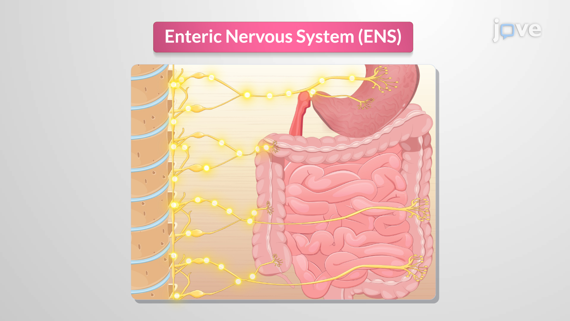
Enteric nervous system
Division of the autonomic nervous system. It embeds in the wall of the digestive tract.
sympathetic chain
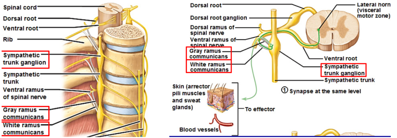
Chain of ganglia on both sides of the vertebral column. Part of the sympathetic nervous system, a division of the autonomic nervous system. Has white rami communicantes — myelinated preganglionic axons, and gray rami communicantes — unmyelinated postganglionic axons. Both types of communicantes attach to the ganglia chain.
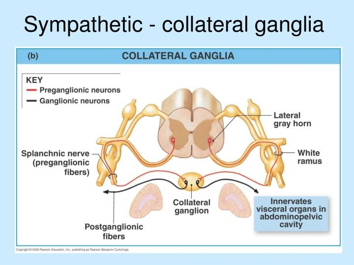
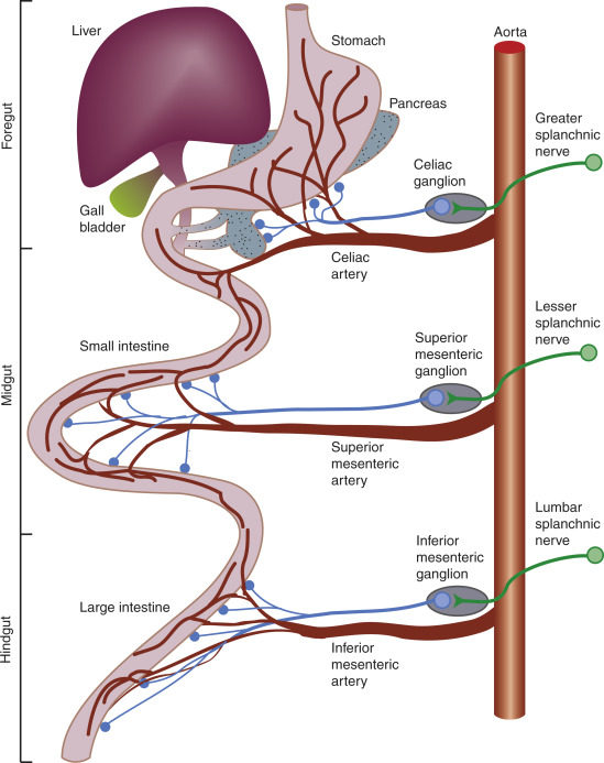
collateral ganglia
sympathetic nerve cell bodies located in the abdominal region that innervates the abdominal organs. They receive preganglionic nerve axons via splanchnic nerves. They contain postganglionic neurons that regulate visceral organ function. It’s ganglia (nerve relay stations that connect nerves) include: celiac, superior mesenteric, inferior mesenteric.
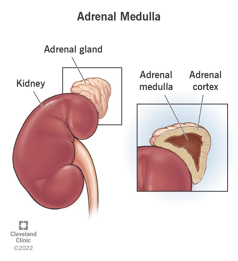
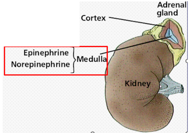
adrenal medulla
Part of the sympathetic system. In the center of each of the 2 adrenal glands. Has modified sympathetic ganglion (nerve relay centers that connect nerves) that secretes hormones (norepinephrine mimics nerve signals).
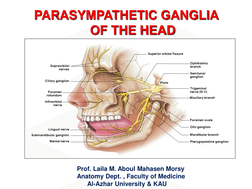
cranial fibers
Nerve fibers. Part of the parasympathetic “rest and digest” nervous system, a division of the autonomic system.
1. oculomotor nerve (III) -- connects to the ciliary ganglion
2. facial nerve (VII) — connects to the pterygopalatine ganglion and the submandibular ganglion
3. glossopharyngeal nerve (IX) -- connects to the otic ganglion
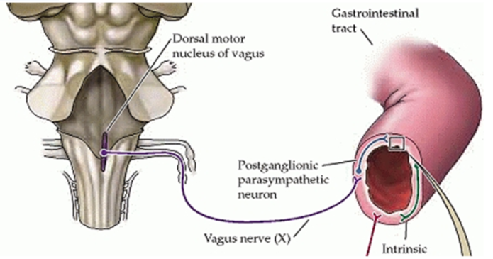
4. vagus (X) -- connects to the intramural ganglia in abdominal organs
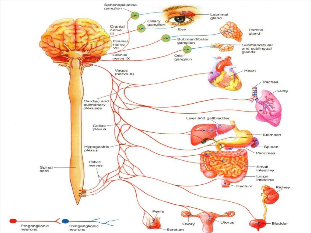
sacral fibers
Nerve fibers. Part of the parasympathetic “rest and digest” nervous system, a division of the autonomic system. Pelvic nerves → lower digestive system → urinary system → reproductive system.

sensory receptors
detect sensory info. types: sensory neurons (for quick, direct info) and specialized epithelial cells (less sensitive). We perceive sensory by sensory impulses going to our cerebral cortex for interpretation: we “sense” with our brains.
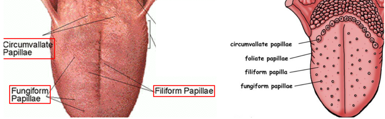
lingual papillae
Part of the gustation sensory system. On the superior surface of the body of the tongue. Provide rough surface for grasping food. Types include: Filiform, Fungiform, and Circumvallate.

Filiform
A type of lingual papillae. Part of the gustation sensory system. Most numerous, slender and pointed, involved with touch, NO taste buds.

Fungiform
A type of lingual papillae. Part of the gustation sensory system. Mushroom shaped, scattered amongst the filiform, has taste buds.

circumvallate
A type of lingual papillae. Part of the gustation sensory system. Largest, round, form an upside down V at the back of tongue body, has taste buds.
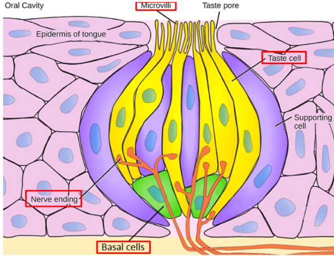
taste buds
1. gustatory cells (hair cells) = specialized epithelial cells –“hairs”: microvilli with chemical receptors, project through taste pore at surface of taste bud. Sensory neuron dendrites synapse (connect) on gustatory cells, transmitting gustatory info to brain.
2. transitional cells -- immature gustatory cells.
3. basal cells -- stem cells.
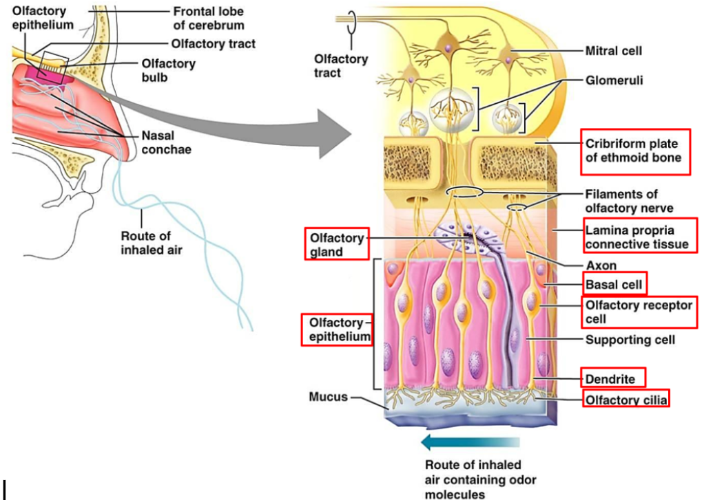
olfactory organs (left and right)
one on each side of the roof of nasal cavity. Control smell. Made of the lamina propria and olfactory epithelium.

lamina propria
In the olfactory organs. Part of the olfactory system. Made of areolar connective tissue. Have olfactory glands which secrete a layer of mucus onto surface.

olfactory epithelium
In the olfactory organs. Part of the olfactory system. Has olfactory receptors, consisting of neurons, dendrites (olfactory knob with cilia, embedded in mucous layer on the epithelium surface), and axons (pass through cribriform plate and form olfactory nerve). Has epithelial cells as support cells. Basal cells form new olfactory receptors.
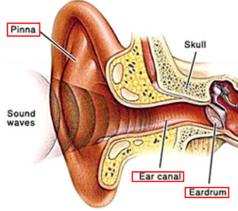
pinna (auricle)
Part of external ear. elastic cartilage, adipose tissue, and skin. Collects sound waves.

external acoustic meatus (ear canal)
Part of external ear. carries sound waves to eardrum. Have ceruminous glands that secrete cerumen (earwax).

tympanic membrane (eardrum)
Part of external ear. Separates external and middle ears, brings sound waves to middle ear.
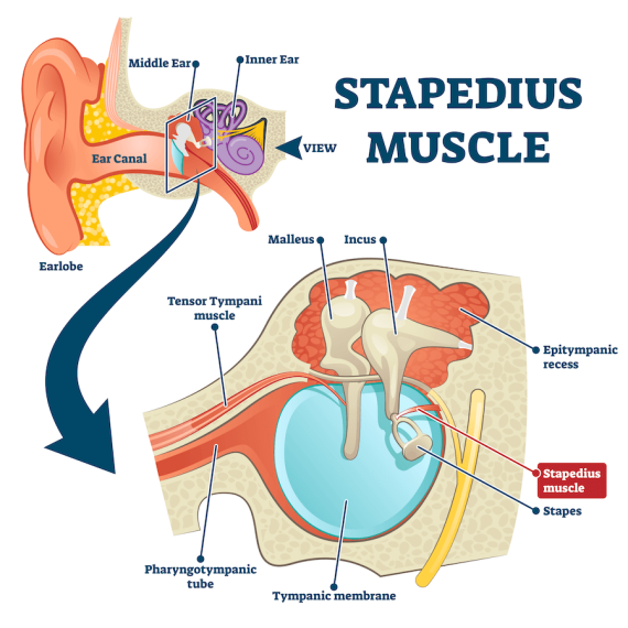
middle ear (tympanic cavity)
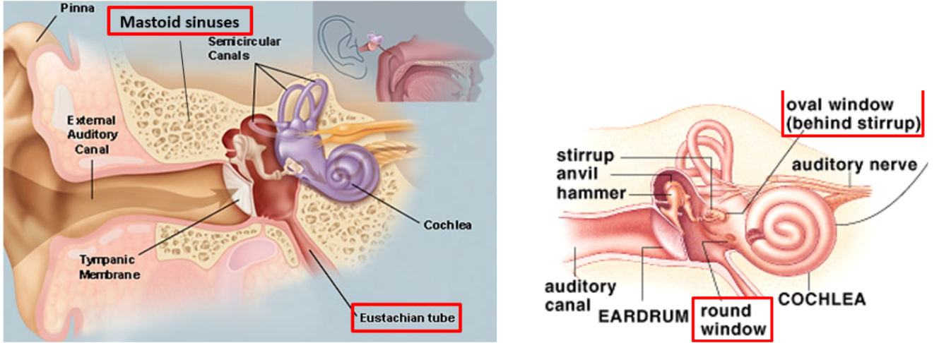
air filled cavity in the temporal bone. Between the inner and external ear. Has auditory ossicles (bones), muscles to protect the ossicles against loud noises, openings, and connections to the inner ear (the oval window and round window).
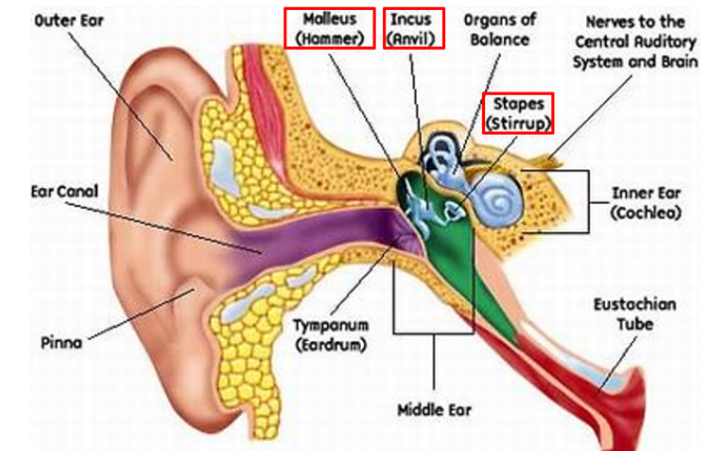
auditory ossicles
Bones. Part of the middle ear (tympanic cavity). Includes the malleous (attached to tympanic membrane), incus (middle bone), and stapes (next to inner ear).
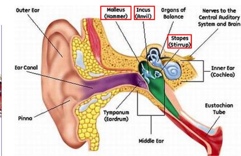
Muscles of the middle ear
protect the ossicles (ear bones) against loud noises. The tensor tympani protects the malleus and the stapedius protects the stapes.
Openings of middle ear
Openings: include the auditory tube (to pharynx) and the mastoid sinuses
connections to inner ear: oval window (covered by stapes, where sound waves enter inner ear) and round window (where sound waves leave inner ear).
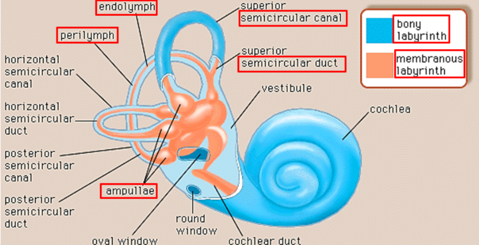
perilymph fluid
fluid between bony and membranous labyrinthes in the inner ear.
endolymph fluid
fluid within the membranous labyrinth in the inner ear.
semicircular canals and ducts
canals: bony labyrinth, 3 canals at right angles to each other, making 3 different planes
ducts: membranous labyrinth inside the canals. Have crista that detect rotational head movements.