T3 Introduction to Clinical Dermatology (Describing Lesions)
1/105
There's no tags or description
Looks like no tags are added yet.
Name | Mastery | Learn | Test | Matching | Spaced | Call with Kai |
|---|
No analytics yet
Send a link to your students to track their progress
106 Terms
4 types of lesions
raised, depressed, flat, fluid-filled
6 types of raised lesions
papule, nodule, cyst, comedo, wheal, plaque
what lesion is depicted
papule
what lesion is depicted
nodule
what lesion is depicted
cyst
what lesion is depicted
comedo
what lesion is depicted
wheal
what lesion is depicted
plaque
papule size
<1 cm
plaque size
>1 cm
4 types of papule
pedunculated, dome-shaped, flat-topped, umbilicated
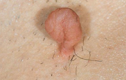
what lesion is depicted
pedunculated
what lesion is depicted
dome-shaped
what lesion is depicted
flat-topped
what lesion is depicted
umbilicated
important component of peduncle
stalk
peduncle example
skin tag
flat-topped papule example
lichen planus

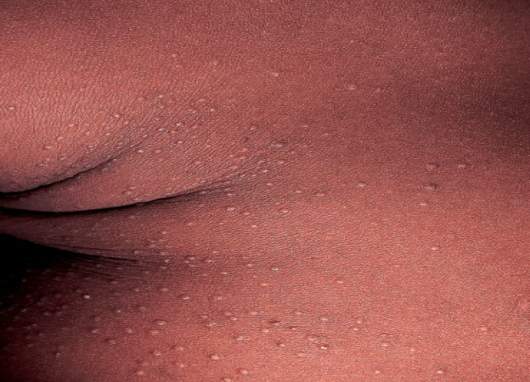
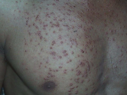
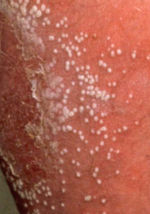
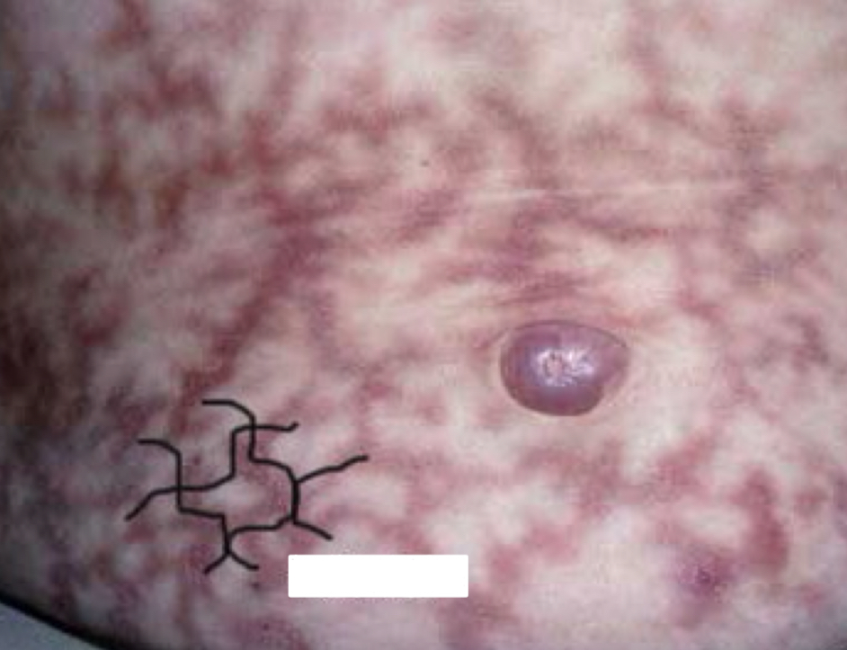
what type of lesion and condition is depicted
dome-shaped papule; pityrosporum folliculitis
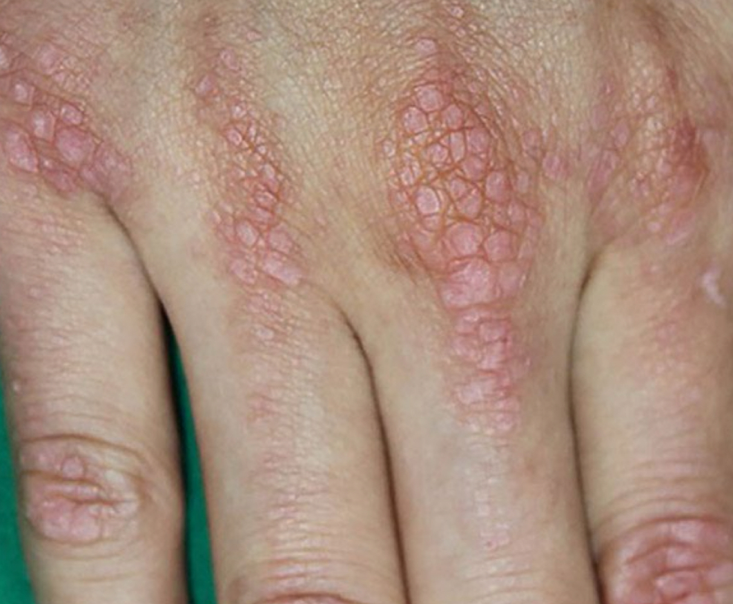
what type of lesion and condition is depicted
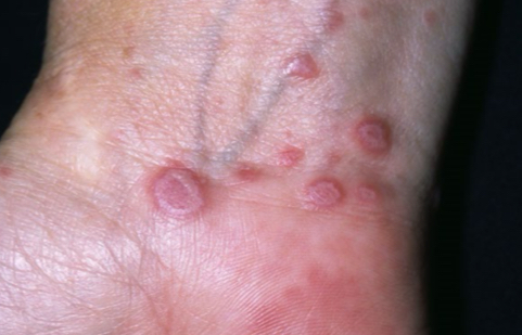
flat-topped papule; lichen planus
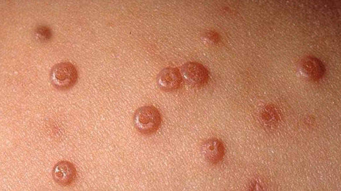
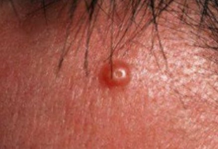
what type of lesion and condition is depicted
umbilicated papule; molluscum contagiosum
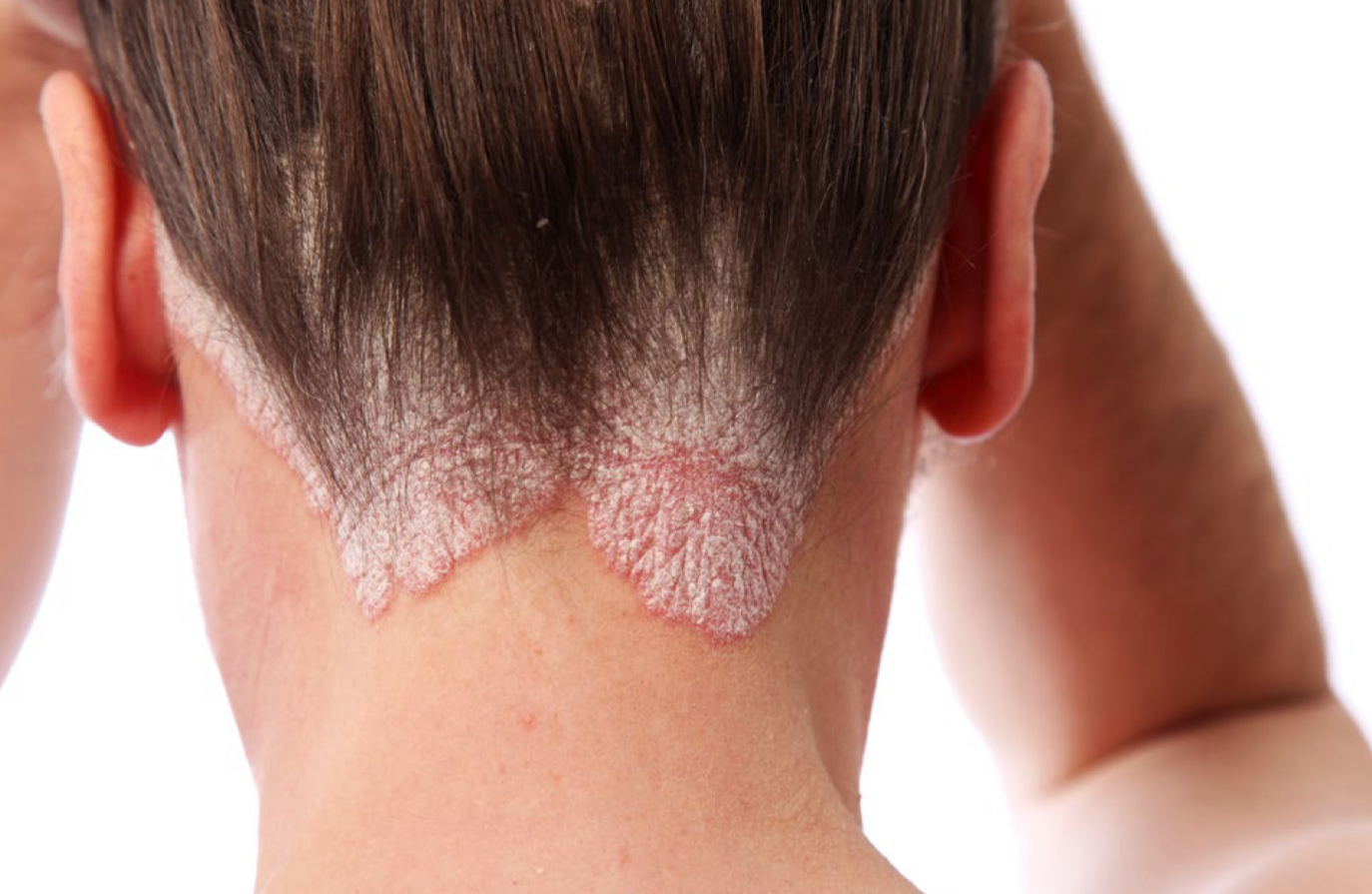
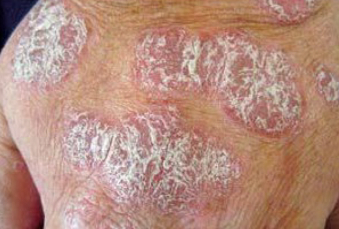
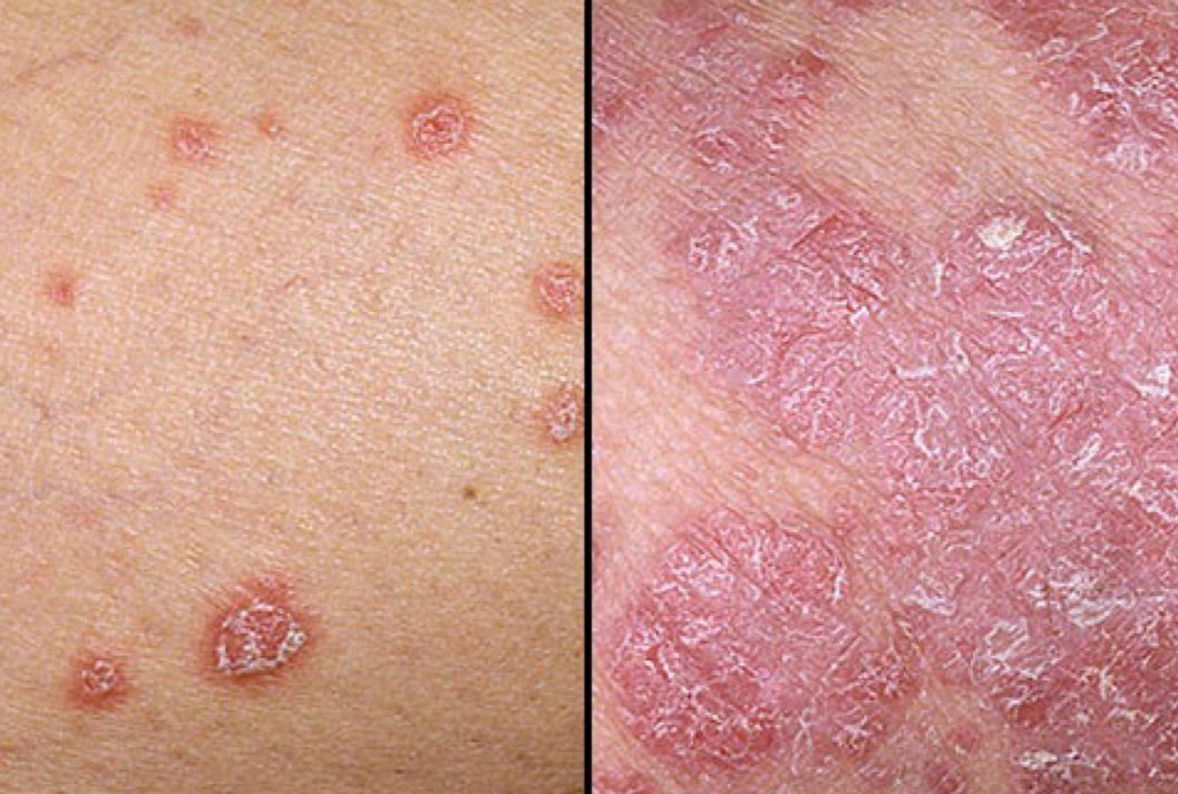
what type of lesion and condition is depicted
plaque; psoriasis
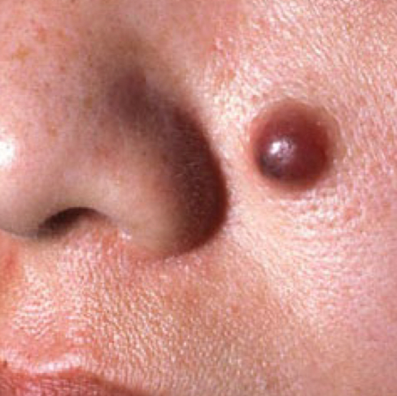
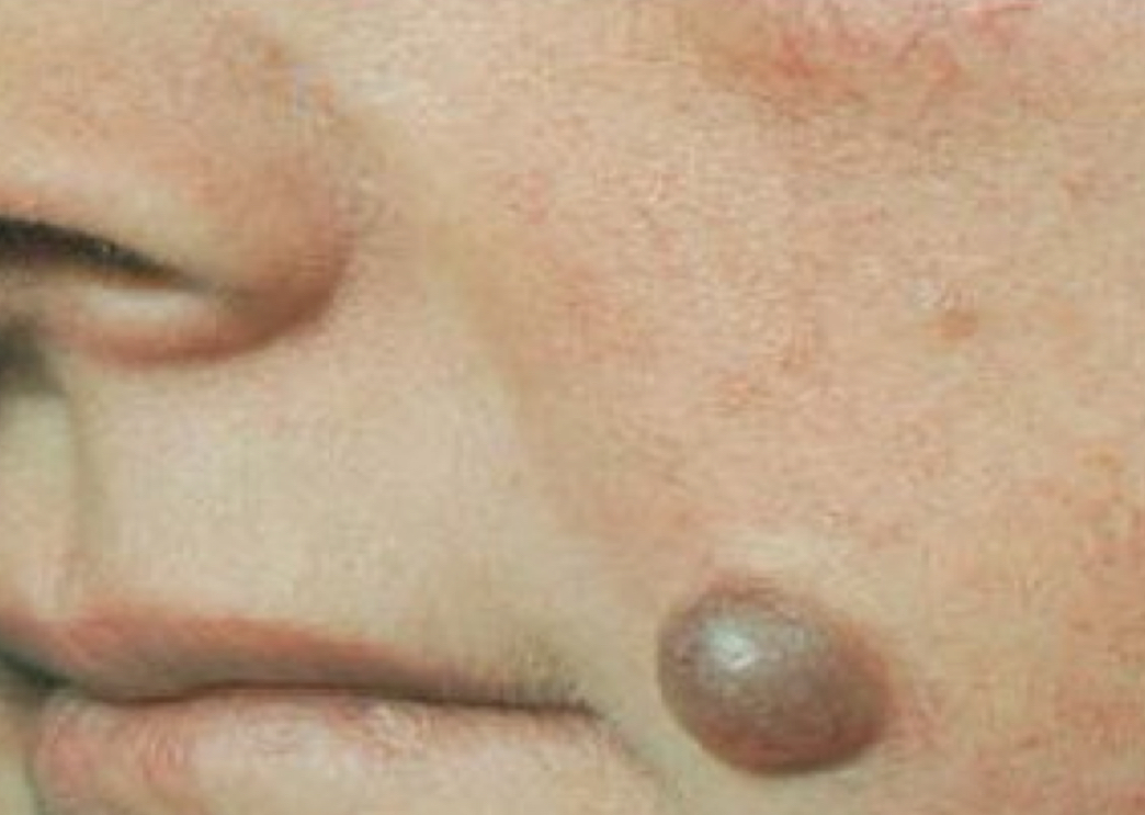
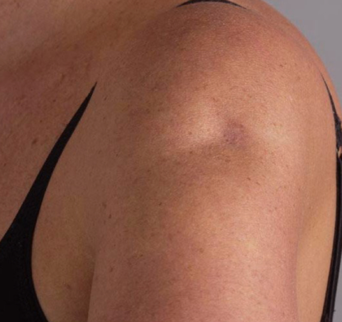
what type of lesion and condition is depicted
nodule; compound nevus
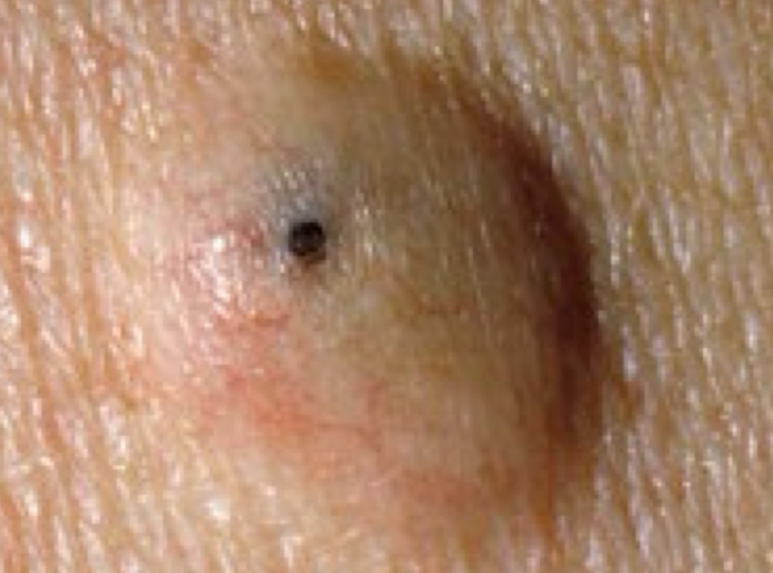
what type of lesion and condition is depicted
cyst; epidermal inclusion cyst
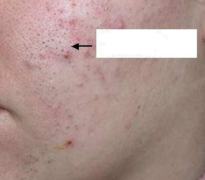
what type of lesion and condition is depicted
open comedo; blackhead
what type of lesion and condition is depicted
closed comedo; whitehead
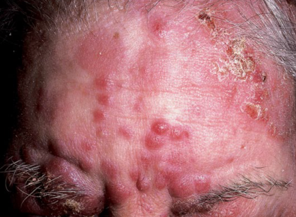
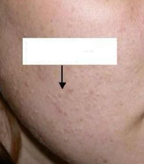
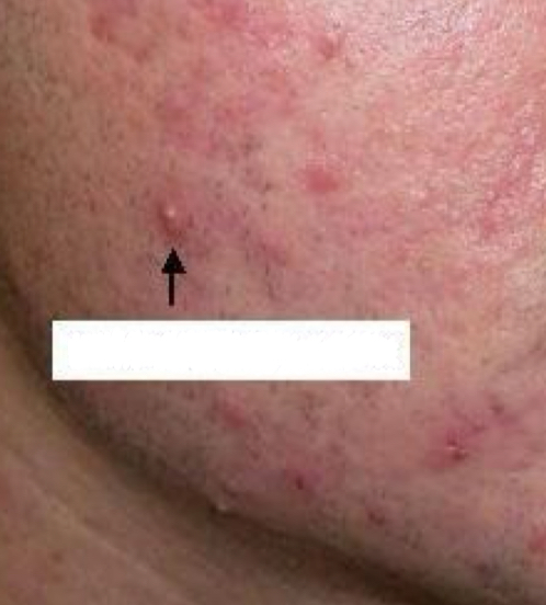
what type of lesion is depicted
inflammatory papule
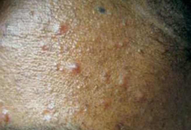
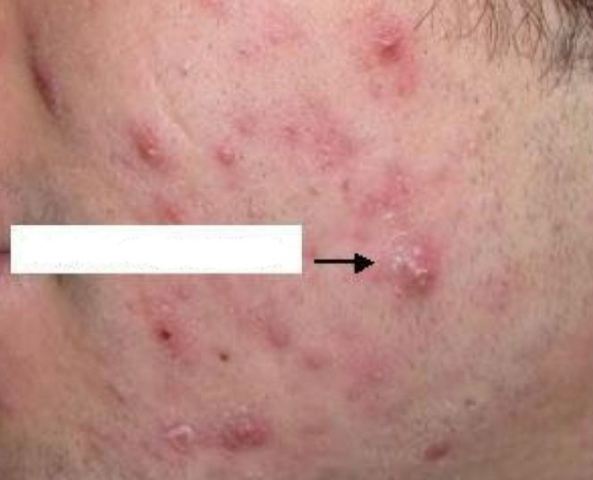
what type of lesion is depicted
inflammatory pustule

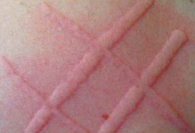
what lesion is depicted by the black arrow
wheal
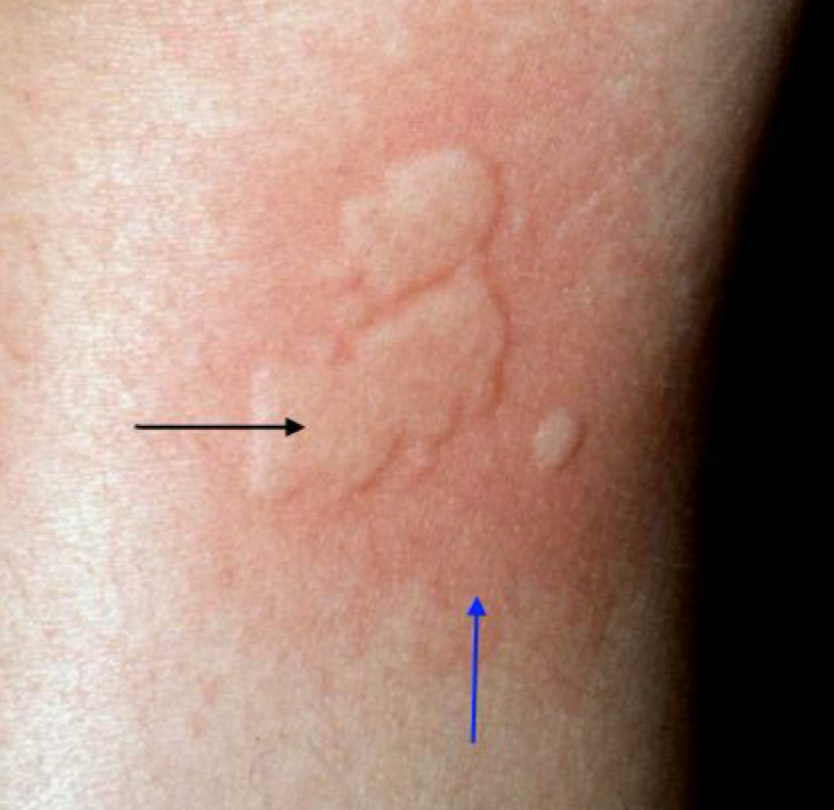
what lesion is depicted by the blue arrow
flare
flat lesion sizes
macule (<1 cm), patch (>1 cm)
blanchable
fade if pressed
types of blanchable flat lesions
erythema, erythroderma, telangiectasia
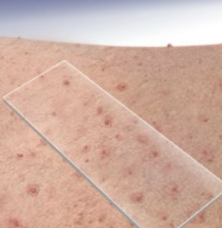
types of nonblanchable flat lesions
petichiae, purpura (palpable), ecchymosis
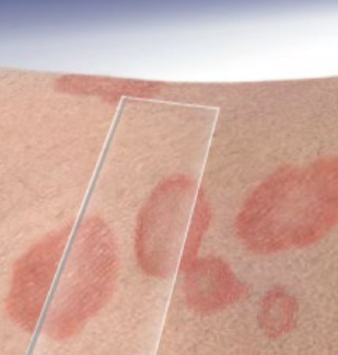
what type of lesion is depicted and what area does it cover
erythema; small area
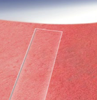
what type of lesion is depicted and what area does it cover
erythroderma; >90% of body
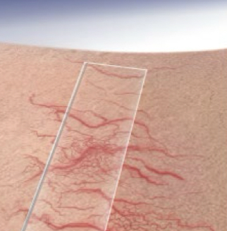
what type of lesion is depicted
telangiectasia
what type of lesion is depicted
petichiae
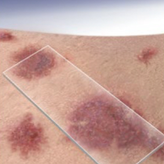
what type of lesion is depicted
purpura (palpable)
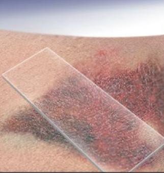
what type of lesion is depicted
ecchymosis
3 types of depressed lesions
fissure, erosion, ulcer
fissure
varying depth, dry skin
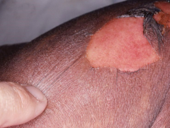
erosion
epidermis, first degree burn
ulcer
dermis
what type of lesion is depicted
fissure
what type of lesion is depicted
erosion
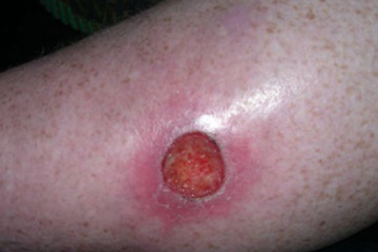
what type of lesion is depicted
ulcer

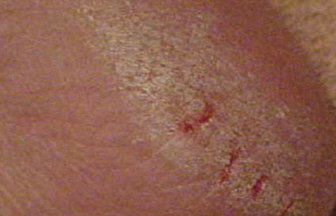
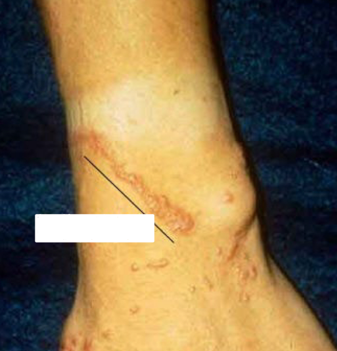
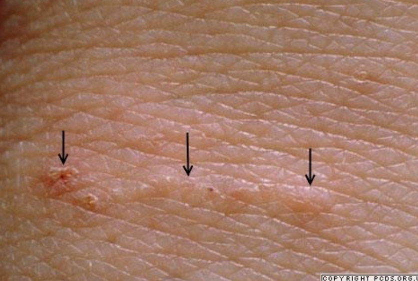
what type of lesion is depicted, describe it, what condition is it
burrow depressed erosion; thread-like linear tunnel; scabies
what type of lesion is depicted
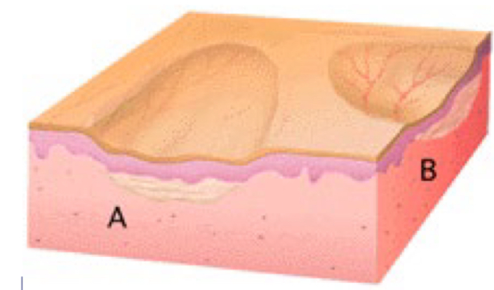
atrophy

what type of condition is depicted by A
epidermal atrophy
what type of condition is depicted by B
dermal atrophy
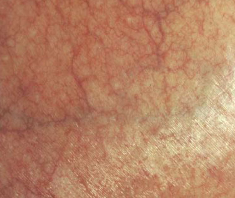
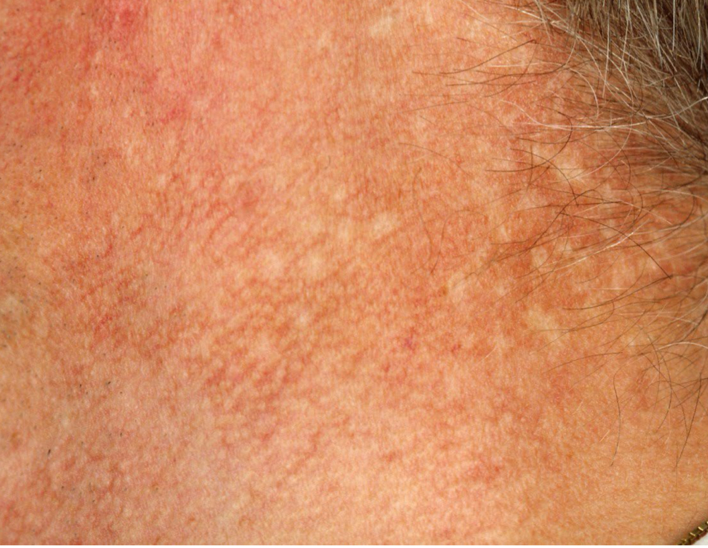
what lesion and what condition is present
telangiectasia, epidermal atrophy
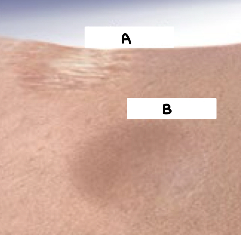
what condition is depicted
dermal atrophy
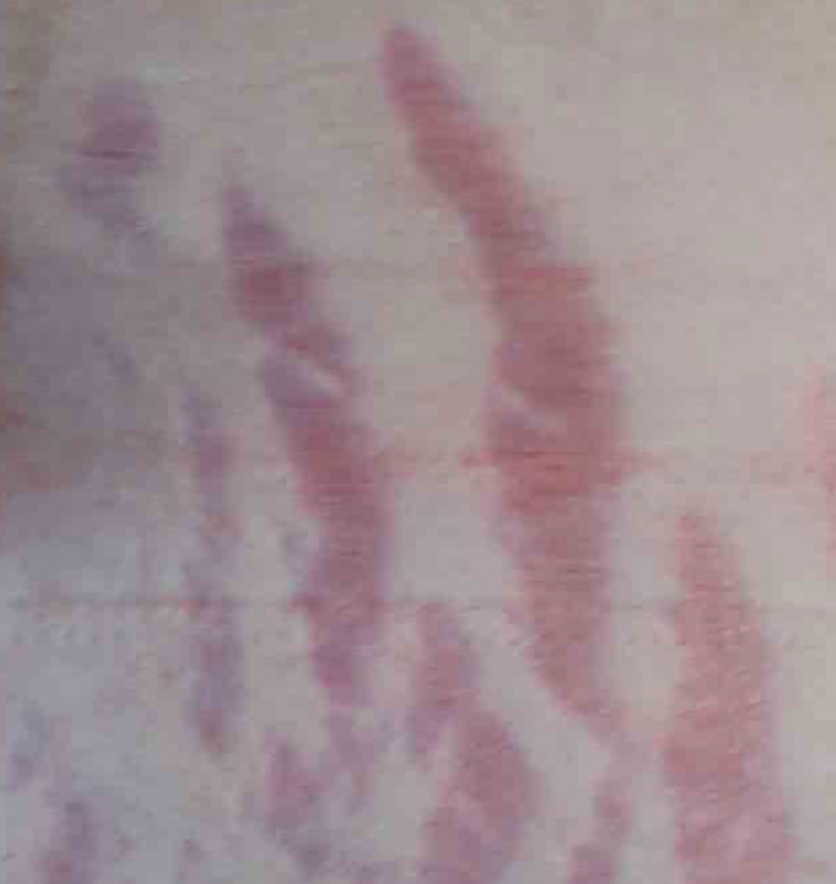
what type of lesion is depicted and what condition is it
striae; stretch marks
components of poikiloderma
telangiectasia, atrophy, hypo/hyperpigmentation
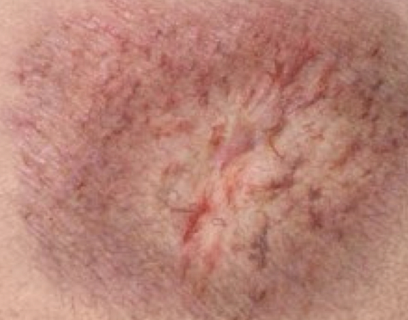
what condition is depicted
poikiloderma
what condition is depicted
poikiloderma
fluid-filled lesion sizes
vesicle (<0.5 cm), bulla (>0.5 cm)
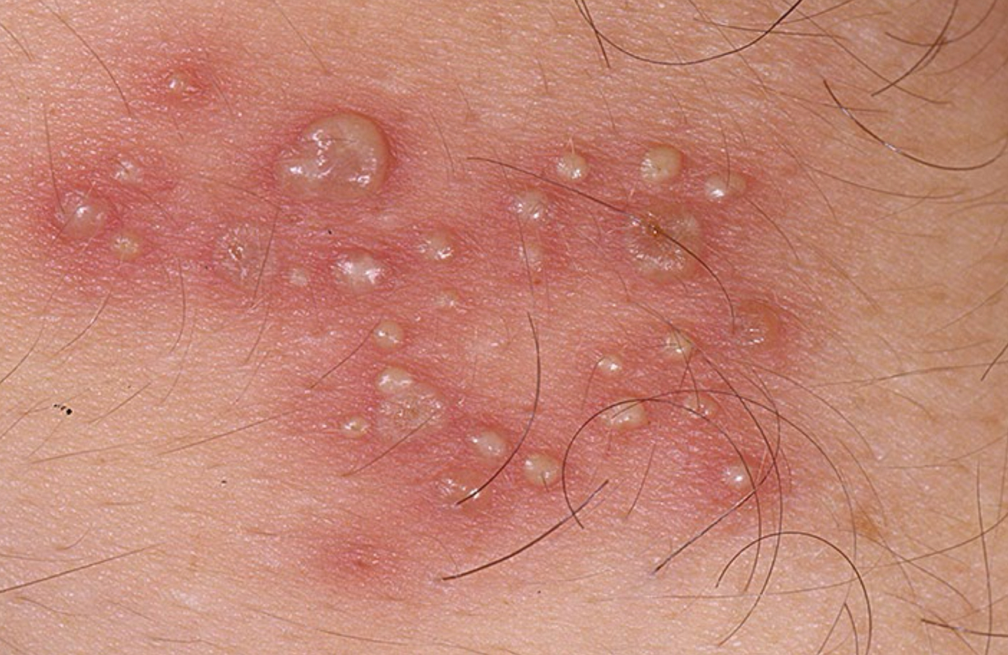
describe the lesion; what condition
group of vesicles on erythema background; herpes
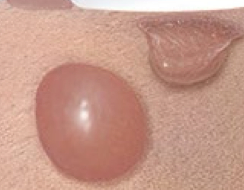
what bulla is depicted on the left
tense bulla

what bulla is depicted on the right
flaccid bulla
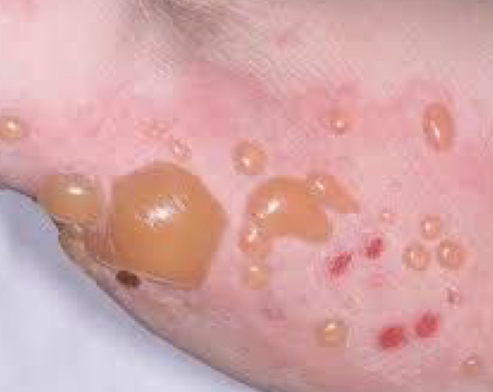
what type of lesion is depicted and what condition
tense bulla; bullous pemphigoid
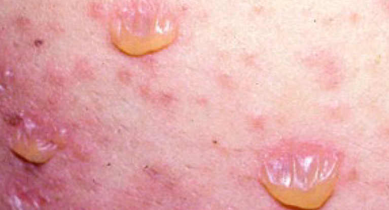
what type of lesion is depicted and what condition
flaccid bulla; pemphigus vulgaris
what type of lesion is depicted
pustule
types of surface change
scale, crust, excoriation, lichenification
what type of lesion is depicted
scales
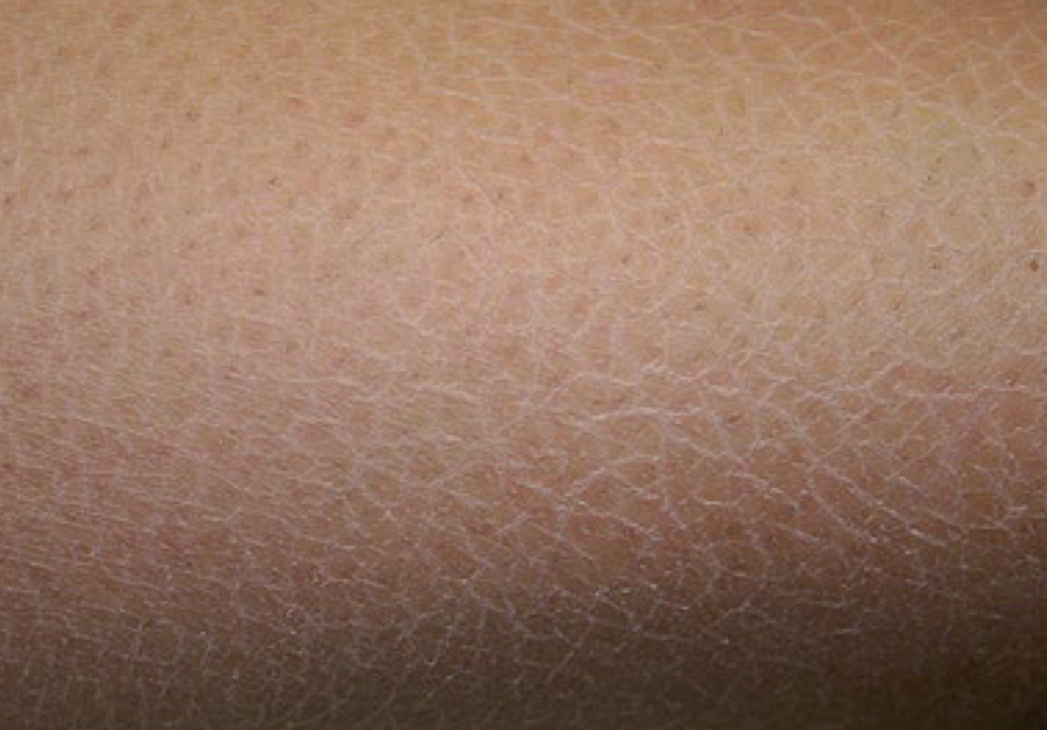
what type of lesion and what condition is depicted
crack-like scale; xerotic eczema
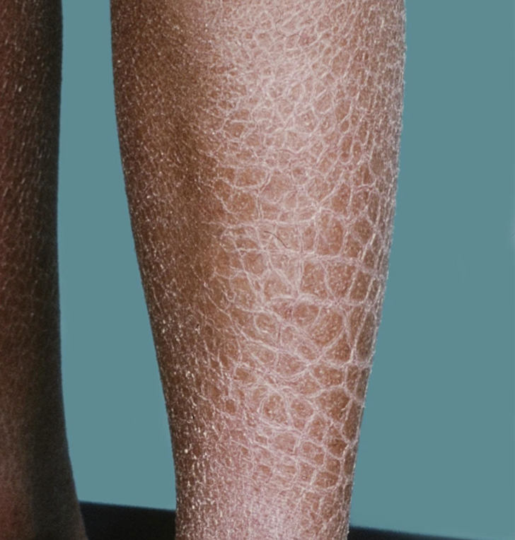
what type of lesion and what condition is depicted
ichthyosiform scale; ichtyosis
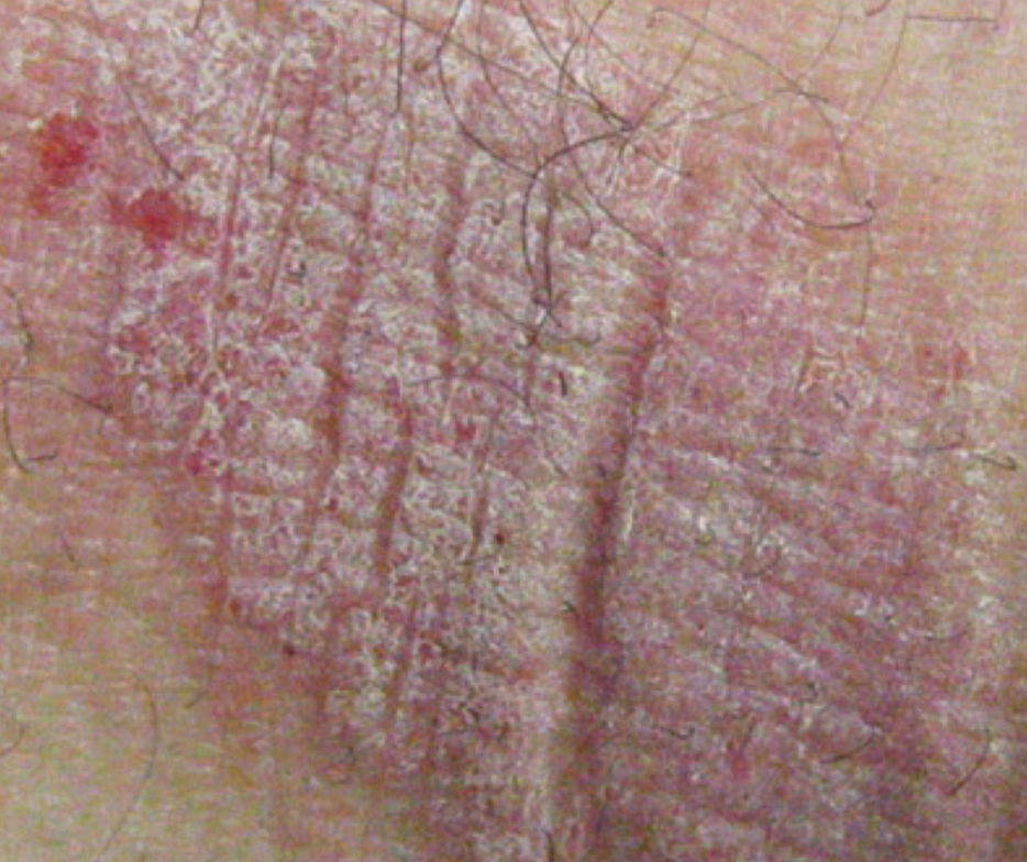
what type of lesion and what condition is depicted
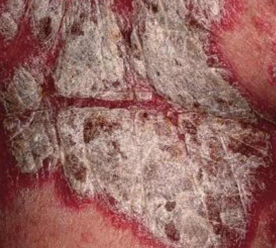
micaceous (silvery) scale; psoriasis
what type of lesion and what condition is depicted
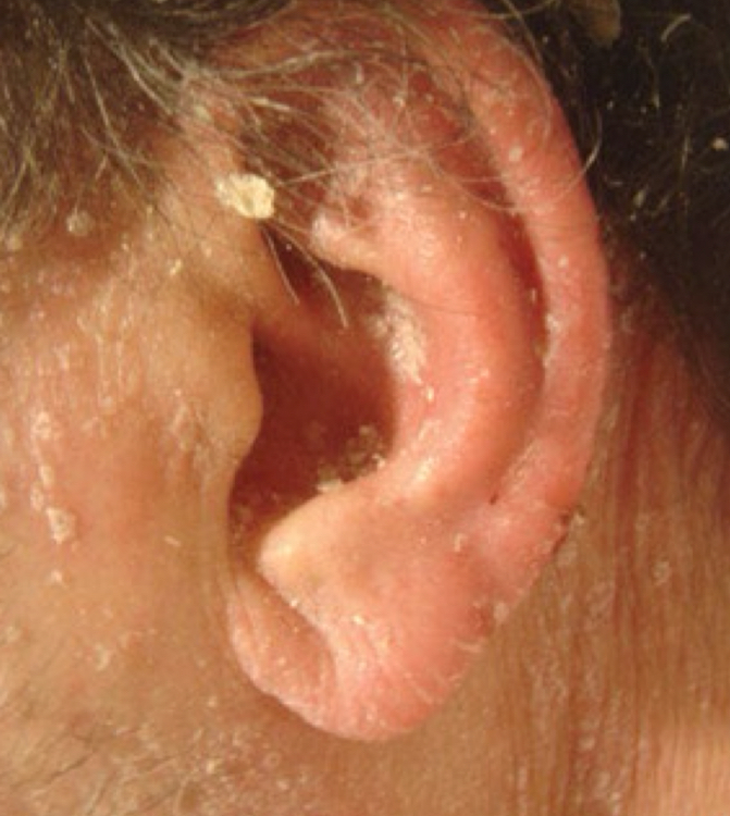
greasy scale; seborrheic dermatitis
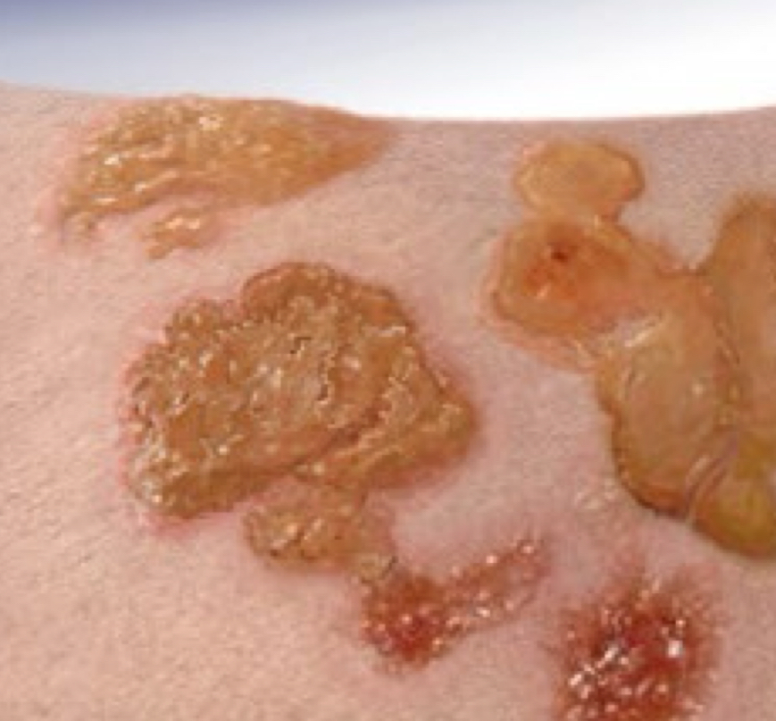
what type of lesion is depicted
crust
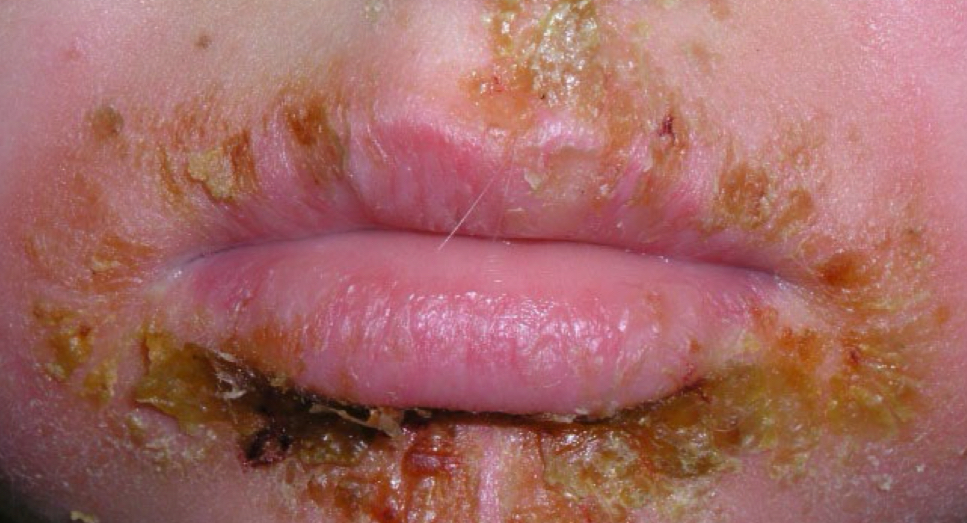
what type of lesion is depicted
crust
what type of lesion is depicted and when does it occur
excoriation (secondary after scratched)
what type of lesion is depicted and when does it occur
lichenification (secondary after rubbing)
what becomes more pronounced in lichenification
skin crease
types of color change in lesions
bright red, violaceous, dusky red, blue-black, hypo-depigmentation, yellow
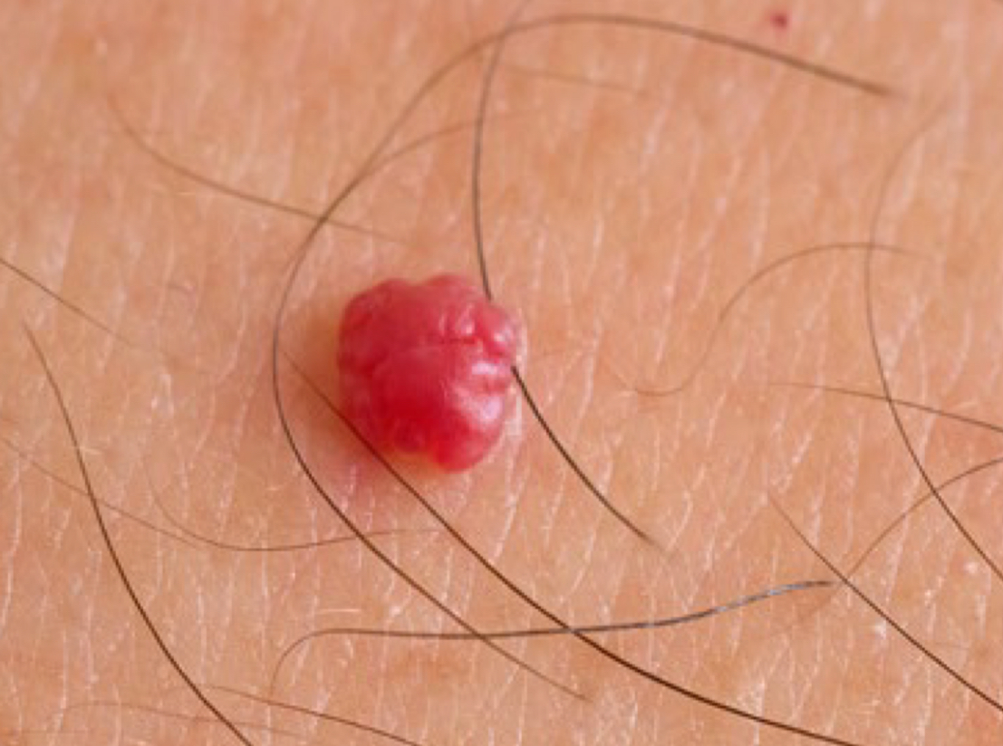
what type of color change and condition is depicted
bright redness; cherry angioma
what type of color change and condition is depicted
violaceous; kaposi’s sarcoma
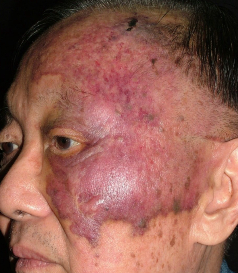
what type of color change and condition is depicted
violaceous; angiosarcoma
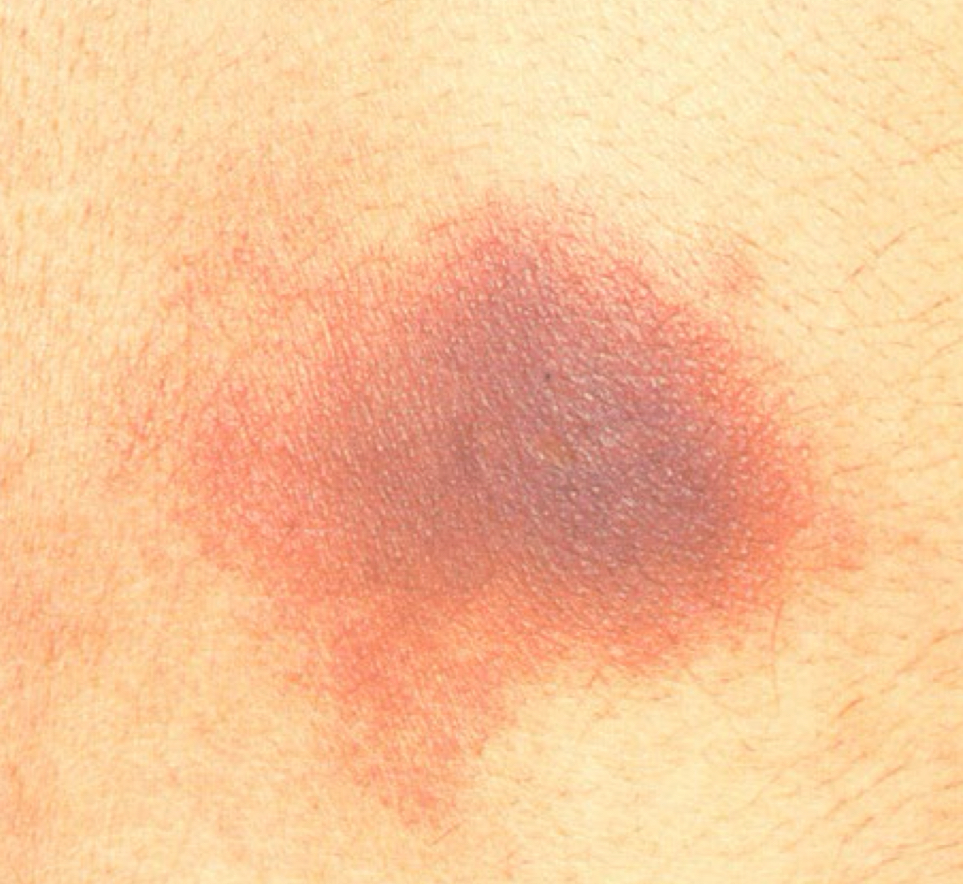
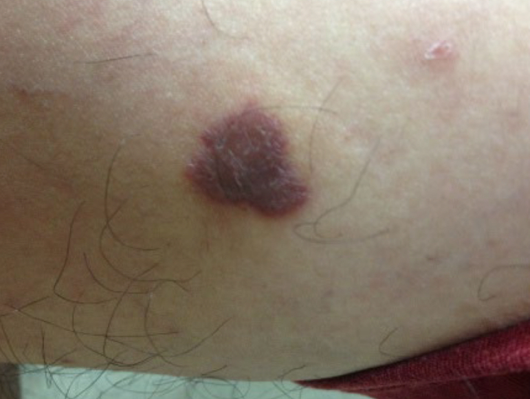
what type of color change and condition is depicted
dusky red; fixed drug eruption
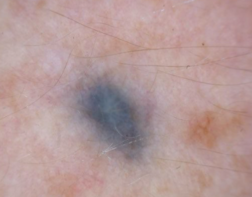
what type of color change and condition is depicted
blue-black; dermal nevus
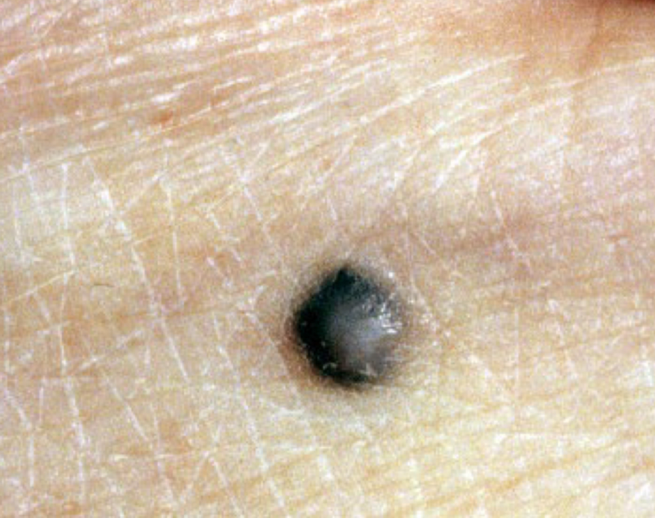
what type of color change and condition is depicted
black; malignant melanoma
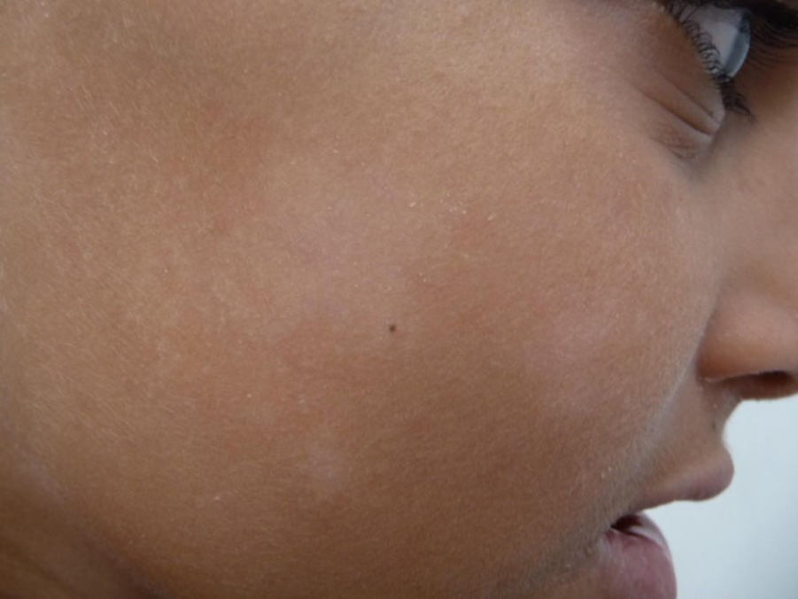
what type of color change and condition is depicted
hypo-depigmentation; pityriasis alba
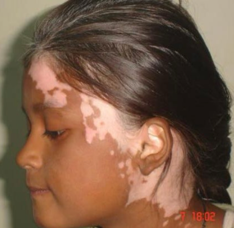
what type of color change and condition is depicted
hypo-depigmentation; vitiligo
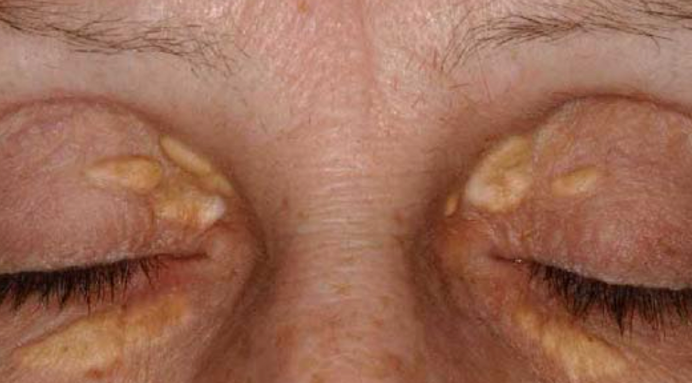
what type of color change and condition is depicted
yellow; xanthelasma
types of shapes and arrangements
annular, nummular (coin), linear, serpiginous, grouped (herpetiform), reticular, target,
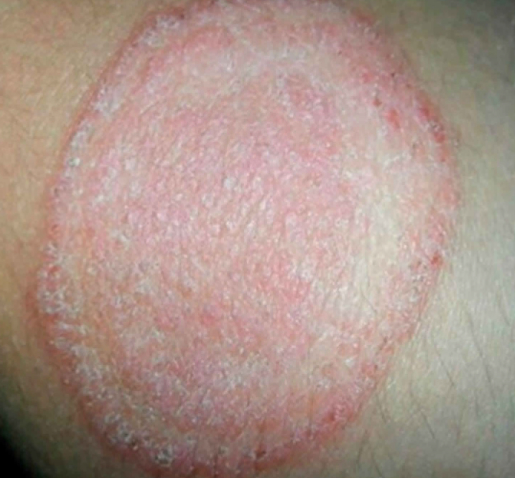
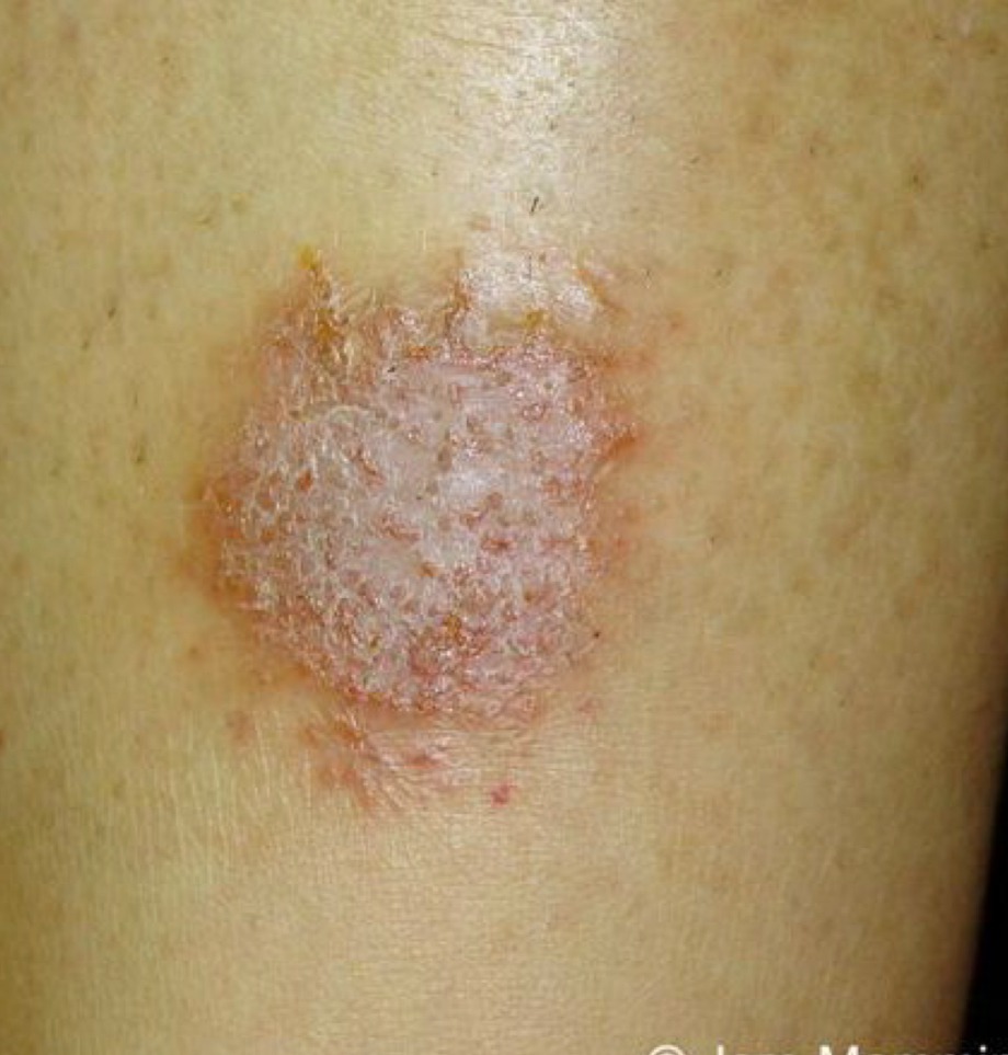
what shape/arrangement and condition is depicted
annular; tinea corporis
what shape/arrangement and condition is depicted
nummular (coin); nummular eczema
what shape/arrangement is depicted
linear
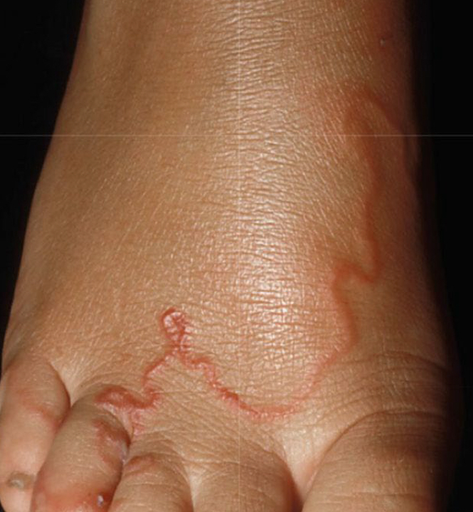
what shape/arrangement and condition is depicted
serpiginous; cutaneous larva migrans
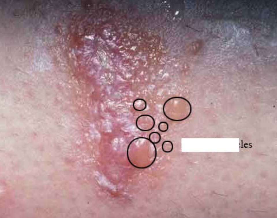
what shape/arrangement and condition is depicted
grouped (herpetiform); herpes
what shape/arrangement and condition is depicted
reticular; erythema abigne
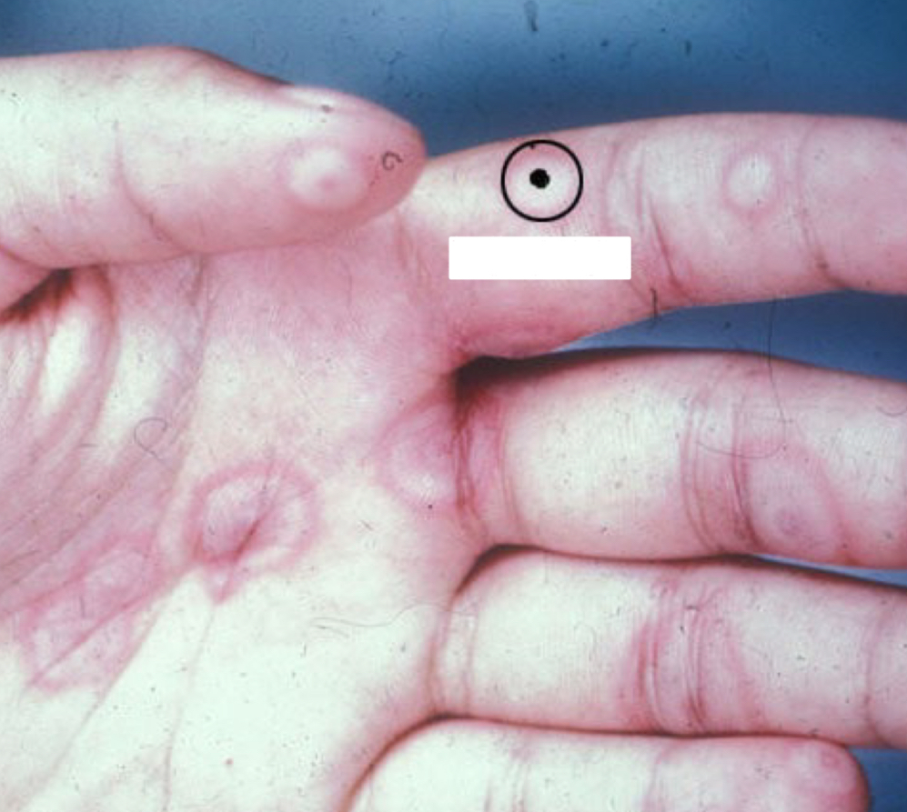
what shape/arrangement and condition is depicted
target; erythema multiforme
what shape/arrangement and condition is depicted
linear; scabies
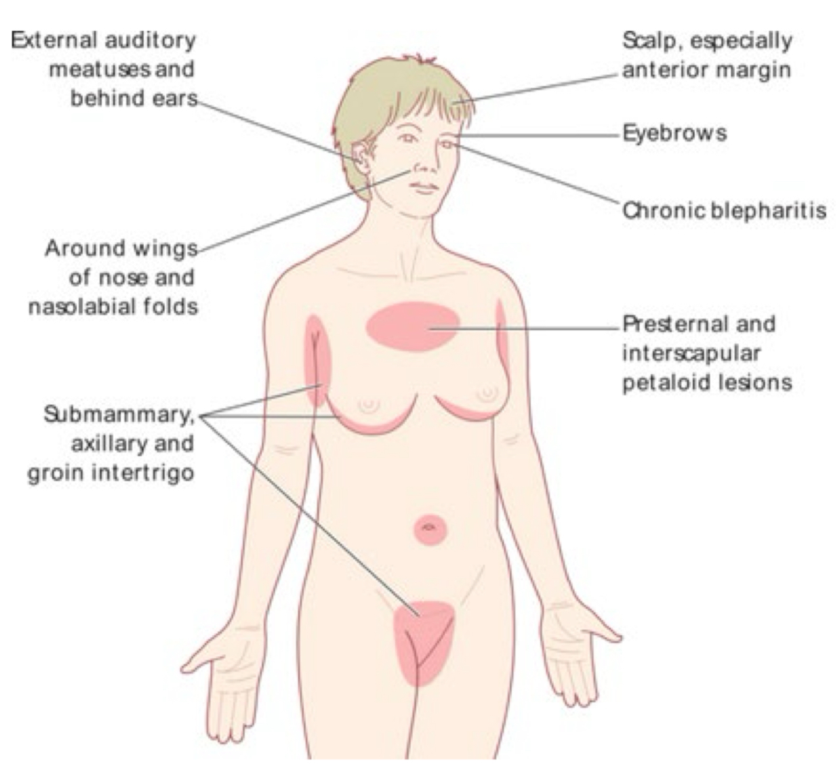
what type of distribution
seborrhea (oily areas)
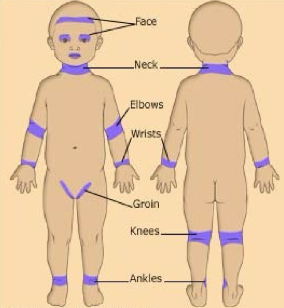
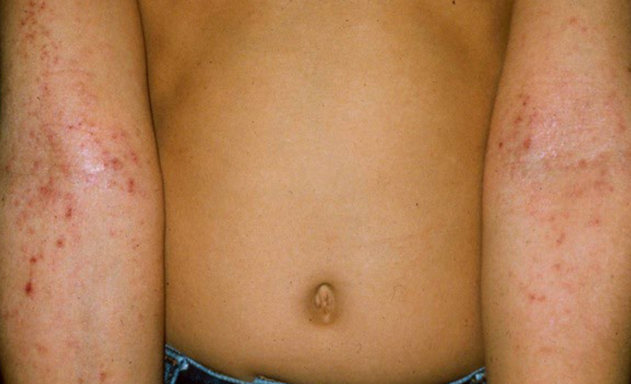
what type of distribution
eczema sites, atopic area
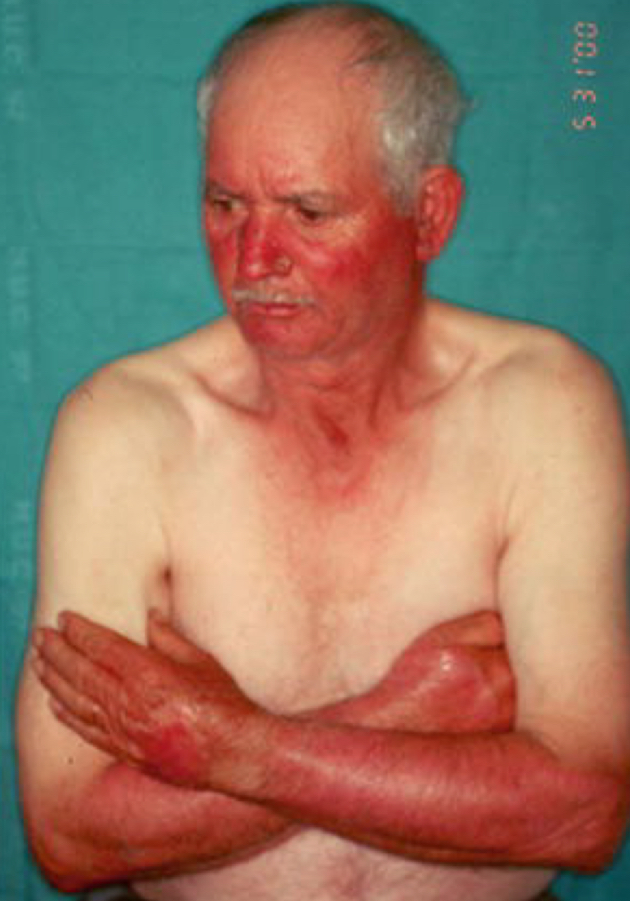
what type of distribution
photodistributed area
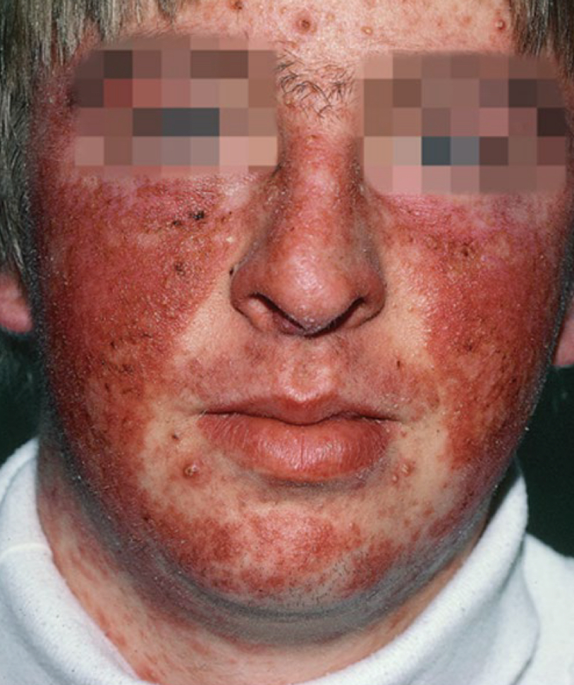
what type of distribution
photodistributed areas
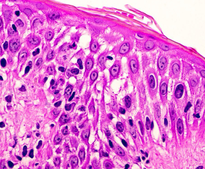
what condition is depicted, describe it
spongiosis; intercellular edema makes spikes more visible but still contact
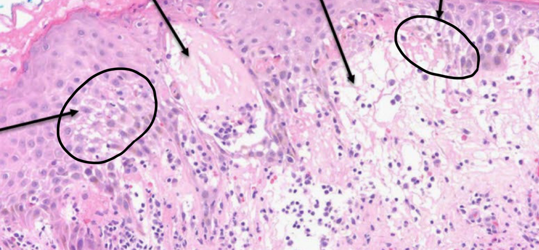
what condition is depicted
spongiosis