Neuro Week 10: Functional Anatomy of the Cerebellum and Basal Ganglia
1/35
There's no tags or description
Looks like no tags are added yet.
Name | Mastery | Learn | Test | Matching | Spaced |
|---|
No study sessions yet.
36 Terms
if you had a lesion on the left hemisphere of the lower thoracic cord of the spinal cord what motor deficits would you experience?
ipsilateral paralysis/paresis to the lower limbs
extrapyramidal systems
include other UMN tracts that do not run within the pyramids of the medulla
reticulospinal tract
reticular formation → through pyramids → spine
what kind of neurons are in the reticular formation
UMN
how do neurons terminate in the reticulospinal tract?
axons terminate bilaterally onto
LMNs in medial regions of ventral horn
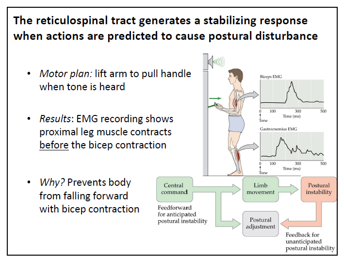
function of the reticulospinal tract
initiates anticipatory, feedforward adjustments that stabilize posture during ongoing movements
what happens when we are about to pull on something strong standing up right? (think legs)
what are the correlations between SMA, Cerebellum, PMC, and Basal ganglia?
The basal ganglia works with motor cortex to initiate and terminate movements, while the cerebellum and SMA monitor ongoing activity to produce smooth movements
roles of the cerebellum
Detects the difference or “motor error” between intended
and actual movements
makes corrective adjustments via efferents to UMNs
Important for balance and coordinated movements
Functions ipsilaterally
cerebellar ataxia
• Jerky and imprecise trajectory
• Overshoots/undershoots target
• Requires frequent corrective movements
lobes of the cerebellum
anterior lobe
posterior lobe
flocculonodular lobe (smallest)
what are the two fissures of the cerebellum
primary fissure (in between anterior and posterior lobe)
posterolateral fissure (in between flocculonodular and posterior)
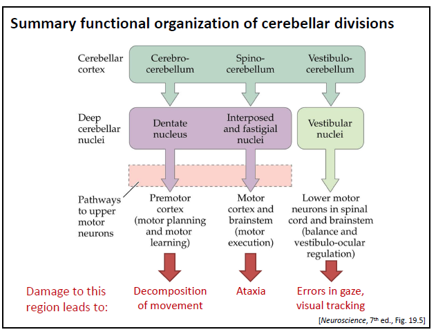
what are the three functional divisions of the cerebellum?
1. Spinocerebellum – control of muscle tone and coordination (mostly vermis and anterior lobe)
2. Cerebrocerebellum – motor planning, learning and memory
3. Vestibulocerebellum – balance, postural adjustments, coordination of eye movements
where do cortical projection to the cerebellum come from?
relay neurons in the pons
what is the path of the cortical motor system?
cerebellar cortex → deep cerebellar nuclei → superior cerebellar peduncle → VL complex (thalamus) → PM and premotor cortex
what is the feedback circuit
cerebellum sends info back to itself to modulate its own activity
fastigial nucleus
is the major efferent output to the brainstem motor systems,
largely through connections to the superior colliculus and reticular
pathways and lesion of the cerebellar divisions
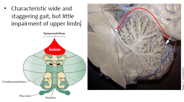
what happens to people with chronic alcohol abuse
degeneration of the anterior portion of the spinocerebellum which is crucial for lower limb activity therefore they have a wide and staggering gait, but little impairment of upper limbs
spiny neurons
excitatory projection neurons
non-spiny neurons
inhibitory interneurons
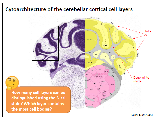
2 layers (white matter isnt a layer) just outermost dark matter split into two layers
what layers can you see in a Nissl stain?
molecular and granule
what layer can you see with fluorescent yellow
purkinji
where do purkinje cells project to?
the molecular layer
where are purkinje cells found?
only the cerebellum
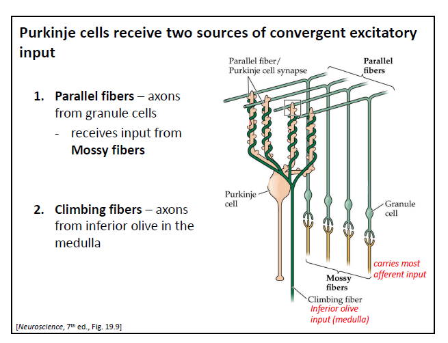
what are the two sources of convergent excitatory input to the purkinje cells?
1. Parallel fibers – axons from granule cells
- receives input from Mossy fibers
2. Climbing fibers – axons from inferior olive in the medulla (strong because its making the most contact on one dendrite (wrapping))
what inhibitory interneurons modulate the activity of Purkinje cells?
Basket cells – found in molecular layer; form an inhibitory “nest” of synapses with
Purkinje cell bodies
• Stellate cell – star-shaped, cell bodies in the molecular layer. Receives input from
parallel fibers and projects inhibitory input to Purkinje cells
• Golgi cells – cell bodies found in granule layer, but dendrites extend into molecular
layer. Also receive input from parallel fibers and provide feedback to granule cells
what is the purpose of having an indirect pathway for basic loopsof cerebellar processing
error correction
if the amount of inhibition does not match the level of excitation, there is an error somewhere that needs to be fixed
which is more active at rest, purkinje cells or deep nuclear cells
purkinje
major input regions of the basal ganglia?
striatum - caudate nucleus and putamen
what are the principle neurons of the striatum
Medium Spiny Neurons
make up 90% of cells of the striatum
Large amount of spines on dendrites
They are GABAergic (inhibitory)
types of Medium Spiny Neurons
2 distinct MSN phenotypes
D1-type MSNs (direct path)
facilitate movement
D2-type MSNs (indirect path)
suppress unwanted movement
dopamine role in basal ganglia
dopamine acts to reduce this inhibitory outputs
parkinson’s disease
Hypokinesia and bradykinesia
• Tremor at rest
• Rigidity
• Postural instability
• Shuffling gait
• Cognitive deficits – decreased motivation and spontaneity,
depression, lack of affect
effects substantia nigra
Huntington’s Disease
• Hyperkinesia: excessive movement
• Chorea
• Cognitive deficits – depression, personality changes (irritability, impulsiveness), deficits in memory and attention