Lecture 13: Cranial Nerves and Special Senses
1/60
There's no tags or description
Looks like no tags are added yet.
Name | Mastery | Learn | Test | Matching | Spaced |
|---|
No study sessions yet.
61 Terms
What are the cranial Nerves (CNs)?
12 peripheral nervous structures
• Originate within cranium
• Enter/exit brain stem directly
CNs primarily
innervate the face and its internal/external structures
One of cranial nerve
innervates thoracic and abdominal organs
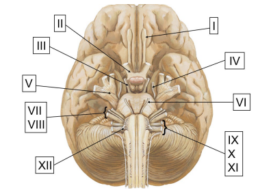
CNs are numbered
from anterior to posterior
I. Olfactory II. Optic III. Oculomotor IV. Trochlear V. Trigeminal VI. Abducens VII. Facial VIII. Vestibulocochlear IX. Glossopharyngeal X. Vagus XI. Spinal Accessory XII. Hypoglossal
Mnemonics for cranial nerves
On, On, On, They Traveled And Found Voldemort Guarding Very Ancient Horcruxes
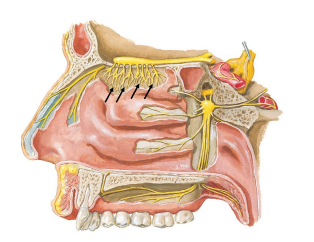
I. Olfactory Nerve
Smell, small nerves in nasal cavity roof
• Pass through cribriform plate of ethmoid

CN I: How do we smell?
Bipolar neurons in mucous membrane, odorants (Scent molecules) bind to olfactory nerves, and can arrive at CN I in 2 ways
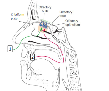
Two ways smell can arrive at CN I
1. Nostrils during breathing
2. Oropharynx during chewing
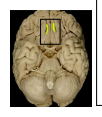
CN I fibers pass through
Cribriform plate of ethmoid
• synapse with two long olfactory tracts
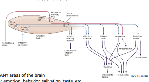
Projections from olfactory bulb have multiple destinations
Olfaction → MANY areas of the brain
• Affects memory, emotion, behavior, salivation, taste, etc
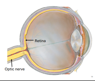
II. Optic Nerve : Vision
Formed by axons of the ganglionic cells in the retina of the eye
Eye structures:
Sclera
Iris
Pupil
Conjunctive
Eyelids

Sclera
White outer layer of eye
• Dense CT

Iris
Pigmented ring of smooth muscle
• Pigment = melanin
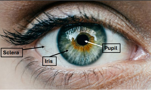
Pupil
Central hole in iris
• Allows light into eye
Conjunctive
Membrane protecting eye and inner eyelid
• Present, but not visible
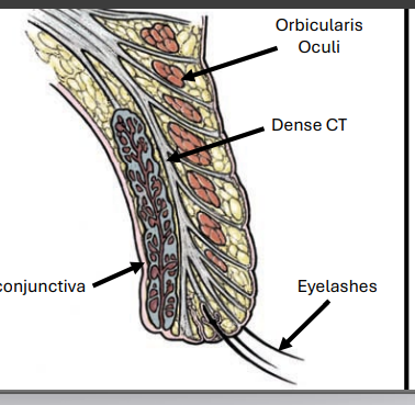
Eyelids
Dense connective tissue core
• Skin on external surfaces
• Conjunctiva on inner surface
• Protect and spread tears
Lacrimal gland
Secretes tears and are spread across the eyes by the eyelids
Drained into the nasal cavity thru a duct
Conjunctivitis: AKA “Pink eye”
Infection and inflammation of conjunctiva
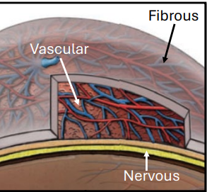
3 major layers (tunics) of Eye
1.Fibrous Tunic
2.Vascular Tunic
3.Nervous Tunic

1.Fibrous Tunic
Sclera, cornea, and lens

2.Vascular Tunic
Major blood supply to/from eye

3.Nervous Tunic
Sensory and autonomics
• Retinal cells

Light must pass through several structures on its way to the retina
1.Cornea → 2. Aqueous humor → 3. Iris (pupil) → 4.Lens → 5.Vitreous humor
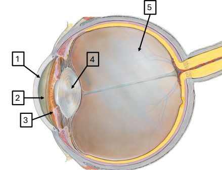
1.Cornea
Dome shaped protective outer layer

2. Aqueous humor
Watery fluid between cornea and lens

3. Iris ( + pupil)
Muscular ring that controls amount of light entering eye
• Pupil: Hole in center of iris

4.Lens
Curved, transparent structure
-Directs light towards retina

5.Vitreous humor
Gel -like substance. Fills most of the eye
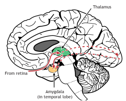
Essentials of optic pathway
Light passes through pupil, lens, humors, and strikes retina
• Optic nerve sends impulses to thalamus, Brodman areas 17, 18,& 19, and other regions of brain
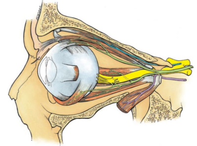
III. Oculomotor Nerve
IV. Trochlear Nerve
VI. Abducens Nerve (6)
Control skeletal muscles that move the eyeball
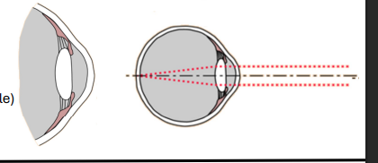
Viewing distant objects
Lens under tension (smooth muscle)
Flatter lens → see farther away
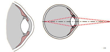
Viewing nearby objects:
Accommodation reflex: Relaxation of lens
• Now more curved → See closer objects in focus
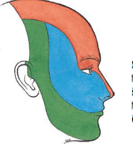
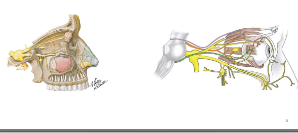
V. Trigeminal Nerve: Three nerve bundles
Opthalmic, Maxillary, Mandibular
Sensation for the bones & skin of the face, teeth & cavities of head
Trigeminal nerve also innervates
The muscles of mastication
-Masseter, temporalis, medial pterygoid, lateral pterygoid
VII. Facial Nerve (7)
Motor to muscles of facial expression
• Taste from anterior 2/3 of tongue
Facial Nerve (7) Parasympathetics Fibers
Lacrimal glands (tears) and Salivary glands (not the parotid)
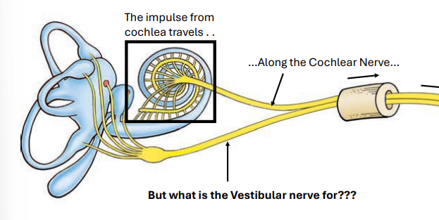
VIII. Vestibulocochlear Nerve
Made of 2 nerves: Vestibular and Cochlear
Balance and hearing
How do we hear ?
The ear (AKA pinna or auricle) receives vibrations in the air (sound waves)
Vibrations pass thru outer ear f
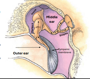
Vibration route
First enter outer ear via ear canal, then strike tympanic membrane (eardrum), and then the vribrations are carried thru 3 tiny ossicles in the middle ear
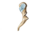
Malleus
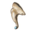
Incus
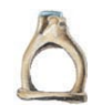
Stapes
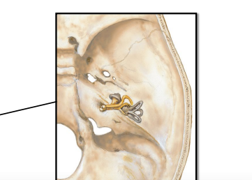
Temporal bone
Contains tiny bony structures that generates sense of hearing and balance
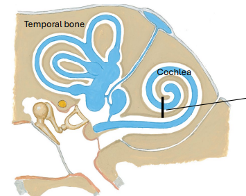
Cochlea
Snail shell shaped spiral filled with fluid called endolymph
Vibration in fluid causes cilia to bend and create an action potential
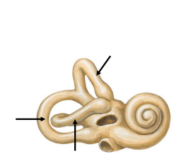
The vestibular system
a sensory system in the inner ear that balance and proprioception
Made of 3 horseshoe-shaped semicircular canals, all oriented differently, and filled with endolymph
Dynamic Equilibrium: Sensing angular movement and velocity
1. Head moves
2. endolymph flows
3. hair cells bend
4. Action potential generated along vestibular nerve to pons
Utricle and Saccule
bulging chambers in vestibule contain fluid & hair cells with suspended crystals - OTOLITHS
Otoliths role
Motion causes crystals to move the hair cells creating a electrical impulse → Important for static equilibrium
IX. Glossopharyngeal Nerve
9, Taste/touch Posterior 1/3 of Tongue
Flavor 5 tastes:
Salty, sweet, bitter, sour, and umiam (savory)
Taste actually comes from
Direct chemical stimulation of tastebuds, stimulated by olfactory receptors by vapors from food
mechanoreceptors, nociceptors and chemoreceptors of oral cavity: Texture, Spice, heat
3 Receptors for taste
Stimulation of mechanoreceptors, nociceptors and chemoreceptors of oral cavity: Texture, Spice, heat
Taste buds
Onion shaped cells on tongue, soft palate, and epiglottis
Have receptors that tastants (molecules from food/drink)
Buds generate action potentials that ride along:
• Facial (CN VII)
• Glossopharyngeal (CNIX)
• Vagus (CN X)
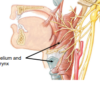
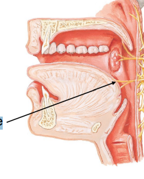
X. Vagus Nerve: Somatic Motor/Sensory
10, Sensory and motor to epithelium and muscles of pharynx and larynx
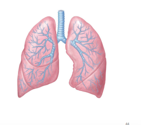
X. Vagus Nerve: Parasympathetics to heart and airway
parasympathetic "rest and digest" nervous system, connecting the brain to organs like the heart and lungs
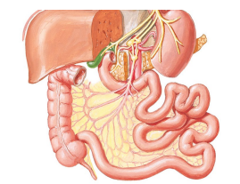
X. Vagus Nerve: Autonomics control
control of smooth muscle and gland secretion in digestive system
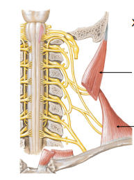
XI. Spinal Accessory Nerve Motor to 2 muscles:
11, Sternocleidomastoid (head movement) and Trapezius (shoulder abd)
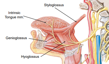
XII. Hypoglossal nerve:
Muscles of the tongue