Anime Trivia Night
1/72
There's no tags or description
Looks like no tags are added yet.
Name | Mastery | Learn | Test | Matching | Spaced | Call with Kai |
|---|
No analytics yet
Send a link to your students to track their progress
73 Terms
Masseter
What muscle is most responsible for protrusive movements?
Facial nerve
The muscle that closes the eyelid is innervated by?
Parietal & frontal lobe
Homunculus motor and somatosensory cortex are in what region of the brain?
Occipital lobe
What areas of the brain process visual information & perception of motion?
Lateral & Medial geniculate of the thalamus
The midbrain's superior colliculus and inferior colliculus is associated with what thalamus nuclei?
a. Is covered by the parotideomasseteric fascia
Parotid gland:
a. Is covered by the parotideomasseteric fascia
b. Is supplied by the retromandibular artery
c. Has its sympathetic innervation provided by the otic ganglion
d. Penetrated by the auriculotemporal nerve
e. Has lymph drainage to submandibular lymph nodes
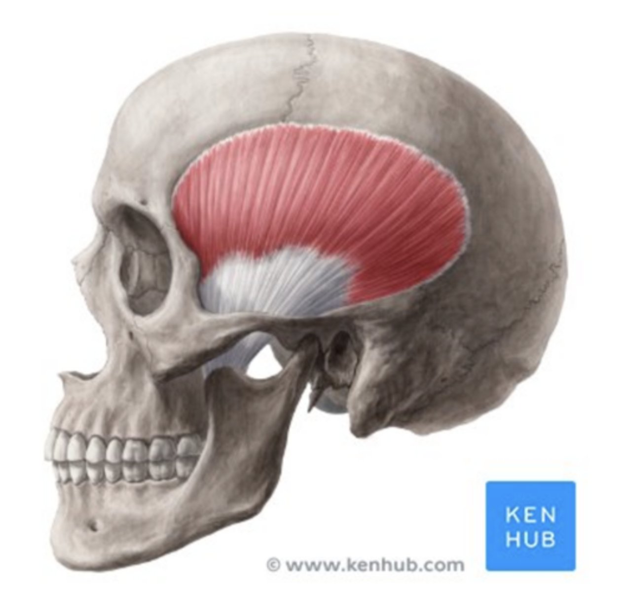
Temporalis muscle
What is the labelled structure?
d. All
The TMJ is mainly innervated by which nerves?
a. Auriculotemporal nerve
b. Masseteric nerve
c. Deep temporal nerves
d. All
Submasseteric space/masseteric space
What is the name of the space that is between the lateral side of the mandible and medial to the masseter, and tends to be infected when there is mandibular third molar infection?
Auriculotemporal nerve
A patient comes to your office complaining of jaw pain. To rule out the possibility of TMJ pain, you inject lidocaine (an anesthesia) extraorally to the nerve that is posterior to the condyle neck. What is the name of the nerve?
Globus pallidus
Which is the most medial structure of the cerebral ganglion?
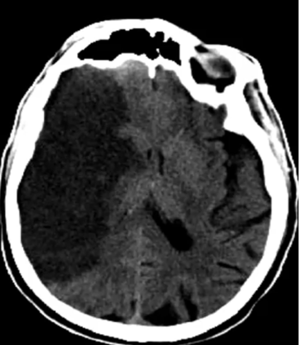
Middle cerebral artery (MCA)
What vascular area is affected by this stroke?
a. Receptor density
MRI can detect all of the following EXCEPT:
a. Receptor density
b. Grey matter
c. White matter
d. Oxygenation level
e. Tumor
a. Palatine Tonsils
Which is not drained by the submental lymph node?
a. Palatine Tonsils
b. Frenulum and apex of tongue
c. Mandibular incisors
b. Palatine- floor
Medial wall of orbit is formed by all the following bones except:
a. Ethmoid
b. Palatine- floor
c. Maxilla
d. Sphenoid
e. Lacrimal
e. All of these structures
The periorbita is continuous with the:
a. Dura at the optic canal
b. Fascial sheaths of the extraocular muscles
c. Periosteum covering the skull
d. Orbital septum
e. All of these structures
Sulcus & Gyri
Brodmans area are based off of?
Abducens nerve
The lateral rectus muscle is innervated by the:
3rd
What ventricle lies medial to the thalamus?
Infraorbital nerve
Either bleeding or an expanding aneurysm in the cavernous sinus may cause a loss of sensation in the skin just below the eye. Which nerve would be affected to cause this symptom?
Oculomotor
Posterior communicating artery aneurysm would cause compression of what nerve?
Trigeminal nerve
Which nerve comes from the metencephalon?
Temporal lobe
What is the inferior border of the lateral fissure?
Frontal, temporal
What lobes are anterior to central sulcus & ventral to lateral fissure?
Arthralgia
A 49 year old patient comes oftenly to your office. She recently complains of a new pain, occurring when yawning and eating. When you examine her TMJ, you find that her joint movement is normal but there is tenderness and palpation when pressing to the area superior to the condyle. What is your diagnosis?
Oculomotor
What nerve innervates the muscle extorting the eye?
Hypoglossal
What is the nerve coming from between the pyramid and the olive of medulla?
GVE
The superior salivary nucleus has cell bodies of neurons that control secretion from the submandibular salivary gland. These neurons are which functional type?
Nasopalatine
Which nerves innervate the gingiva medial to upper canine teeth?
Pterygopalatine
Which parasympathetic postganglionic ganglion is responsible for controlling mucus secretions of the nasal cavity?
Cornea
Which structure serves as the main refractive medium for light entering the eye?
Internal carotid artery
The ophthalmic artery is a branch of the:
Facial vein, angular vein, ophthalmic vein, cavernous sinus
Infection near the corner of the eye is a danger area because of the route it takes which is:
Levator palpebrae superioris muscle
Ptosis, a low positioned upper eyelid, is due to the paralysis of what muscle?
III, VII, IX, X
All of the cranial nerves in which group contain preganglionic parasympathetic fibers?
a. They have a central process that is part of the olfactory tract
Which is not true about olfactory neurons?
a. They have a central process that is part of the olfactory tract
b. They have a process that passes through the cribriform plate
c. They synapse in the olfactory bulb
d. They have cilia that carry the odor receptor molecules
e. They are bipolar neurons
Otic ganglion
Cell bodies of postganglionic parasympathetic neurons controlling secretion from the parotid gland are located in which ganglion?
Petrotympanic fissure
A portion of CN VII, in the chordae tympani, passes through which fissure?
Superior and inferior of glossopharyngeal
What ganglion has nerve fibers innervating the posterior portion of the tongue and responsible for taste?
Lacrimal nerve, frontal nerve, trochlear nerve
Which nerves pass through the orbital fissure, but pass outside of the Annulus of Zinn in order to reach orbit?
Corpus callosum
Which structure contains the vast majority of commissural fibers in the cerebrum?
V, IV
Cortical output to the thalamus originates in layer ____, while thalamic input into cerebral cortex arrives in layer _____
Optic disc
The optic nerve leaves retina at:
It helps the brain float/suspend in the cranial space
What is the responsibility of CSF?
Superior ophthalmic vein → cavernous sinus → inferior petrosal sinus → Sigmoid sinus → Internal jugular vein
Blood circulating the dural sinuses is in the pathway:
c. They are between the dura and arachnoid mater
Regarding the dural venous sinuses, which is incorrect?
a. They are between the parietal and meningeal layers of the dura
b. They contain venous blood
c. They are between the dura and arachnoid mater
d. The superior sagittal sinus communicates with the scalp via emissary veins
e. They are between two meningeal layers of the dura
e. The tentorium cerebelli is formed by the dura mater
Which is correct?
a. The dura mater is supplied by the internal carotid artery
b. The dura mater is single-layered strong membrane
c. The dura mater's sensory innervation is provided by the facial nerve
d. The tentorium cerebelli separates the two hemispheres of the cerebrum
e. The tentorium cerebelli is formed by the dura mater
a. It courses superficial to the zygomaticus major muscle
Regarding the facial vein, which is incorrect?
a. It courses superficial to the zygomaticus major muscle
b. It joins the internal jugular vein after forming the common facial vein with the retromandibular vein and the lingual vein
c. It may take blood to the superior ophthalmic vein
Abducens
Which nerve leaves the brainstem along the inferior border of pons (pons-medulla junction)?
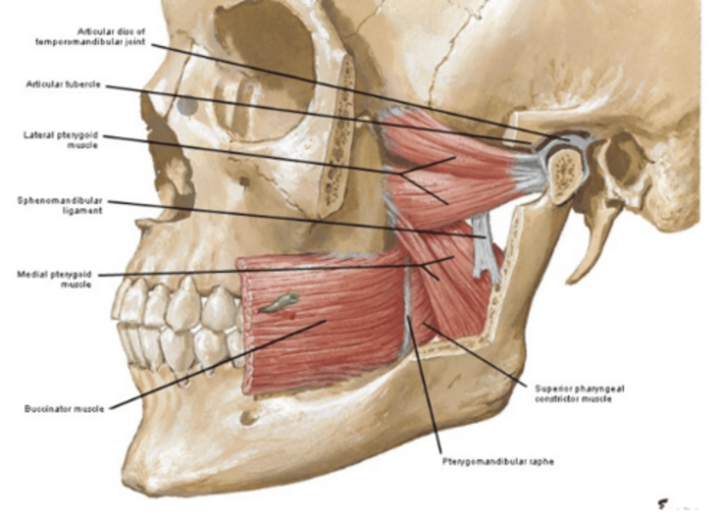
Laterotrusive movement
What movement does the lateral pterygoid help with?
c. Innervate the stylopharyngeus muscle
Special visceral efferent neurons:
a. Innervate the superior oblique muscle of the eye
b. Are preganglionic parasympathetic neurons
c. Innervate the stylopharyngeus muscle
d. Innervate the parotid gland
e. Innervate the genioglossus muscle of the tongue
Parieto-Occipital sulcus
Which structure separates the parietal and occipital lobes?
b. Hypoglossal nerve
Which does NOT contain nerve fibers that innervate taste buds?
a. Chorda tympani
b. Hypoglossal nerve
c. Glossopharyngeal nerve
d. Facial nerve
e. Lingual nerve
Anterior division of V3
The deep temporal nerves are direct branches of which structure?
c. Putaman
Which is not a part of the deep nuclei of the cerebellum?
a. Dentate
b. Fastigial
c. Putaman
d. Globose
e. Emboliform
Mental nerve
What nerve innervates the labial gingiva of the mandibular incisors?
b. Insula
Which structure of the brain cannot be seen in the mid-sagittal section?
a. Hypothalamus
b. Insula
c. Thalamus
d. Corpus callosum
Petrosal sinuses, sigmoid sinus
Cavernous sinus drains to _______ which drains out of the skull via _________:
Trochlear
What nerve innervates the muscle to intort the eye?
Facial
Which nerve arises from the pons-medulla junction?
Ciliary ganglion
Cell bodies of postganglionic parasympathetic neurons that adjust the lens of the eye for close focusing are located in which ganglion?
Tympanic nerve
Preganglionic parasympathetic fibers that control secretion from the parotid gland are found in the:
Internal carotid artery, Oculomotor nerve, abducens nerve, ophthalmic nerve and, maxillary nerve
What structures pass through the cavernous sinuses?
Pyramid
What structure contains corticospinal tracts?
a. SSA: glossopharyngeal nerve, posterior tongue
Which pairing of modalities: cranial nerves is not correct?
a. SSA: glossopharyngeal nerve, posterior tongue
b. SSA: optic nerve, eye
c. GVE: vagus nerve, smooth muscle of stomach wall
d. GSA: glossopharyngeal nerve, pharynx
e. SVE: glossopharyngeal nerve, stylopharygenus muscle
Mandibular incisors
The submental lymphoid nodes collect lymph from?
Frontal lobe
What is superior to lateral fissure?
Dilator pupillae
In horner's syndrome, which muscle is paralyzed, causing miosis?
Orbital septum
Which structure is most responsible for limiting the spread of infection to and from the orbit?
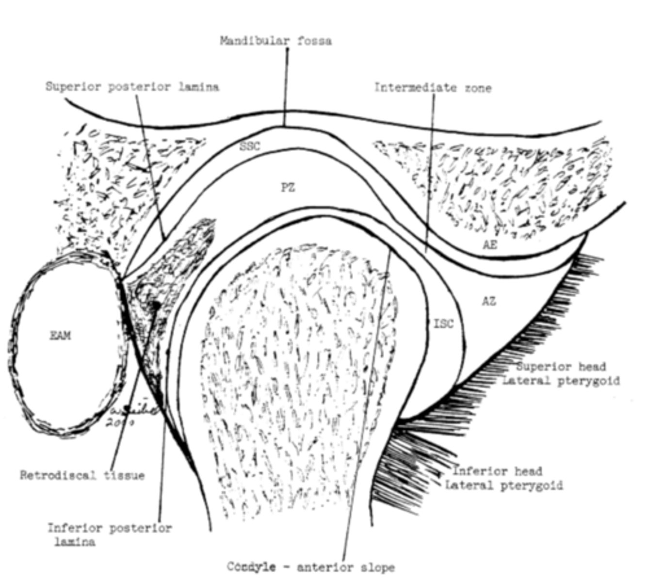
Hinge movement
What movement happens at the inferior joint space?
a. Trigeminal neuralgia only in mandibular branch of trigeminal nerve
A patient feels pain when a doctor presses the infraorbital region. Which is the least likely diagnosis?
a. Trigeminal neuralgia only in mandibular branch of trigeminal nerve
b. Parotitis
c. Swelling of lymph nodes near the parotid gland
d. Root inflammation of a upper molar
Horizontal, sagittal
In which planes (MRI) could you view Both the genu and splenium of the corpus callosum on the same slice?
Superior and inferior ganglia of glossopharyngeal nerve
Fibers that carry sensation of taste from posterior ⅓ of tongue have cell bodies where?