Cardiovascular System
1/36
There's no tags or description
Looks like no tags are added yet.
Name | Mastery | Learn | Test | Matching | Spaced | Call with Kai |
|---|
No analytics yet
Send a link to your students to track their progress
37 Terms
Cardiovascular System
The system that comprises blood, blood vessels, and the heart, responsible for transporting materials throughout the body.
Blood
A fluid connective tissue that transports oxygen, nutrients, and waste products throughout the body.
Arteries
Blood vessels that carry blood away from the heart under high pressure.
Veins
Blood vessels that carry blood toward the heart, possessing valves to prevent backflow.
Capillaries
Tiny blood vessels where gas and nutrient exchange occurs with tissues.
List the 3 functions of the cardiovascular system
transport (gases, nutrients, wastes, regulatory and processed molecules)
protection (inflammation, phagocytosis, antibody production, platelets for clotting)
regulation (fluid balance, pH, temp, blood pressure)
What are the functions of the heart
Pumps blood, generates pressure, separates pulmonary/systemic circulation, regulates blood supply.
How is the heart protected
by rib cage, protective membranes and fluid surrounding it
what is the location of the heart
In the thoracic cavity obliquely situated in the mediastinum, medial to the lungs and superior to the diaphragm.
Pericardium
FIBROUS pericardium: tough outer layer, prevents distention and serves as an anchor for the heart
SEROUS pericardium: thin, inner layer composed of simple squamous epithelium
parietal: lines the fibrous outer layer
visceral: covers the hearts surface
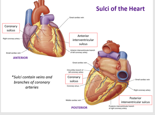
What are sulci
Coronary sulcus, anterior interventricular sulcus and posterior interventricular sulcus. Function is to separate different chambers of the heart.
Where is there fat in the heart
pericardium: between visceral and parietal pericardium
epicardial: between outer layer of myocardium and visceral layer of pericardium
What are trabeculae carnae
muscular ridges and columns on inside walls of ventricles. Function is to assist the contraction of the heart and facilitate blood flow within ventricles.
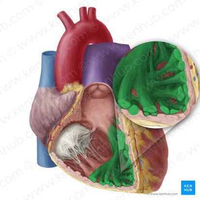
what are the pectinate muscles
muscular ridges in auricles and atrial wall

Epicardium
visceral serous membrane, smooth outer surface, simple squamous epithelium

Myocardium
The thick middle muscular layer of the heart responsible for heart contractions.
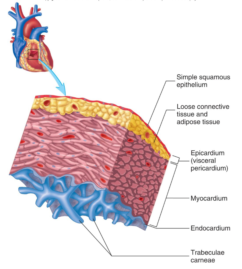
Endocardium
smooth inner surface covering heart cambers and valves, simple squamous epithelium. Covers valve surface and continuous endothelium

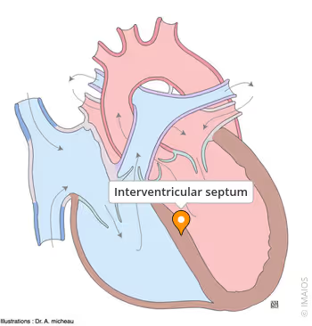
Interventricular Septum
The wall that separates the left and right ventricles of the heart.
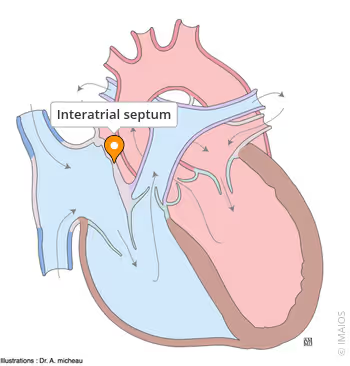
Inter atrial septum
wall between the atria. Contains a depression, fossa ovalis: a remnant of the fetal
opening (foramen ovale) between the atria
Right atrium
Thin-walled, receives deoxygenated blood from the body through three openings: Superior vena cava, inferior vena cava, and coronary sinus.
○ Contains pectinate muscles for large force of contraction
○ Auricles are extensions to increase volume of atrium
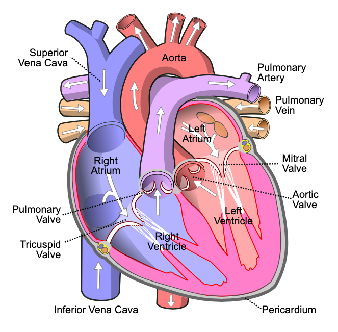
Right ventricle
Pumping chamber, thicker walls, receives deoxygenated blood from the right atrium and opens to pulmonary trunk.
○ Features trabeculae carnae.

Left atrium
Receives oxygenated blood from lungs through four pulmonary veins; forms heart’s base.
○ Also contains pectinate muscles.
○ Auricles are extensions to increase volume
○ Thin walled

Left ventricle
Thickest-walled chamber, pumps oxygenated blood into the aorta.
○ Contains trabeculae carnae and forms the apex of the heart.

Atrioventricular (AV) Valves
Valves located between the atria and ventricles that prevent backflow of blood. They are attached to papillary muscles by tendons called chordae tenineae
Semilunar Valves
Valves located at the base of the pulmonary trunk and aorta that prevent backflow into the ventricles.
Cardiac Cycle
The sequence of contraction (systole) and relaxation (diastole) of the heart chambers.
Cardiac Output
The amount of blood the heart pumps in a given time, calculated as heart rate multiplied by stroke volume.
Sinoatrial Node (SA Node)
The primary pacemaker of the heart, initiating electrical impulses.
Erythrocytes (RBC)
Red blood cells that carry oxygen in the blood; they are biconcave and lack a nucleus.
Leukocytes (WBC)
White blood cells involved in the immune response and protection against infection.
Plasma
The liquid component of blood, making up about 55% of its volume, containing water, proteins, and solutes.
Capillary Exchange
The process by which substances move into and out of capillaries, providing nutrients to tissues.
Edema
Swelling due to excessive fluid accumulation in tissues, often caused by capillary issues or heart problems.
Pericardium
The fibrous and serous membrane that surrounds and protects the heart.
Trabeculae Carnae
Muscular ridges and columns found on the interior walls of the ventricles.
Hemoglobin
A protein in red blood cells that binds to oxygen for transport.
Lymphatic System
A network of vessels and nodes that help maintain fluid balance and contribute to the immune system.