Lesson 9: Spermatogenesis and Oogenesis
1/27
There's no tags or description
Looks like no tags are added yet.
Name | Mastery | Learn | Test | Matching | Spaced | Call with Kai |
|---|
No analytics yet
Send a link to your students to track their progress
28 Terms

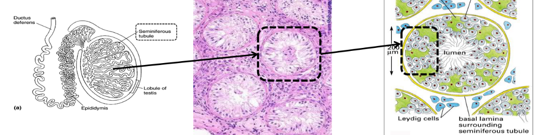
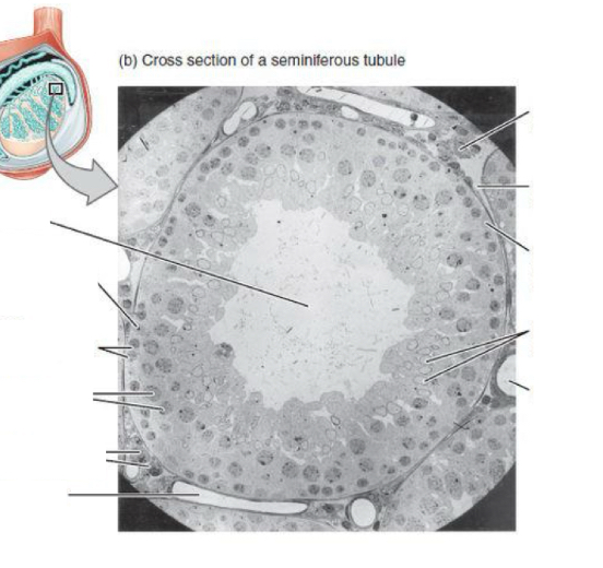
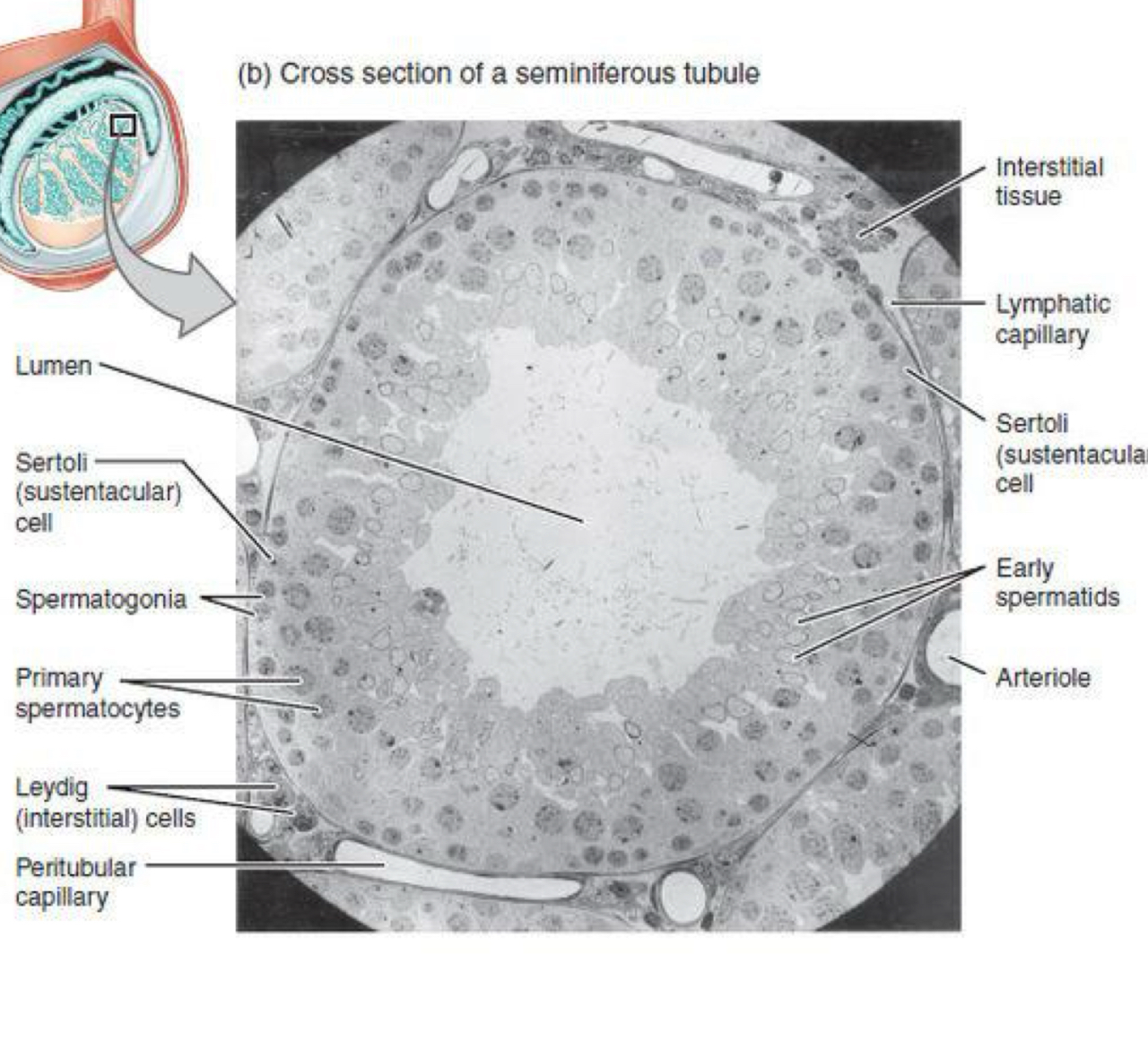
What is spermatogenesis and where does it take place?
-Formation of spermatozoa (sperm cells)
-formed inside the testes, in the seminiferous tubules
-mature by ascending towards the lumen of tubule where they will be released
What are the characteristics of spermatogonia?
-large and round cells with homogenous cytoplasm
-divide by mitosis
-grow and transform in primary spermatocytes and many undergo meiosis to produce spermatozoa
What are supportive cells for sperm and what is their function?
Sertoli cells provide nutrients
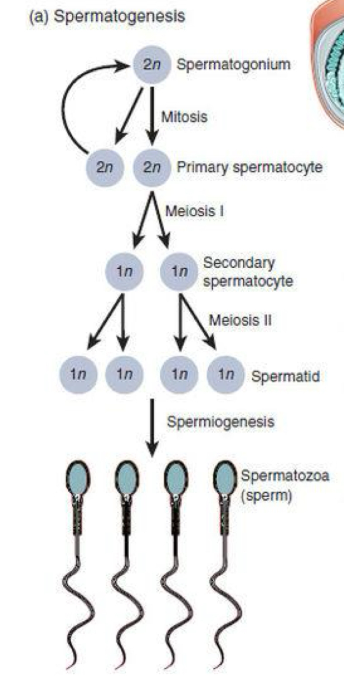
What are the stages of spermatogonia?
Growth of spermatogonia
Meiosis
Spermiogenesis
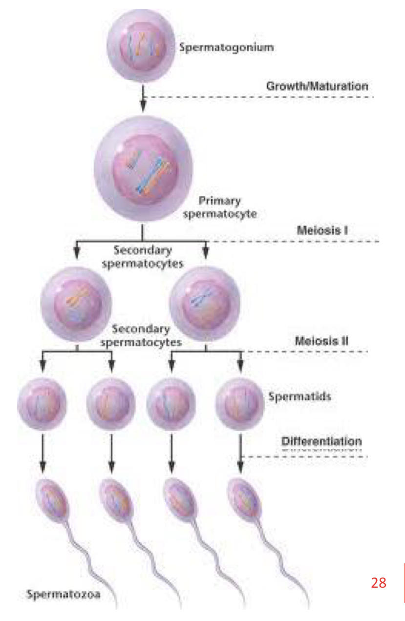
Growth of spermatogonia
A spermatogonium grows (26 days) to become a primary spermatocyte

Meiosis
➢ This primary spermatocyte will enter the first
meiotic division, producing two secondary spermatocytes.
➢ These will quickly go through the second
meiotic division, giving rise to 4 haploid cells called spermatids.

Spermiogenesis
Finally, each spermatid enters spermiogenesis
(metamorphosis) to form mature spermatozoa.
Spermatids formation and structure
Become mature spermatozoa by a differentiation (maturation) process, there are no more cell divisions.
-round, spherical or polygoonal shape
-dispersed chromatin
Structural changes in spermiogenesis: Nucleus, Golgi complex and Acrosome
Nucleus is enlarged and flattened
→ Chromatin condenses
→ histones disappear
→ substituted by protamines.
Golgi complex
-increases its size and forms vesicles that will fuse to form the acrosome
The acrosome binds to the outer membrane of the nuclear envelope
→ contains carbohydrates and several hydrolytic enzymes
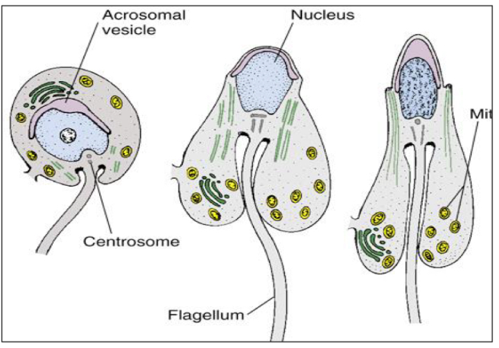
Structural changes in spermiogenesis: Centrioles, Mitochondria and Cytoplasm
Centrioles
→ move towards the opposite side of the head.
- The centriole that is nearest to the nucleus (proximal) remains inactive
- the distal centriole starts to form the flagellum.
Mitochondria
-migrate within the cytoplasm, and are concentrated at the base of the flagellum
Cytoplasm
-migrates towards the base:
o A part surrounds the intermediate area of the spermatozoon.
o the rest forms the residual cytoplasm, which will be phagocytized by Sertoli cells
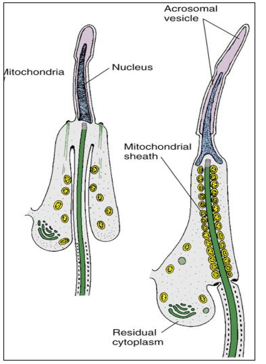
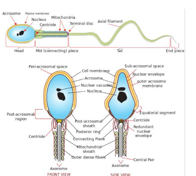
Structure of spermatozoa
Head (nucleus and acrosome)
• Middle or connecting piece (AXONEME)
• Principal piece or tail
• Terminal or end piece
Oogenesis
Synthesis of egg or ovum
→ ocurs in ovaries from germ cells called oogonia
When does Oogenesis start?
starts during fetal development, before birth:
Migration of primordial germ cells towards the gonads: at the beginning of embryogenesis, primordial germ cells migrate to the ovary in formation, becoming oogonia.
Proliferation: oogonia proliferate by mitosis (before meiosis begins).
Growth: oogonia grow, transform into primary oocytes and begin meiosis
Oogenesis in humans
- All primary oocytes are formed between the 4th and 7th month of embryonic development.
-In the 7th month, oogenesis stops and some oogonia start degenerating.
- At birth, there will be only be 500.000 oogonia.
- At puberty (when maturation will continue), there
will be 500 oogonia left.
Steps of oogenesis
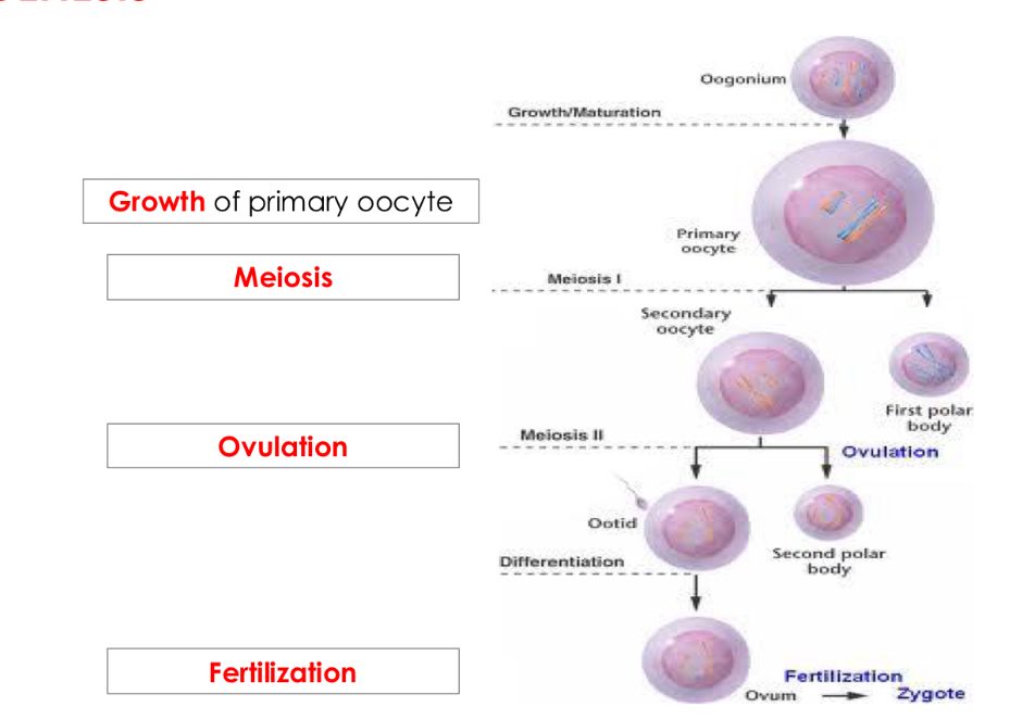
Oogonia growth
Between 4th and 7th month oogonia enter proliferation (7 days) and become primary oocytes, right before entering meiosis.
- Diameter changes from 30 to 140µm.
- Intense metabolic activity (anabolism) and incorporation of exogenous molecules.
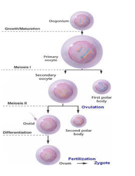
Oogenesis in Meiosis 1
• Primary oocyte enters meiosis I and is ‘paused’ in diplotene of prophase I (7th month of development).
• After puberty and until menopause, every menstrual cycle (28 days) an oocyte will complete the maturation process:
• Primary oocyte will end up first meiotic division forming two types of cells:
- Secondary oocyte (precursor of the egg).
- First polar body.
Oogenesis during Meiosis 2:
• The secondary oocyte starts the second meiotic division.
Resulting in:
- An egg, also called ovum or ovotid
- Another polar body
The oocyte will be arrested in metaphase II until it is fertilized
by an spermatozoon:
o If fertilisation occurs, the secondary oocyte completes
the 2nd division, giving rise to the egg.
o If there is no fertilization, ovotid will degenerate without
having completed the second meiotic division.
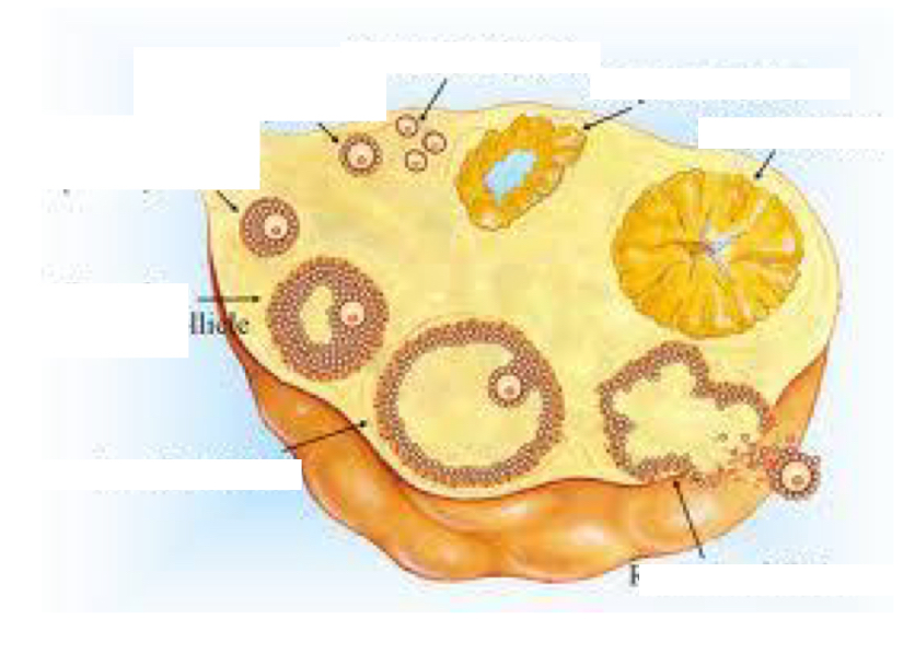
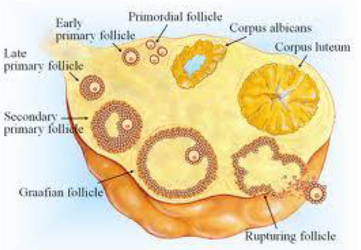
Follicular maturation
In the ovary, oocytes in development are surrounded by specialized
accessory cells, that help in their protection and nutrition: follicle cells forming the follicle.
▪ Follicles undergo several stages of maturation. Follicular maturation is characterised by an increase in the number and thickness of follicular cells.
Follicular maturation in newborn girls
Primary oocytes are surrounded by a single layer of cells forming the primordial follicle, a single layer of flattened follicle cells.
Follicular maturation in pubtery
In puberty, some primordial follicles grow and evolve
to:
▪ Primary follicle: a single layer of cubic follicular cells.
▪ Secondary follicle: several layers
▪ Tertiary follicle: several layers, containing a cavity, called an antrum.
▪ Graafian follicle: large tertiary follicle, ready for ovulation
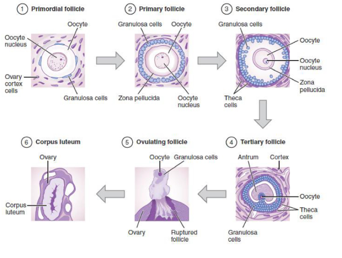
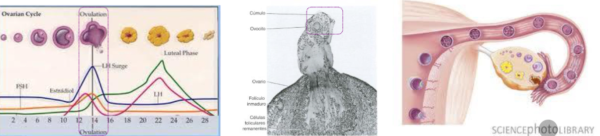
Follicular maturation after ovulation
The follicular cells remaining in the ovary become the corpus luteum
Ovalution
the ovarian follicle ruptures and the secondary oocyte and the first layer of follicular cells are released.
- Ovulation corresponds to the hormonal peak on day 14.
- The oocyte goes to the uterine tubes (or fallopian tube)
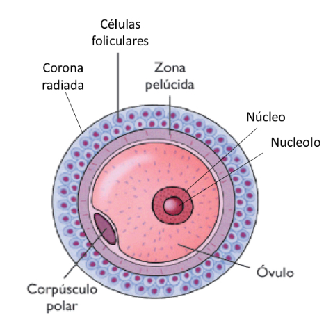
Characteristics of the oocyte/egg
• Large functional gamete.
• Spherical or ovoid shape.
• Contains reserve material: nutrients rich in lipidsand proteins.
Covered by:
o zona pellucida: extracellular matrix that protects from mechanical aggressions.
o Corona radiata: layer of follicular cells around the oocyte.
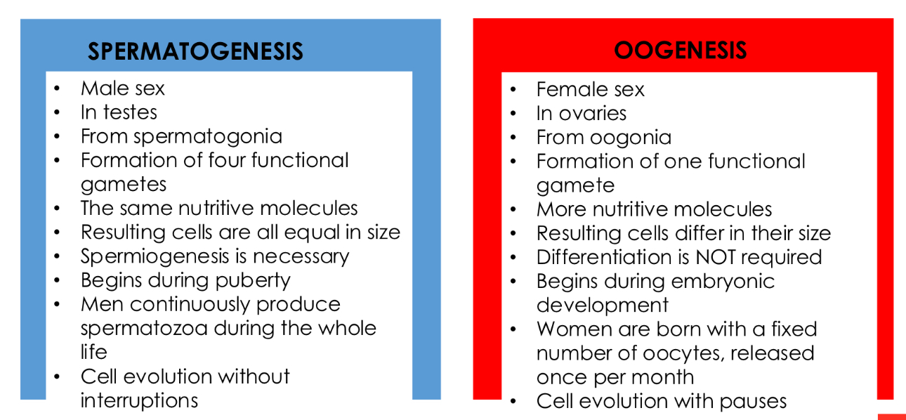
Spermatogenesis and Oogenesis similarities
• Cell production through MEIOSIS. However, both processes initiate their phases from germ cells produced by mitosis.
• Gametes are produced from GERM CELLS (different from somatic cells).
• The process is carried out in GONADS or reproductive organs.
• Generation of GAMETES or sexual cells.
• Progression from DIPLOID TO HAPLOID CELLS.
• Cell fate is FERTILIZATION.
Spermatogenesis and Oogenesis differences