WSU Biology 315 Lab Exam #1. week 1
1/150
There's no tags or description
Looks like no tags are added yet.
Name | Mastery | Learn | Test | Matching | Spaced | Call with Kai |
|---|
No study sessions yet.
151 Terms
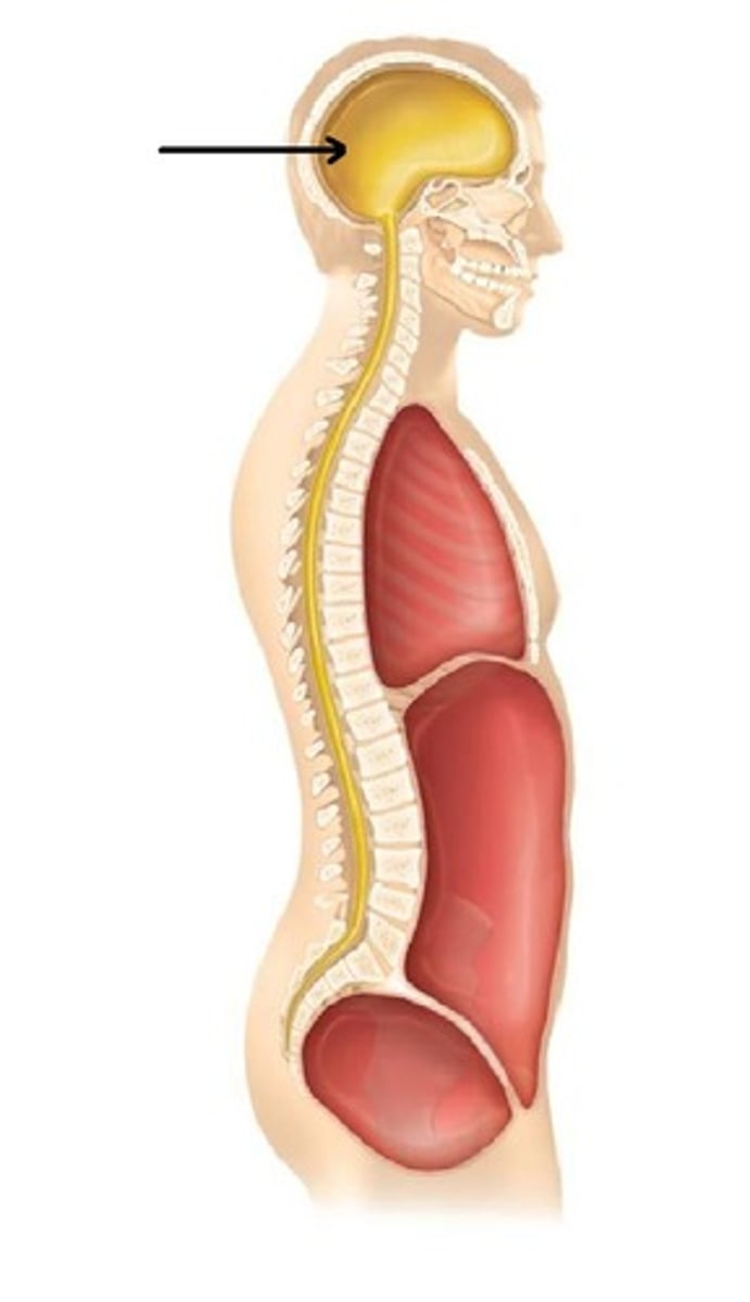
cranial cavity
space
entire space where the brain sits (fist in base)
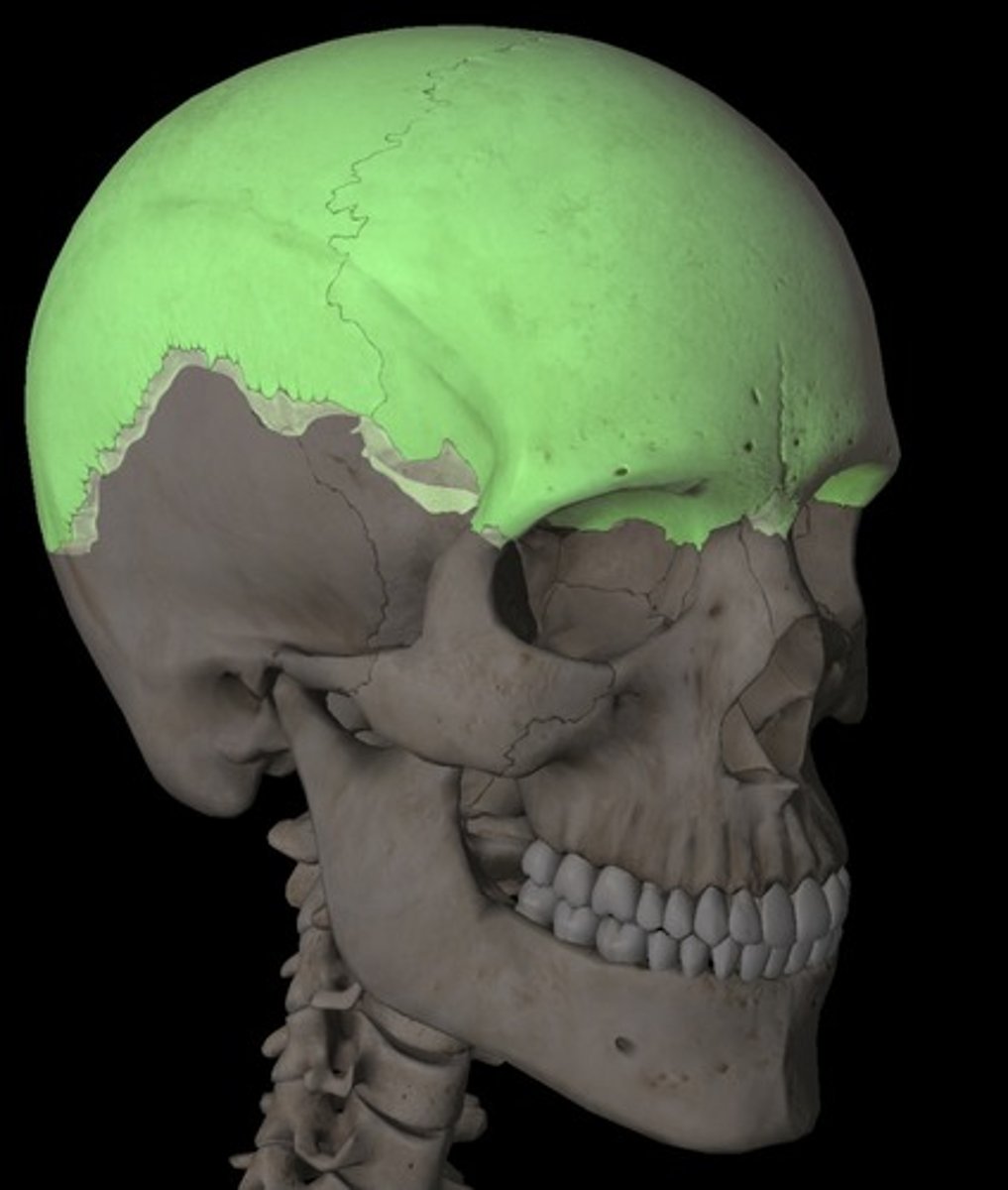
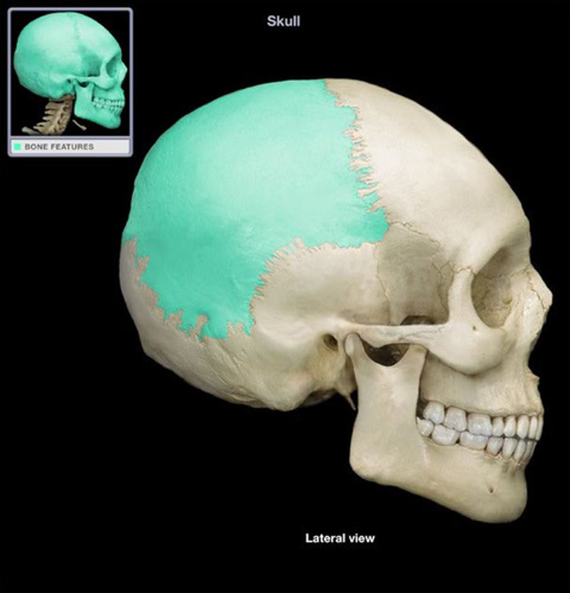
calvaria
structure
top of skull / skull cap (removal of the skull)
cranial base
structure
floor of the cranial cavity, bottom part (hand flat on the inside)
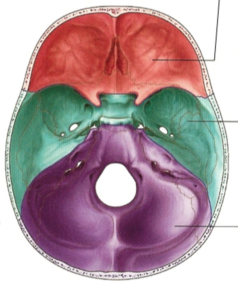
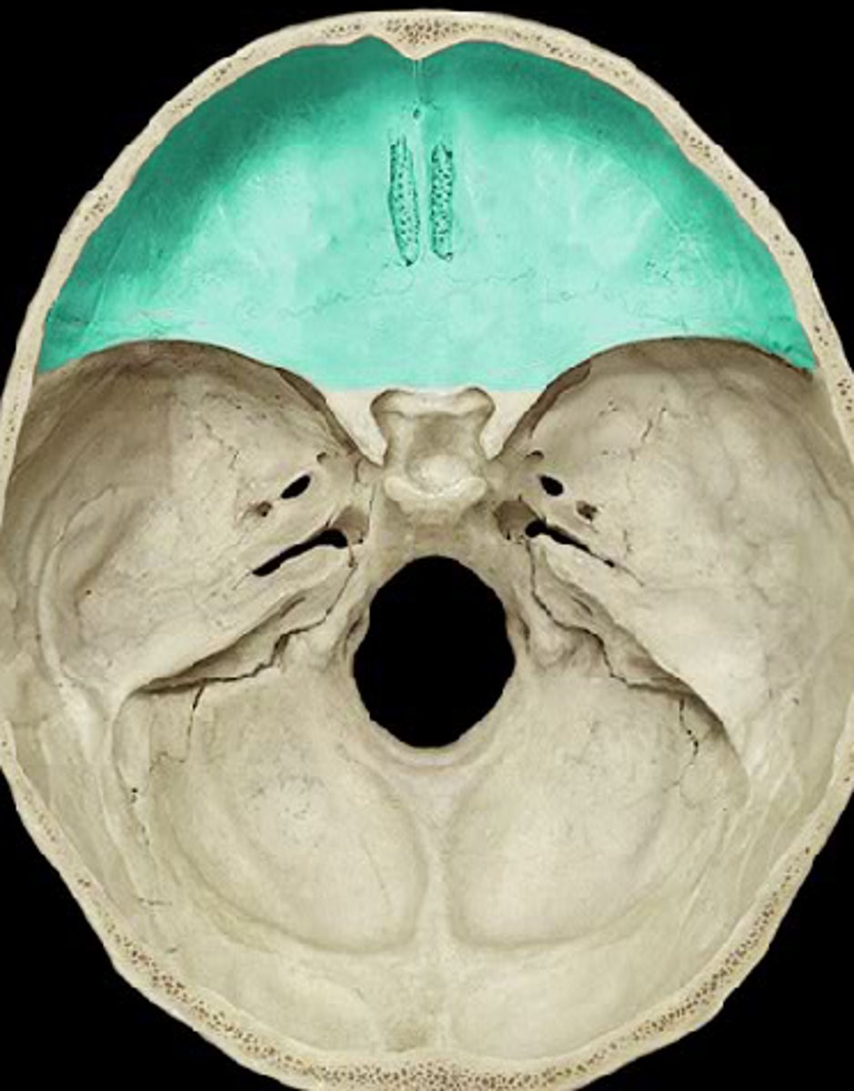
anterior cranial fossa
depression
front part of cranial base
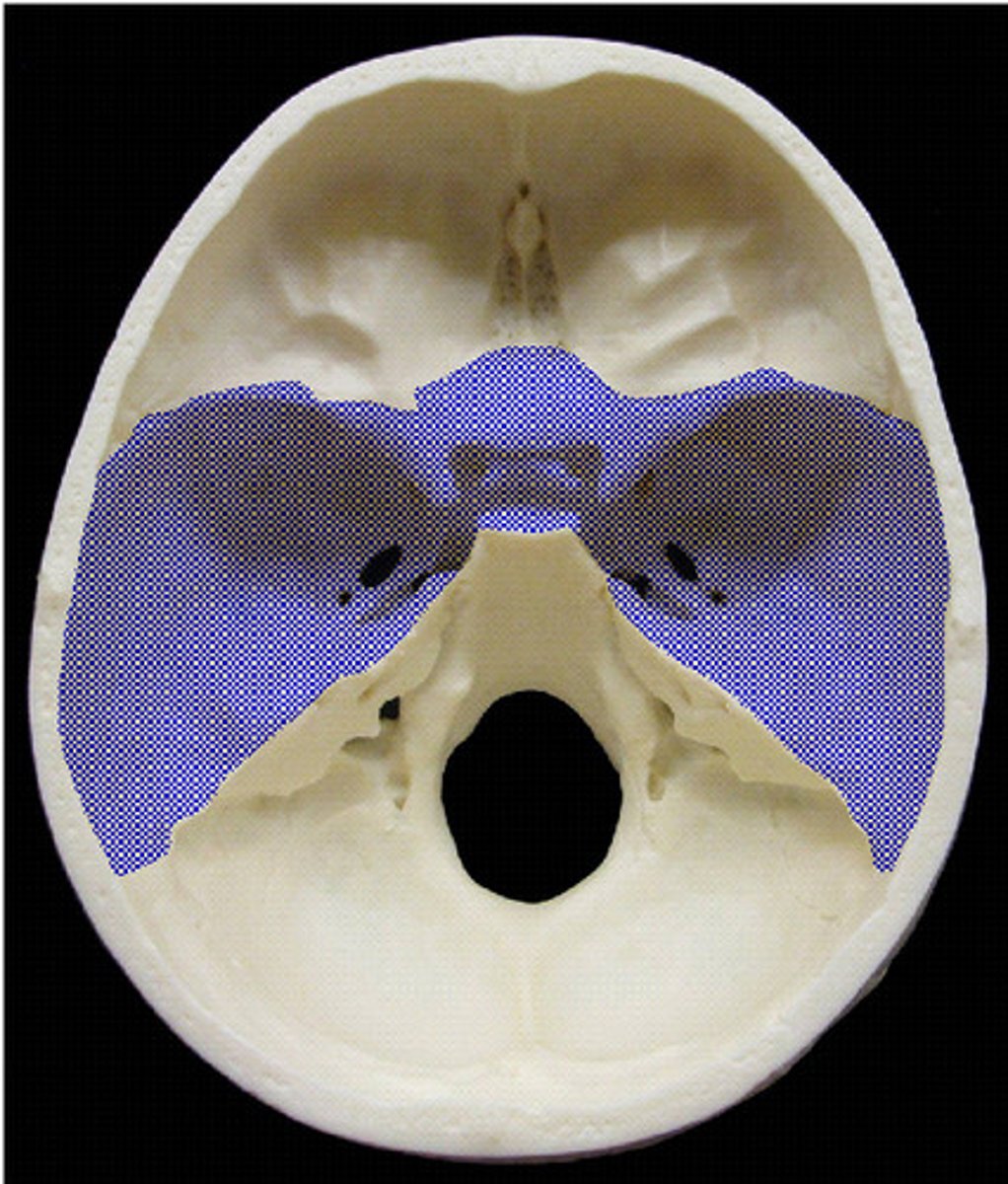
middle cranial fossa
depression
middle part of the cranial base
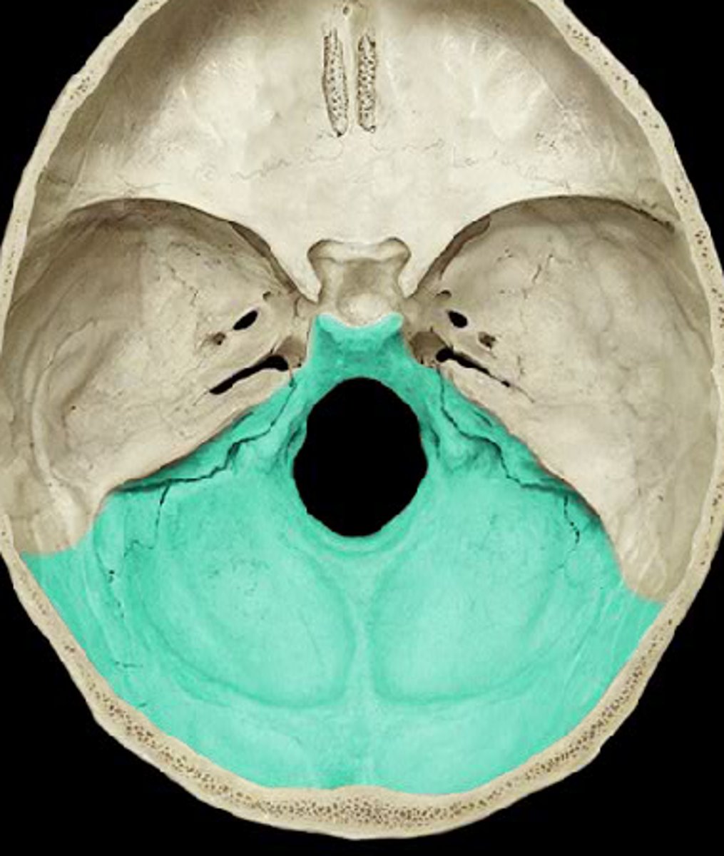
posterior cranial fossa
depression
back side of cranial base
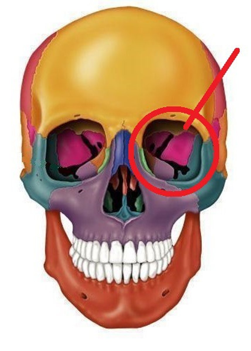
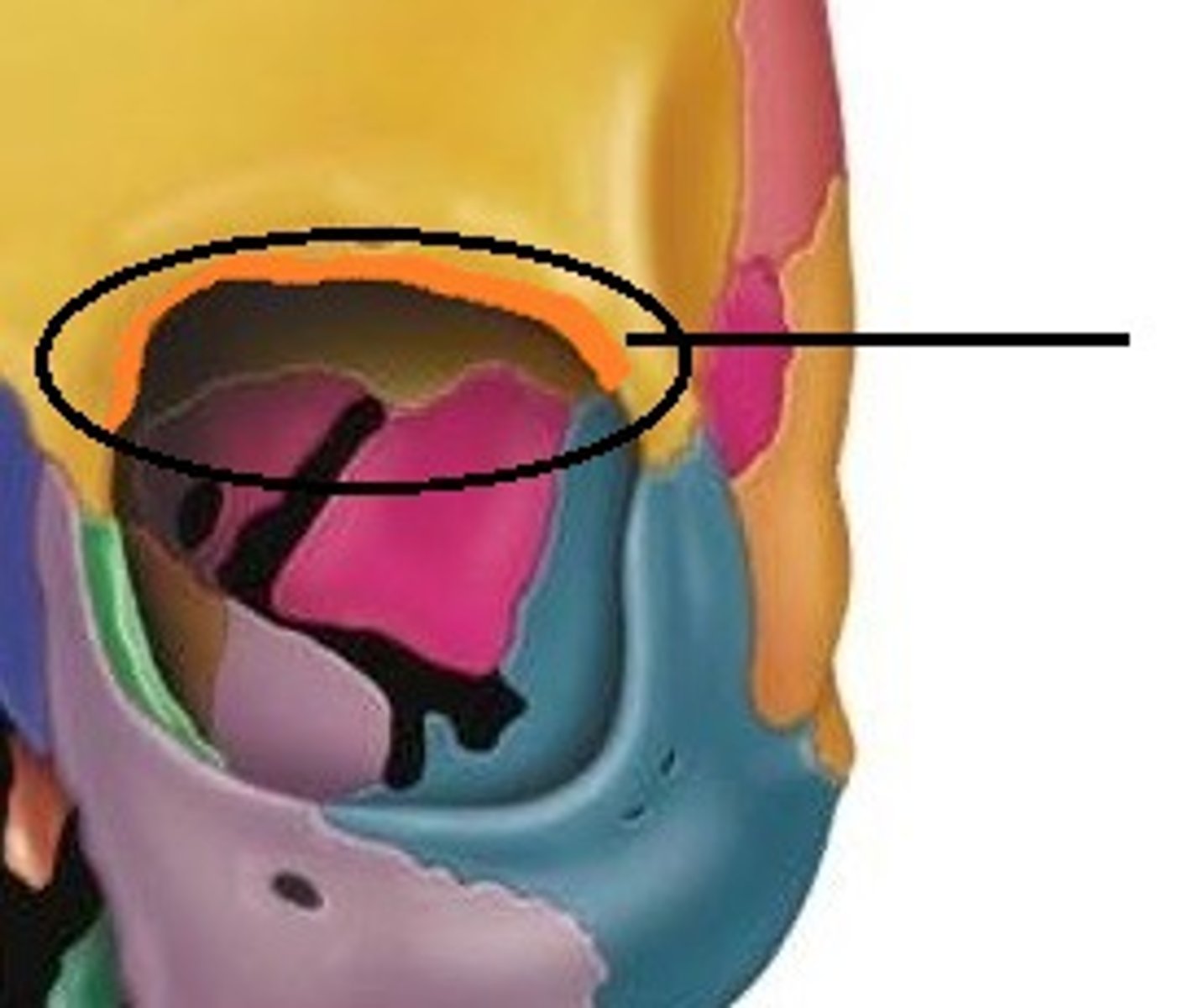
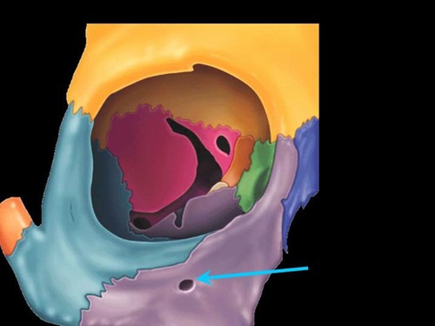
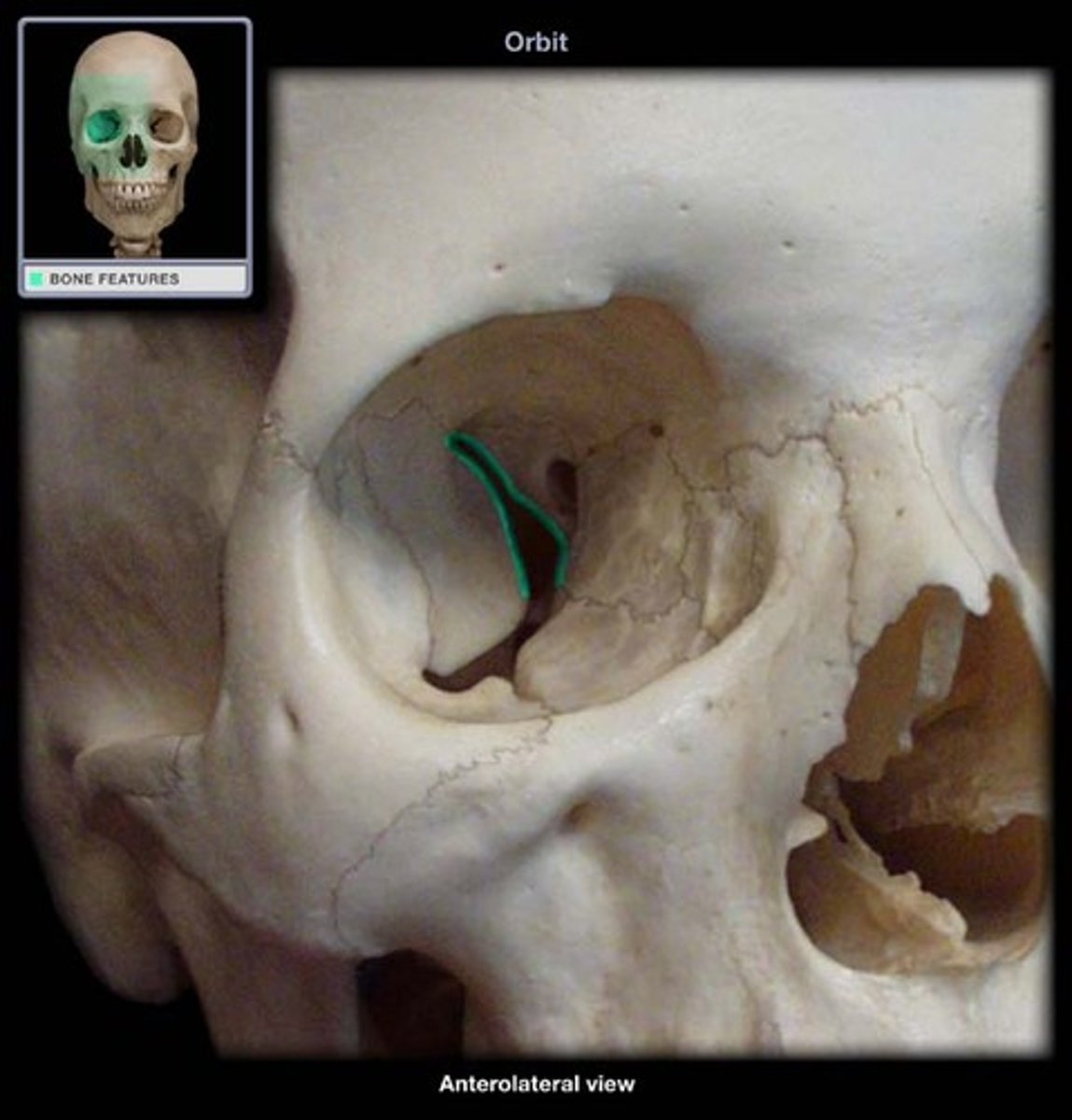
orbits
spaces
eye sockets/ where the eye sits
nasal cavities
3D space
holes of the nose
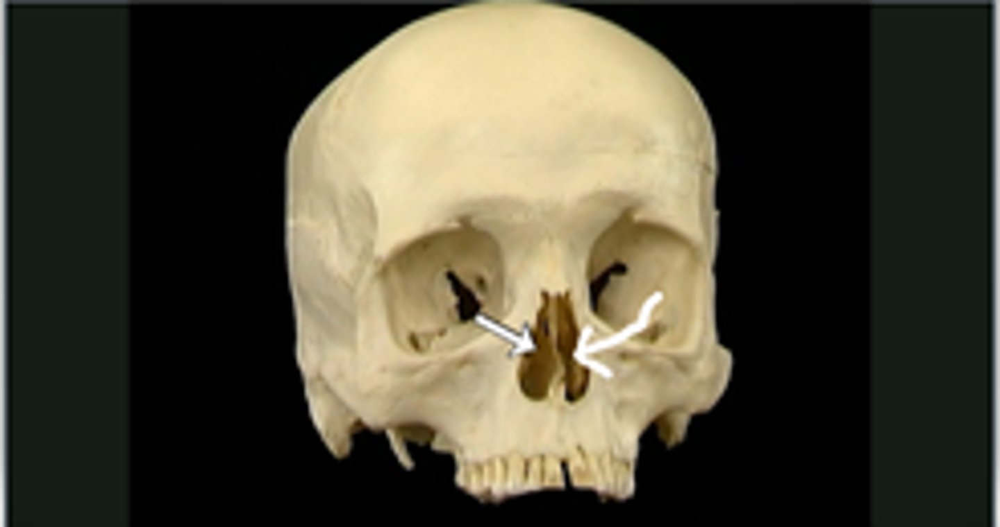
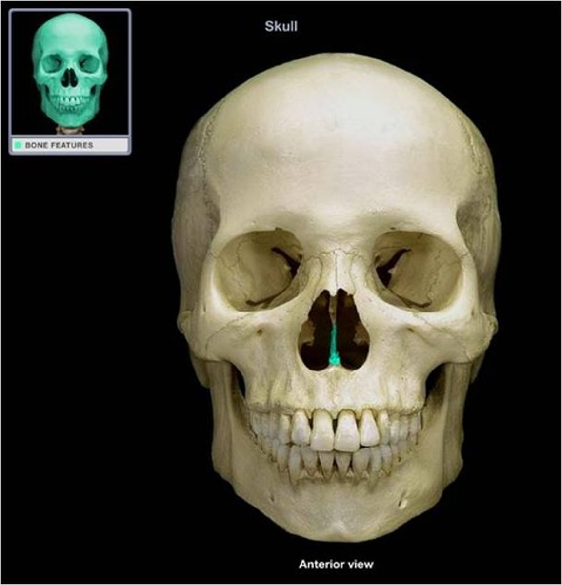
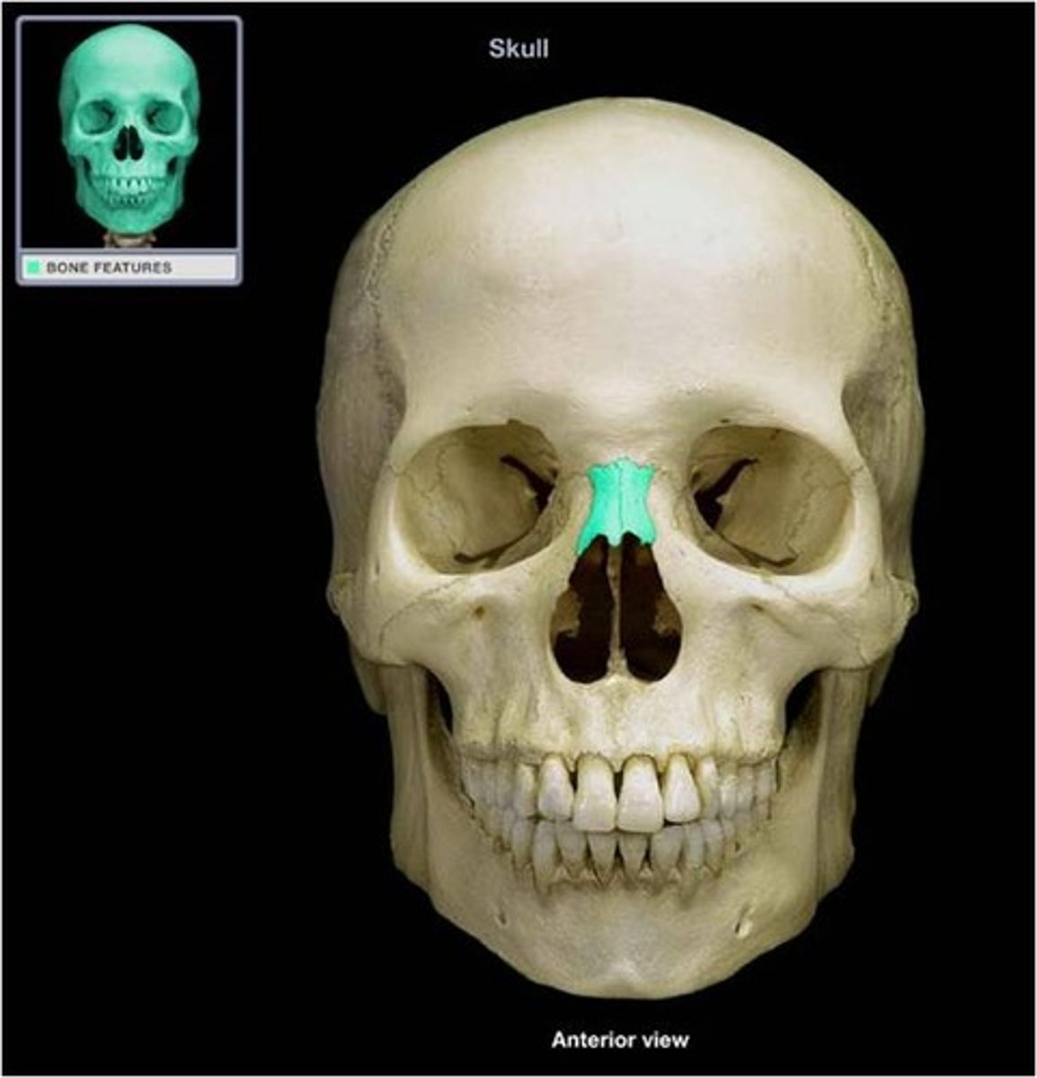
bony nasal septum
structure
down the middle bone of the nose
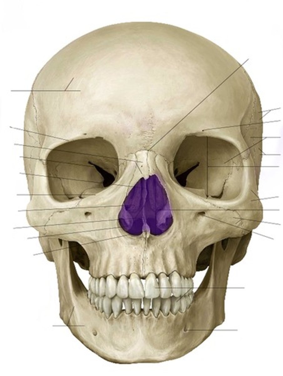
piriform aperture
2D opening
pear-shaped opening (tracing of the nasal cavity)
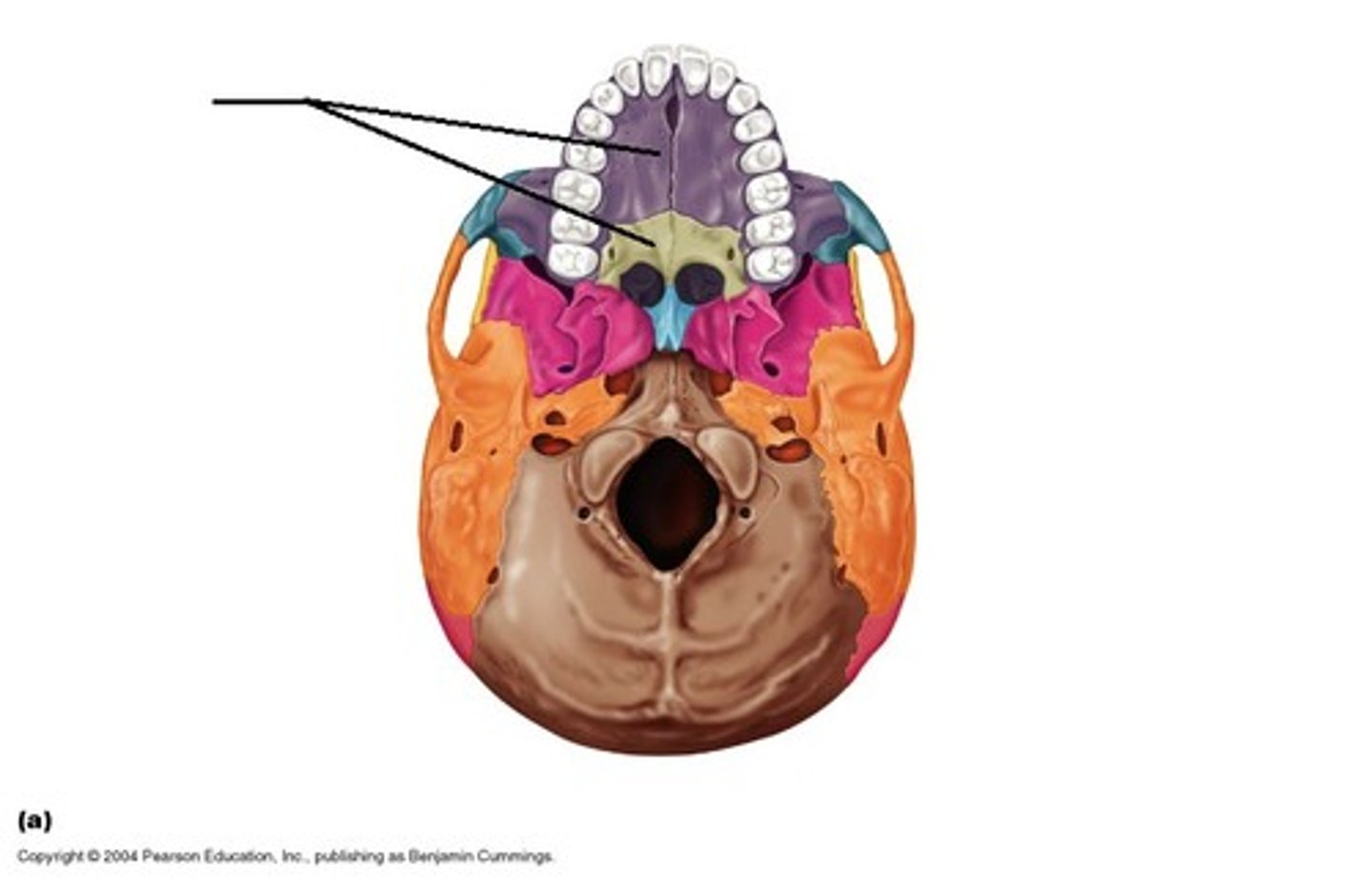
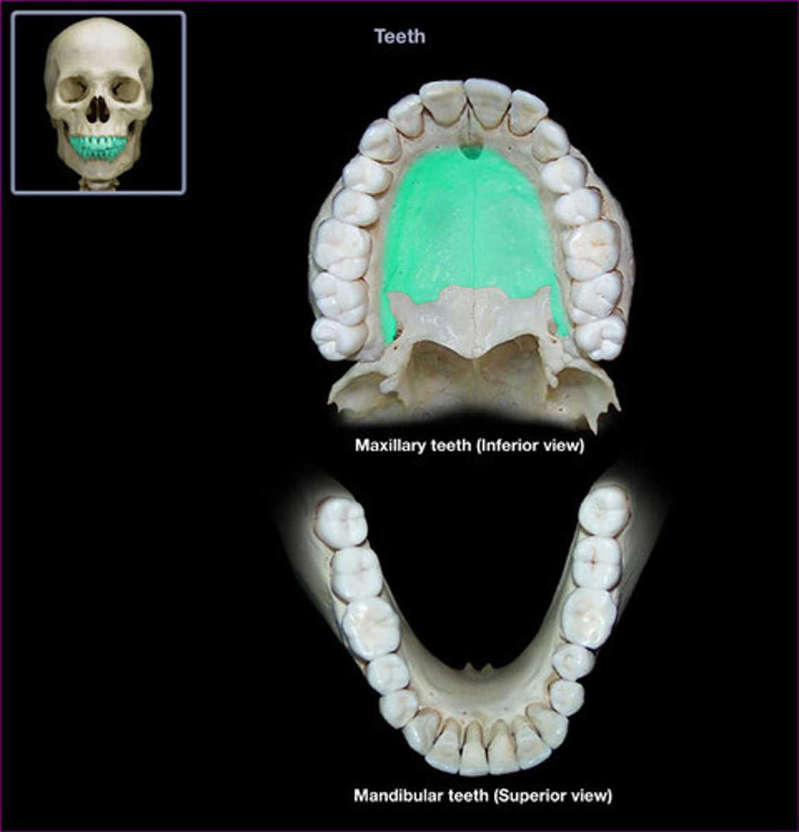
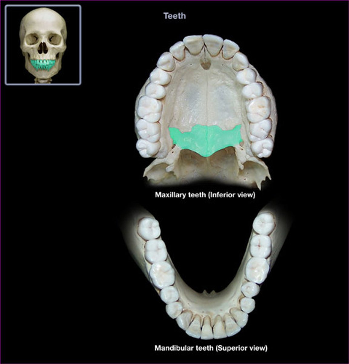
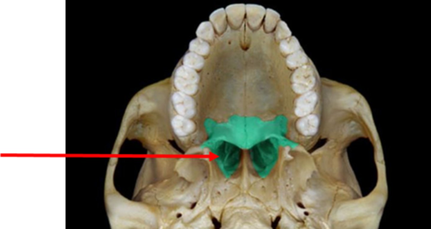
hard palate
collective structure
roof of the mouth (red and purple area)
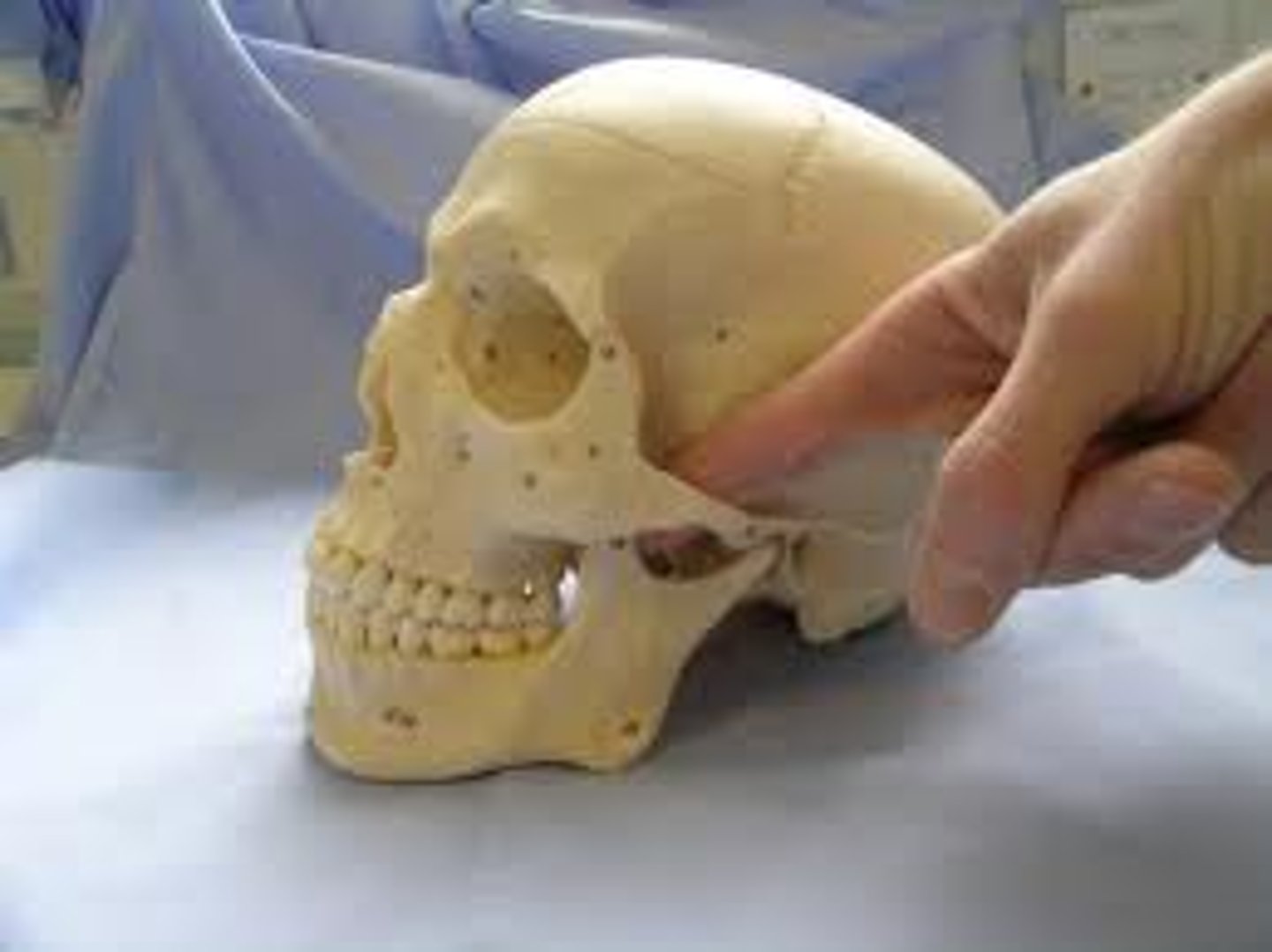
zygomatic arch
structure
cheekbone arch (eye to ear-ish) yellow and brine bone make up the arch.
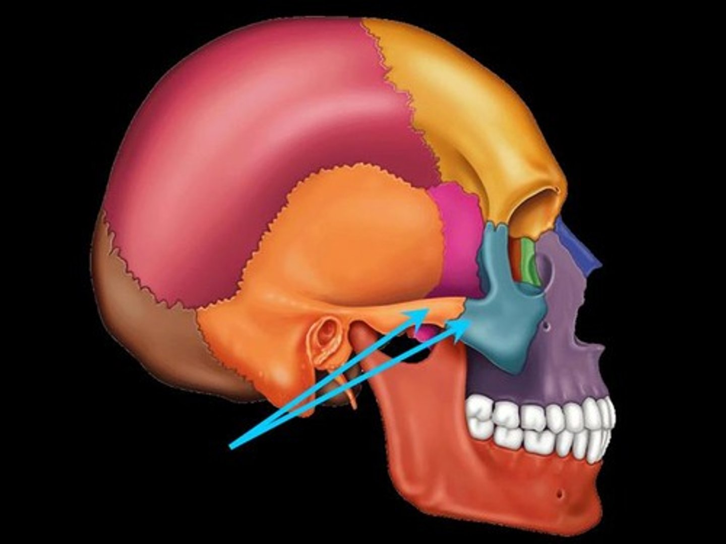
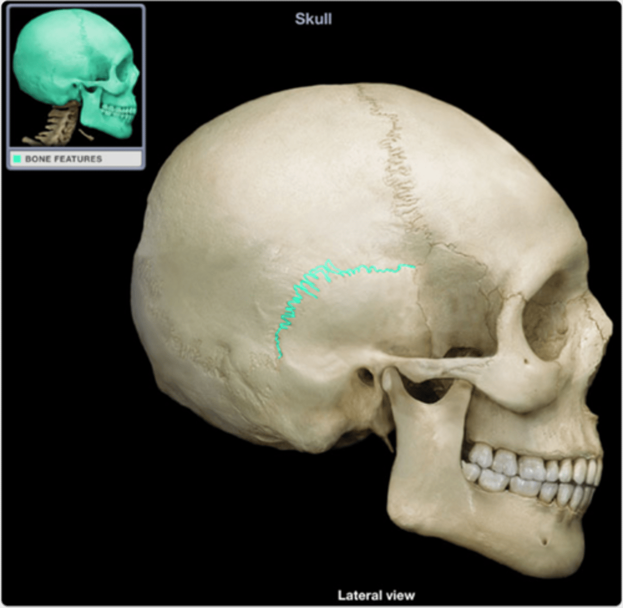
infratemporal fossa
depression
depression behind the zygomatic arch
coronal suture
junction
left to right suture in the front of the head
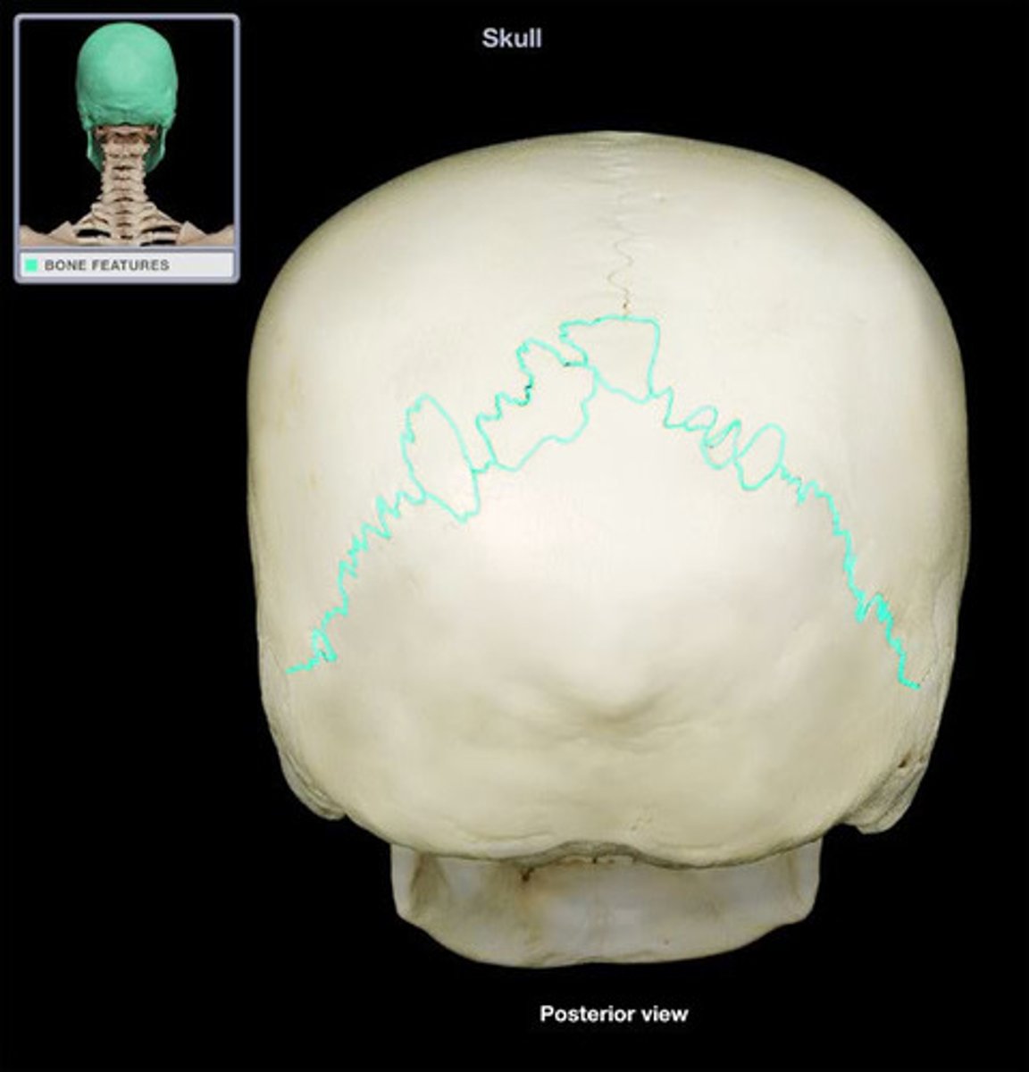
lambdoidal suture
junction
back of the head to suture from right to left
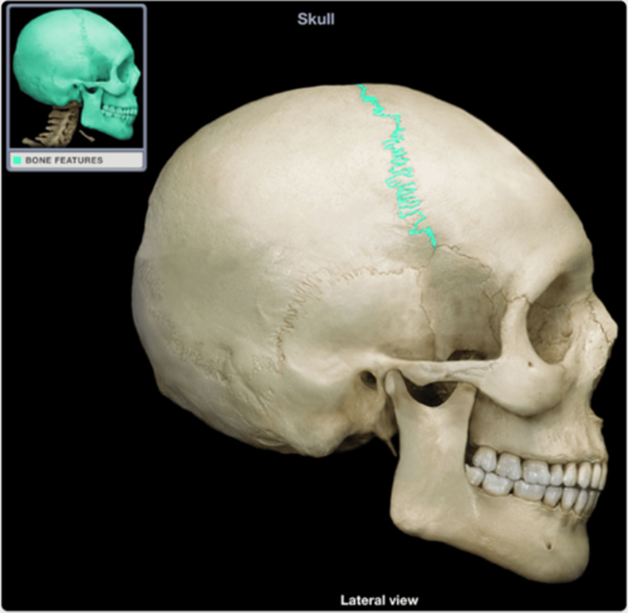
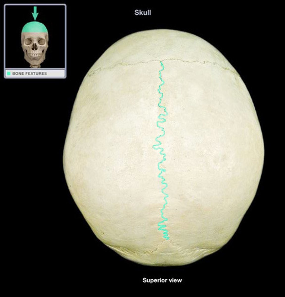
sagittal suture
junction
sutures down the center of the skull
squamous suture
junction
sutures on on both sides of the skull near the temporal bone.
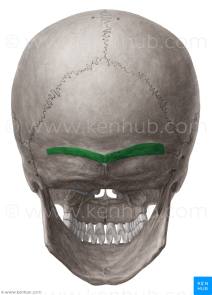
supraorbital margin of the frontal bone
feature
where the eyebrows sit
parietal bones
structure
two bone on the that make up the top of the skull
occipital bone
structure
bone that makes up the back of the skull
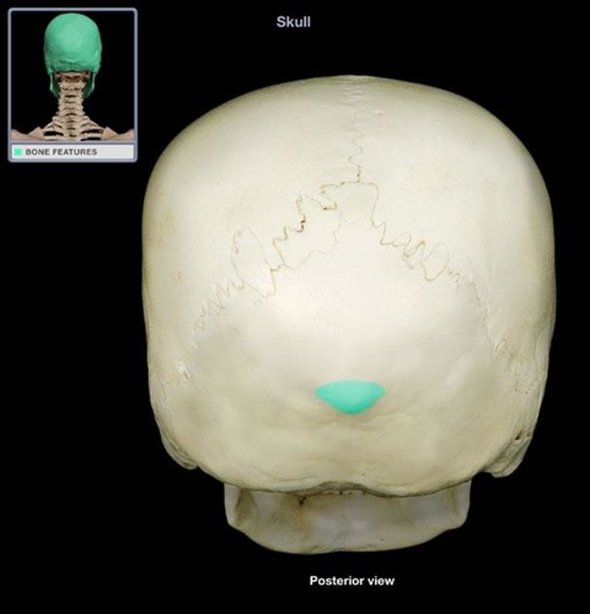
external occipital protuberance of the occipital bone
feature
bump on the back of the head
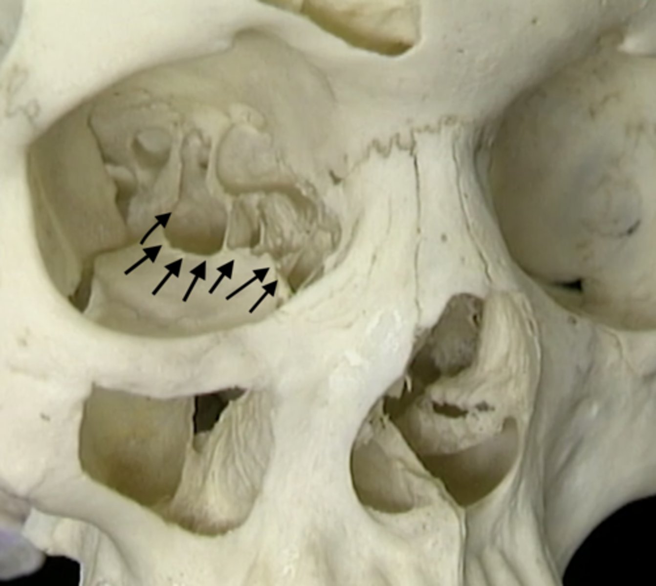
occipital condyles of the occipital bone
feature
flat wedges of the base of the skull

nuchal lines of the occipital bone
feature
lines that come to a point at the base of the skull
temporal bones
structure
sides of the head
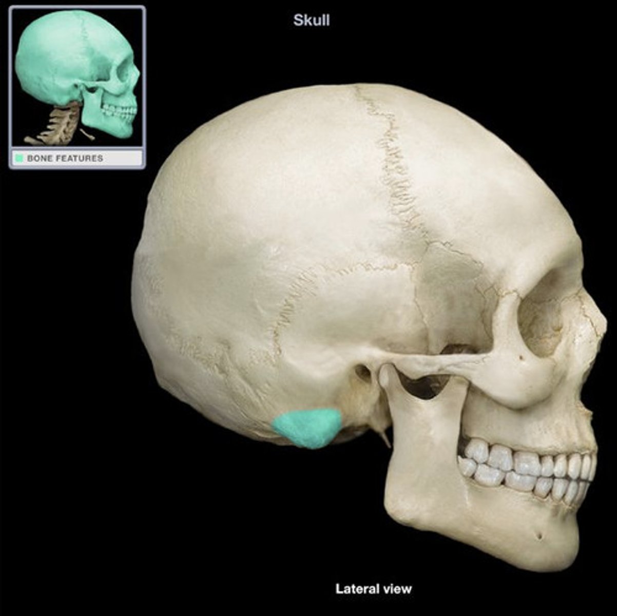
mastoid process of the temporal bones
projection
pointy part of the temporal bone posterior
styloid process of the temporal bones
projection
event pointier part of the temporal bone/ the tip of the point
zygomatic process of the temporal bones
projection
The brown part of the zygomatic arch, posterior to the eye

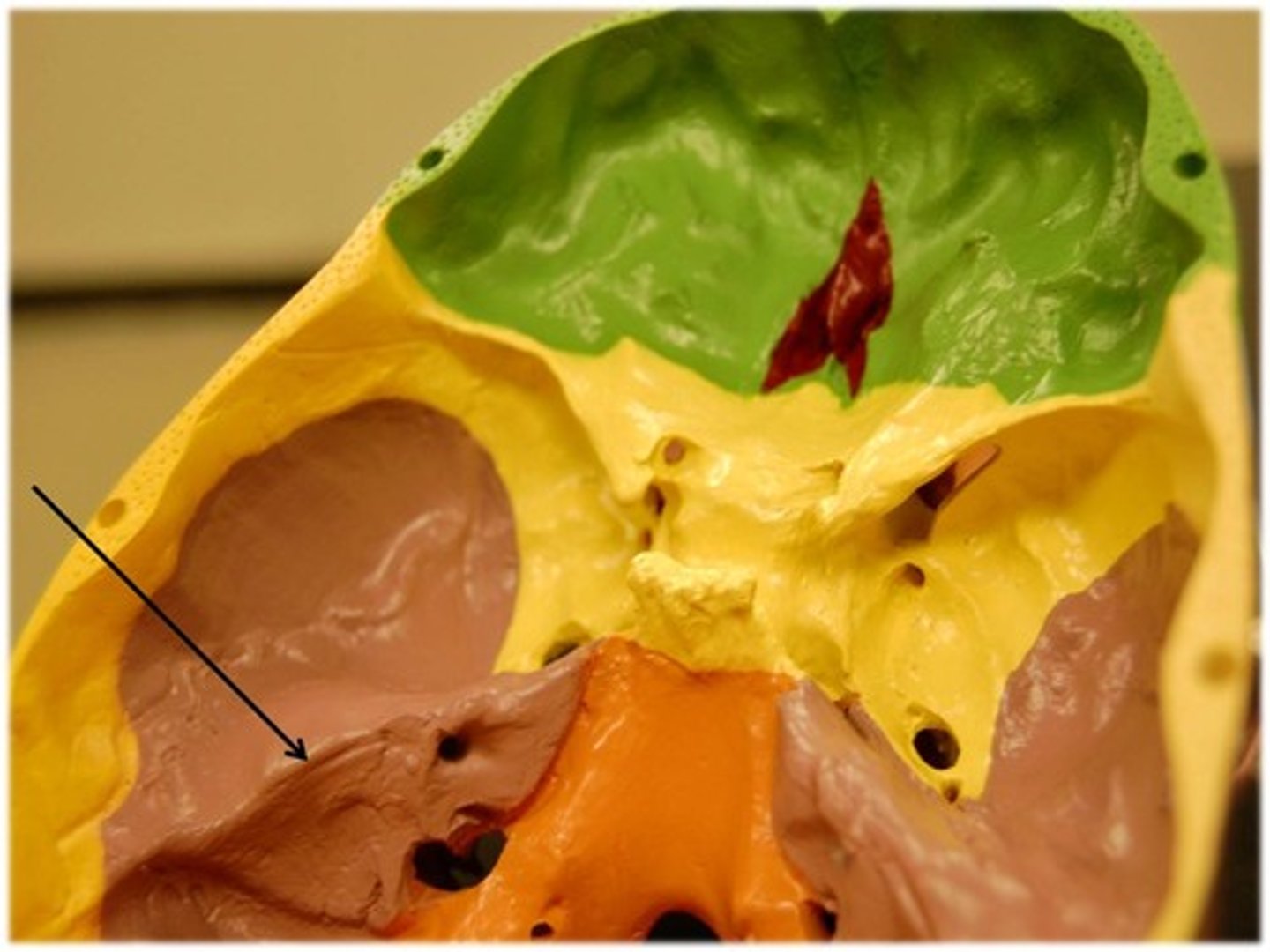
petrous portion of the temporal bones
structure
pinching the middle parts of the center of the cranial base.
mandibular fossa of the temporal bones
depression
sockets where the jaw attaches to both sides of the head
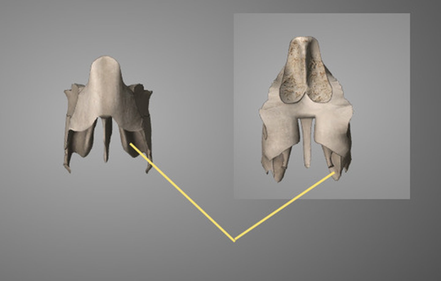
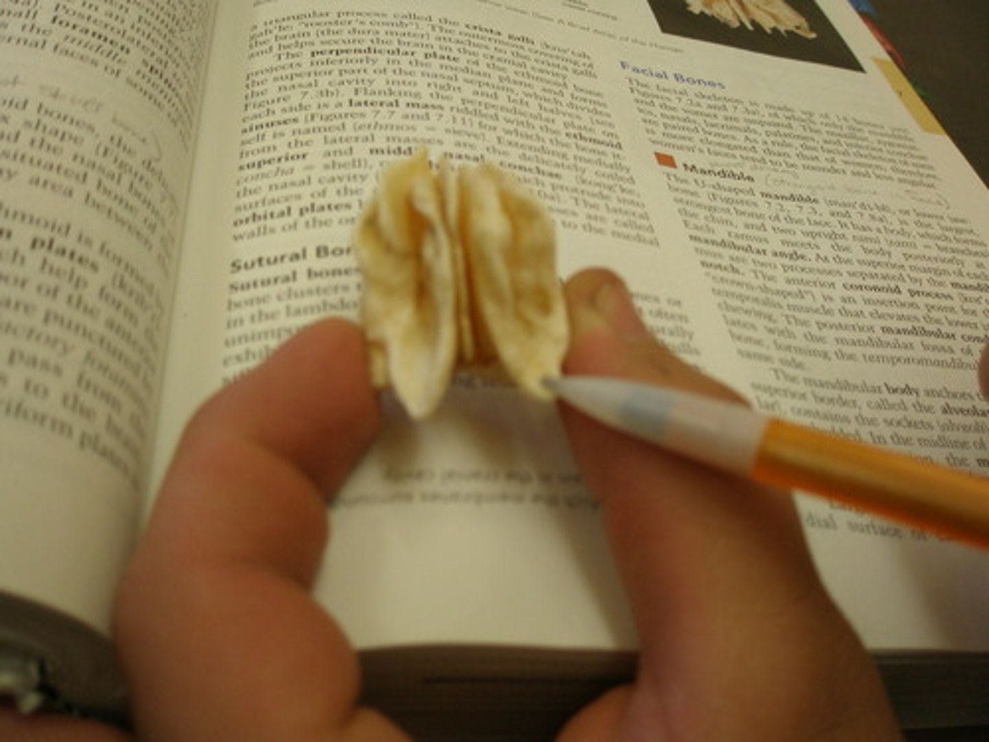
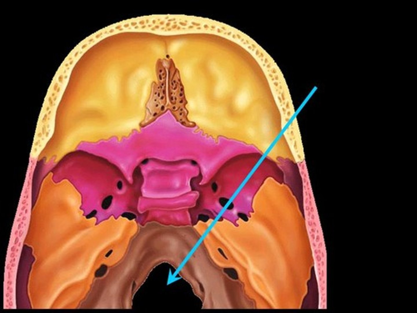
Ethmoid bone
bone that sits in the front of the canical base (can be detached)
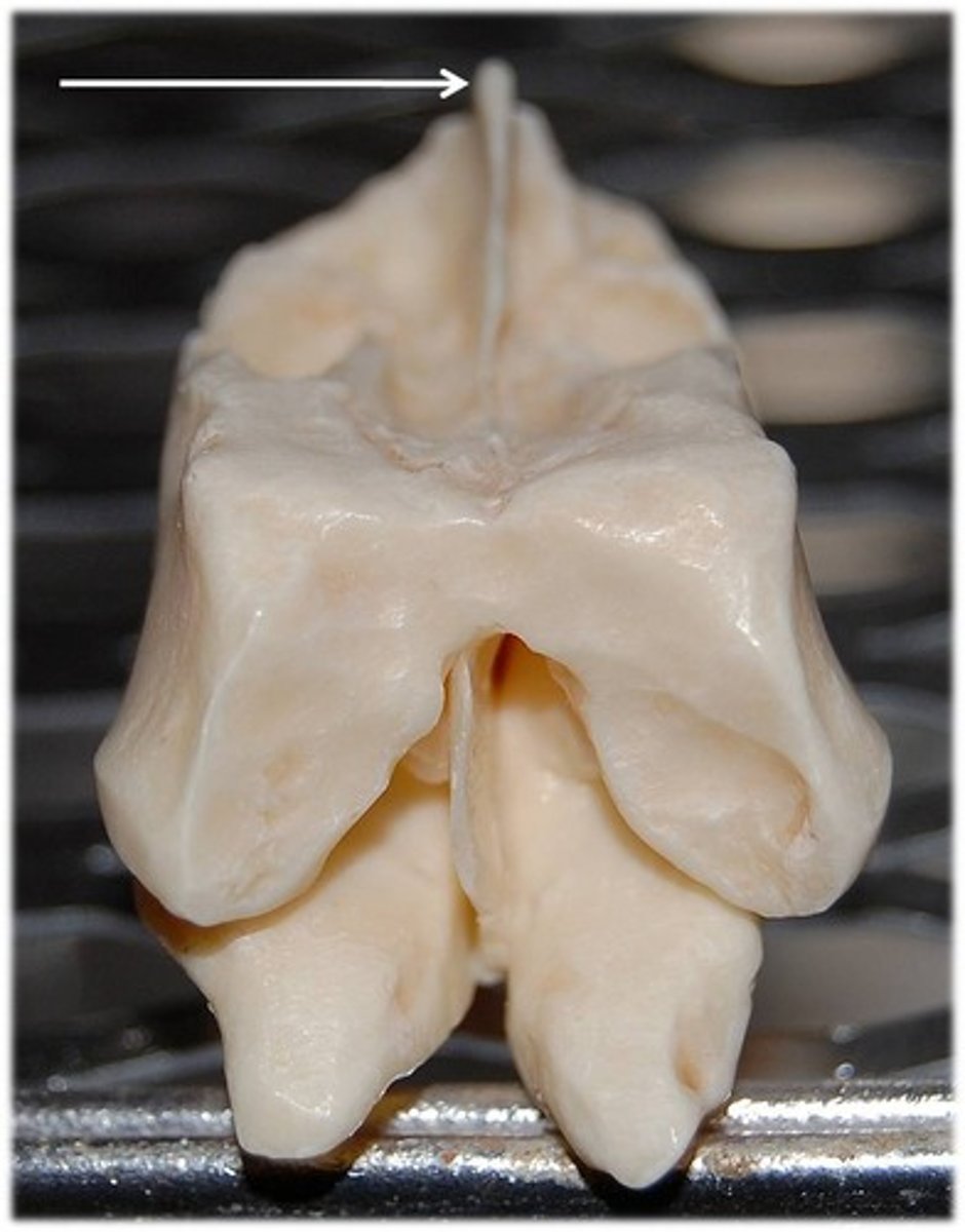
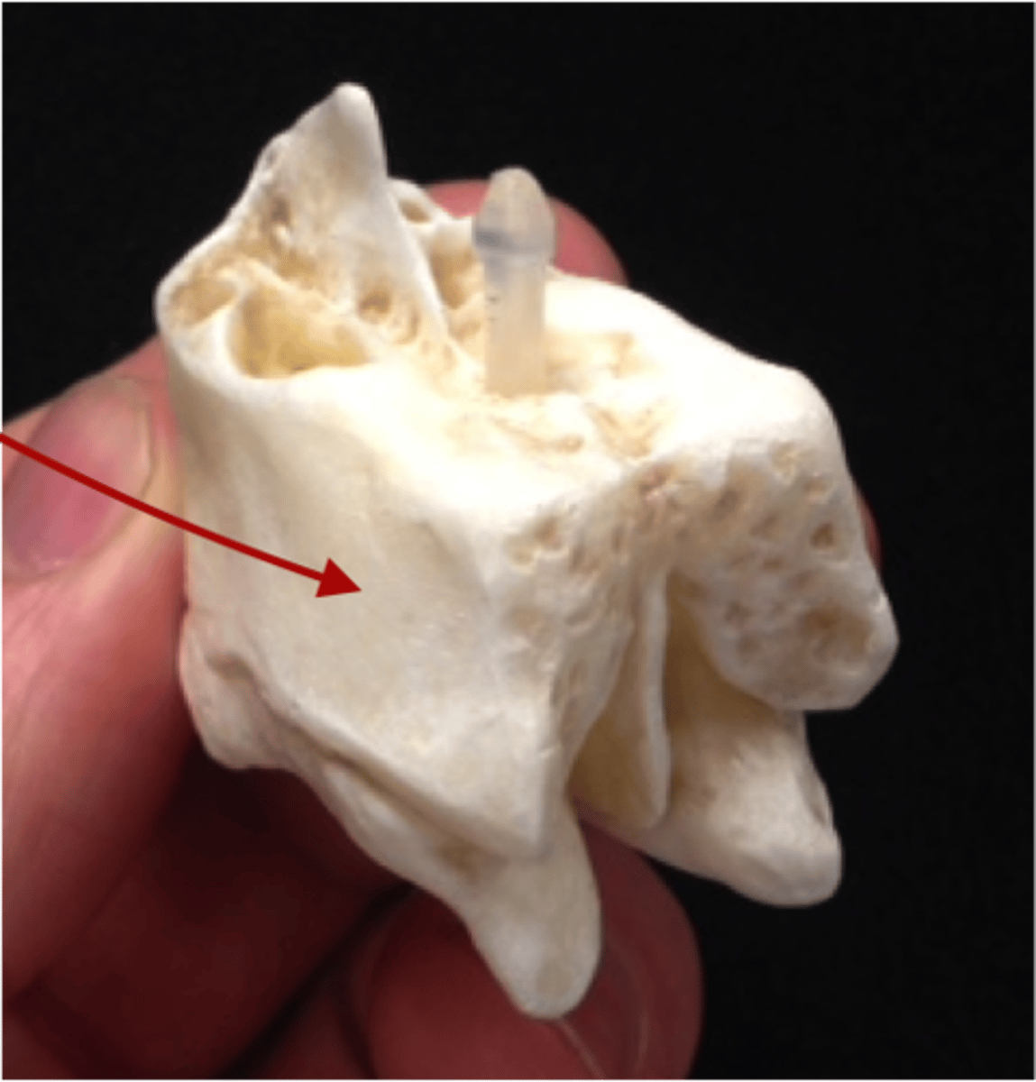
crista galli of the ethmoid bone
structure
top projection from the ethmoid bone
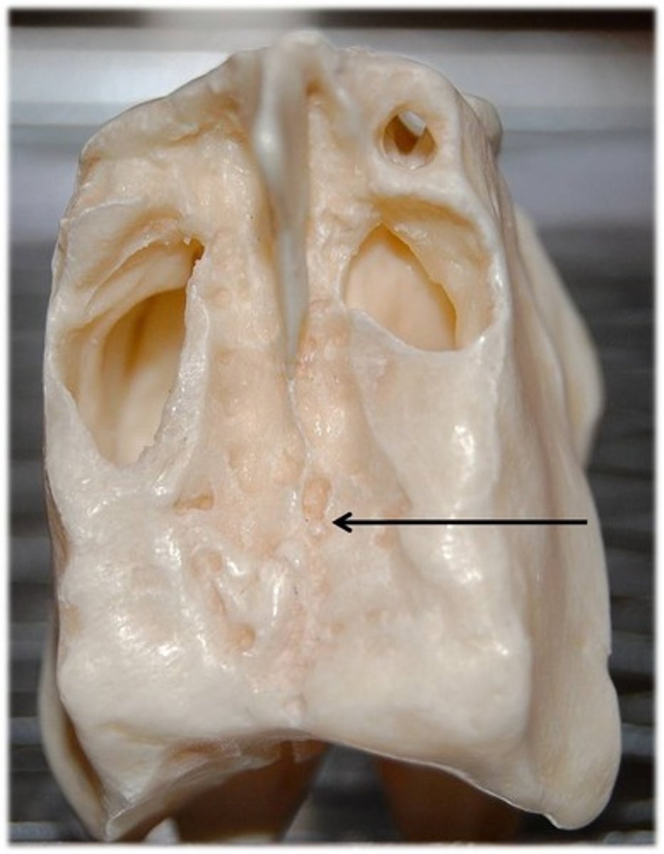
cribriform plate of the ethmoid bone
structure
plate that has many tiny holes on side of crista galli posterior from it
perpendicular plate of the ethmoid bone
structure
anterior ridge to the crista galli sown the center
orbital plate of the ethmoid bone
structure
outside walls with smooth surface of ethmoid bone
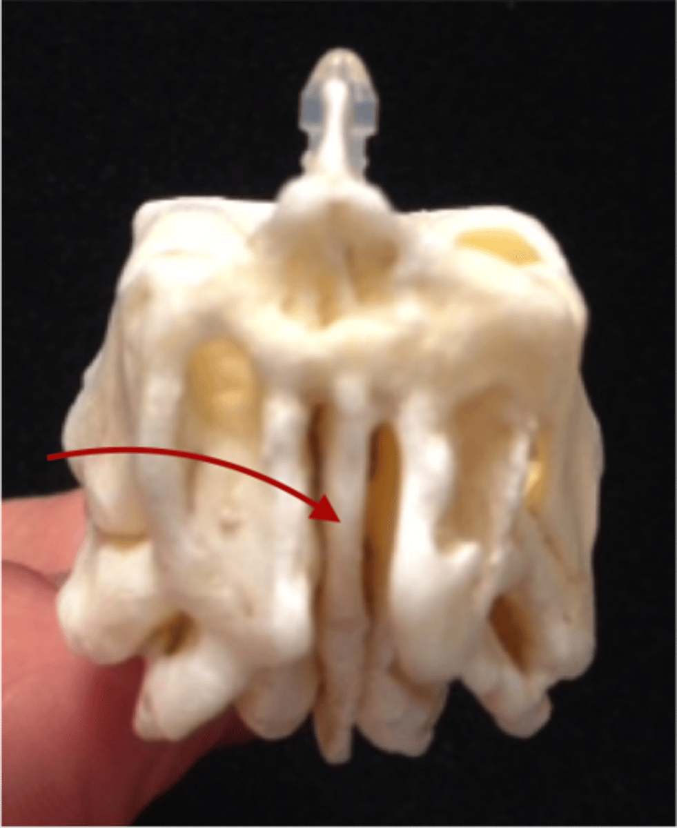
superior conchae of the ethmoid bone
structure
right and left of the perpindicular plate superior part of the sides (inside)
middle conchae of the ethmoid bone
structure
right and left of the perpindicular plate lower to the superior and makes up most of the wall.
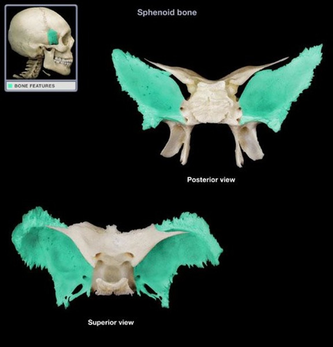
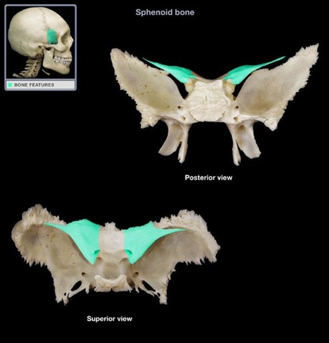
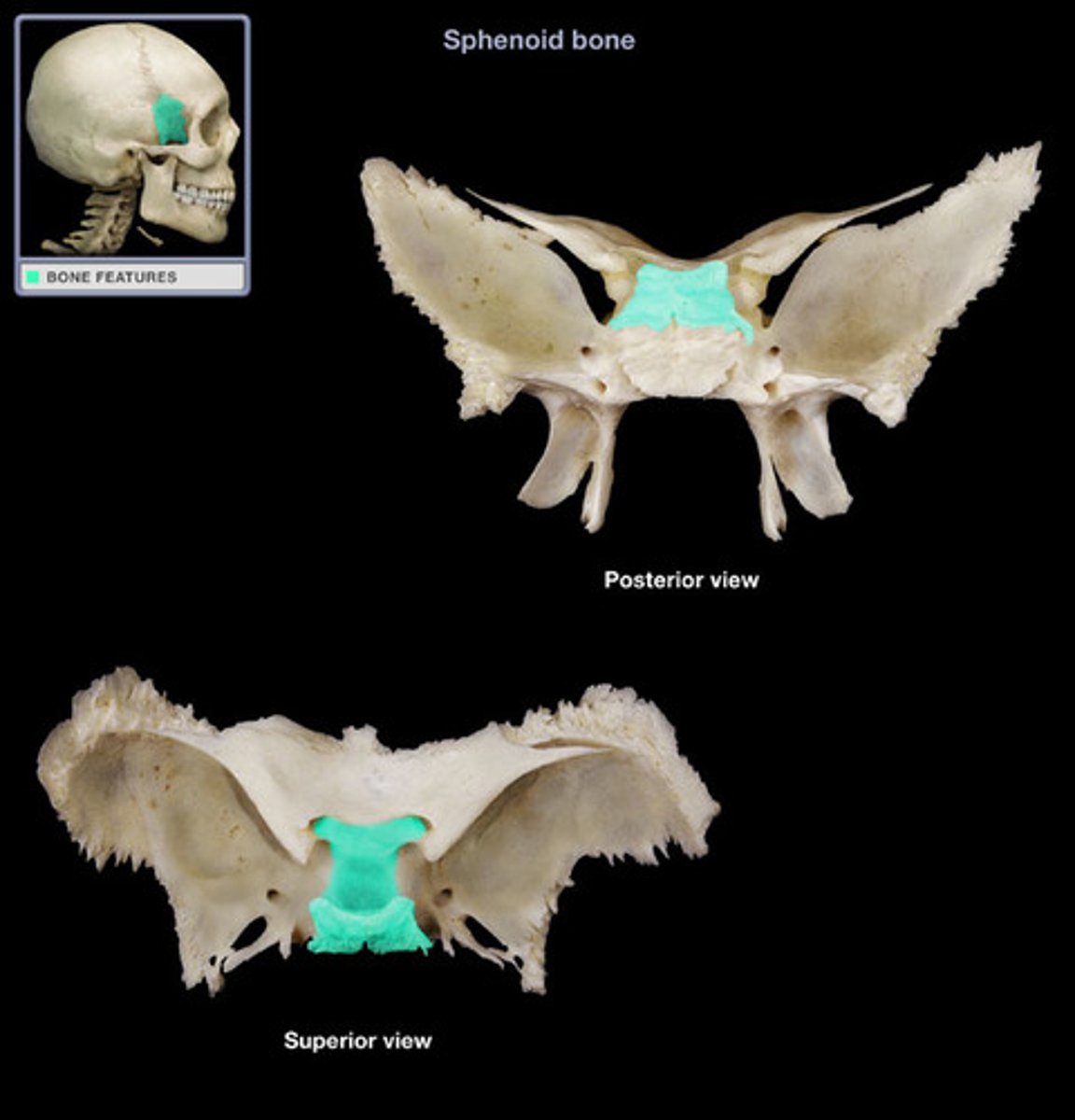
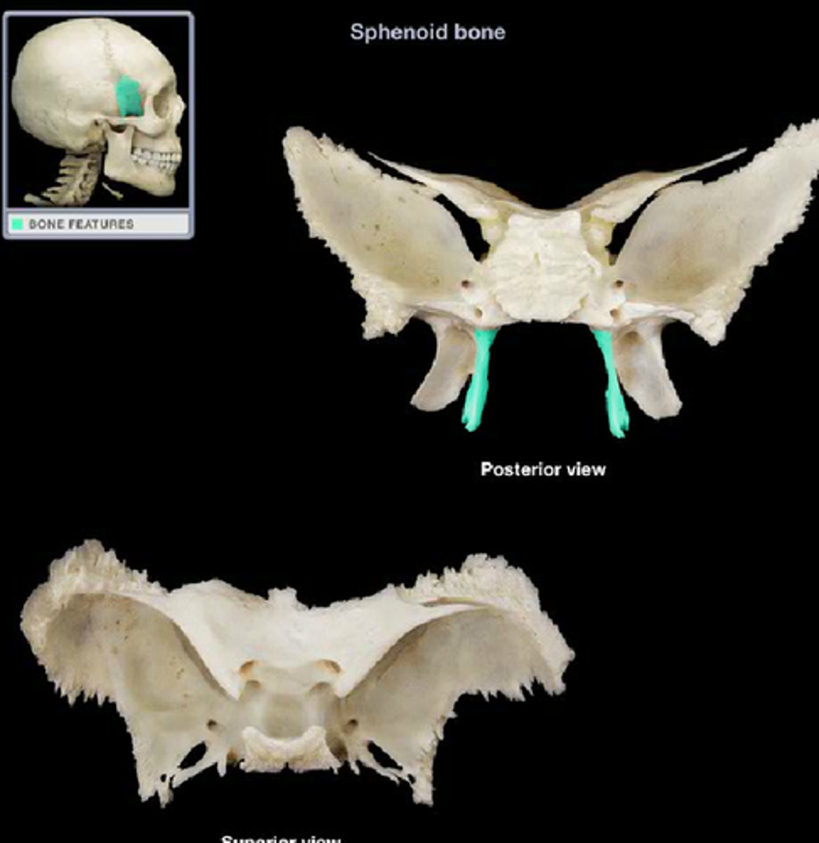
sphenoid bone
structure
sits in the center of the cranial base (looks like butterfly)
greater wing of the sphenoid bone
structure
the larger wings of the sphenoid bone
lesser wing of the sphenoid bone
structure
smaller wings/ inner wings
sella turcica of the sphenoid bone
depression
middle depression of the sphenoid bone
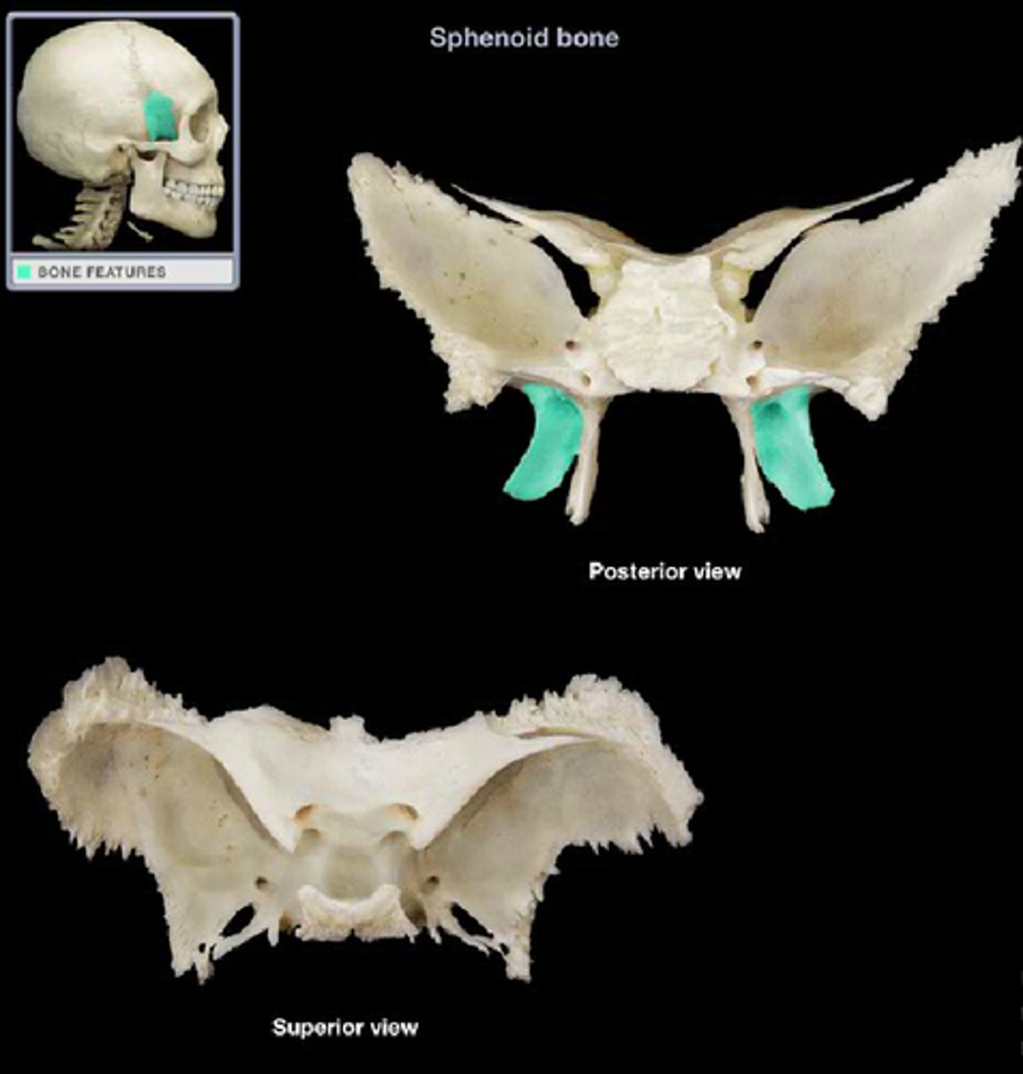
pterygoid processes of the sphenoid bone
projection
legs of the butterfly
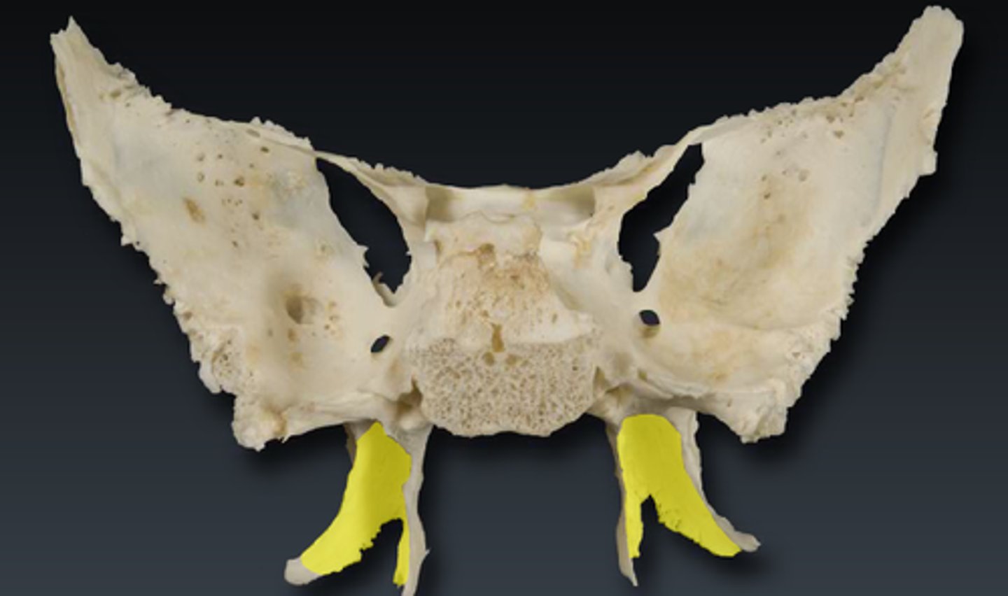
medial plates of the pterygoid processes
feature
The inside part of the legs
lateral plates of the pterygoid processes
feature
outside part of the legs
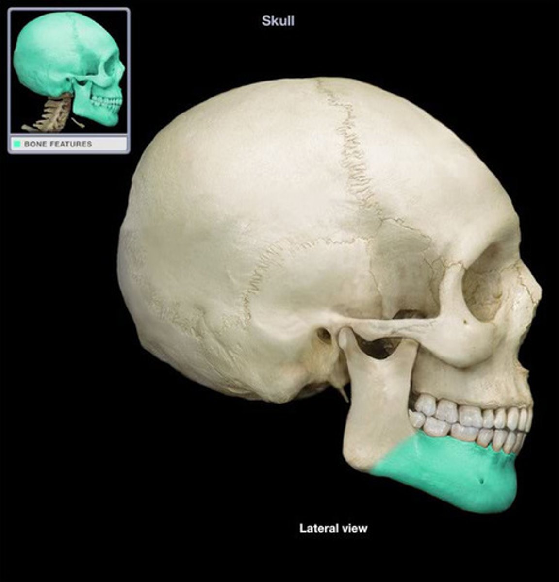
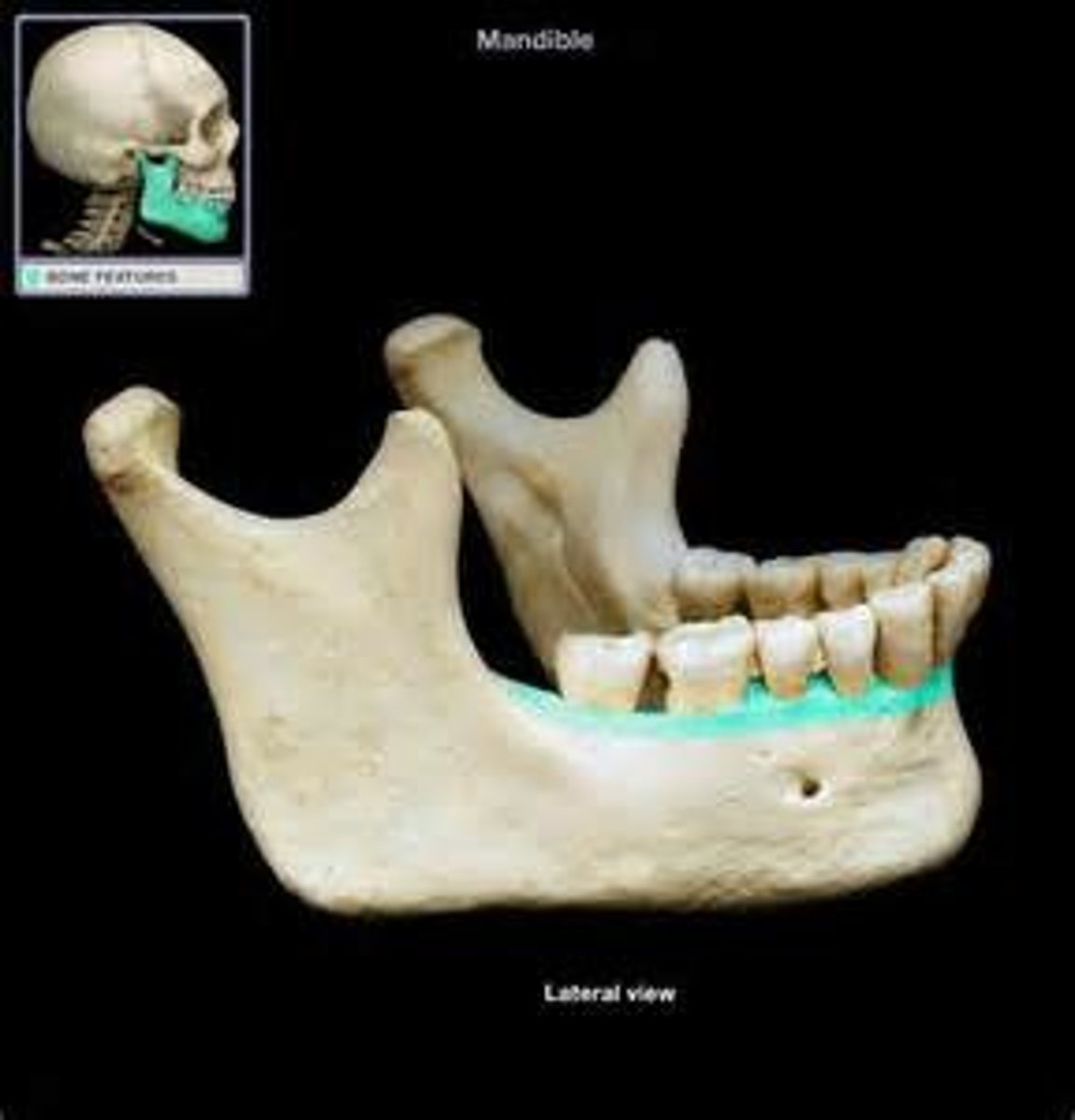
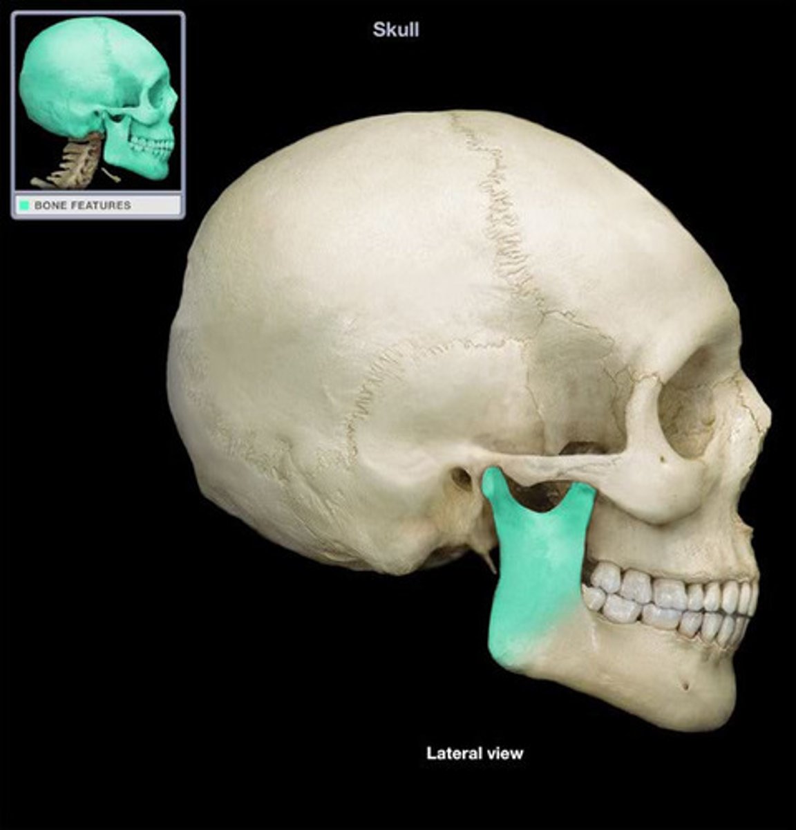
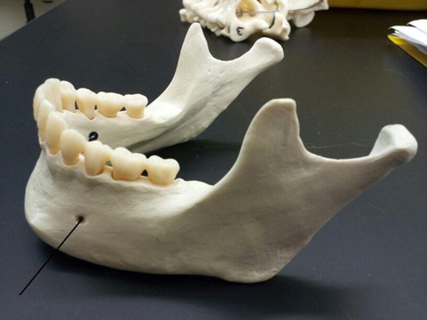
mandible bone
structure
makes up the lower jaw
body of mandible bone
structure
ouside c shape that makes up the mandible
dental alveoli of the mandible bone
depression
any of the tooth sockets
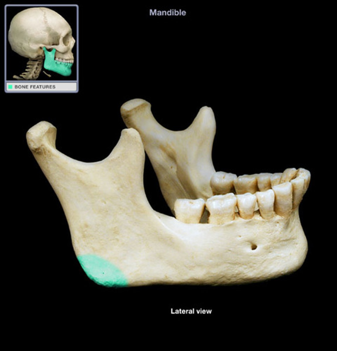
angle of the mandible bone
feature
jawline the angle that the mandible creates most posterior part
ramus of the mandible bone
feature
flat part of manible superior to the angle
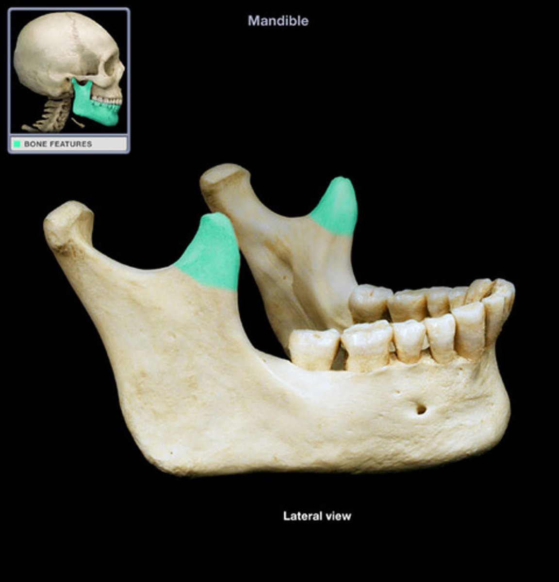
coronoid process of the mandible bone
projection
front two projections of the mandible
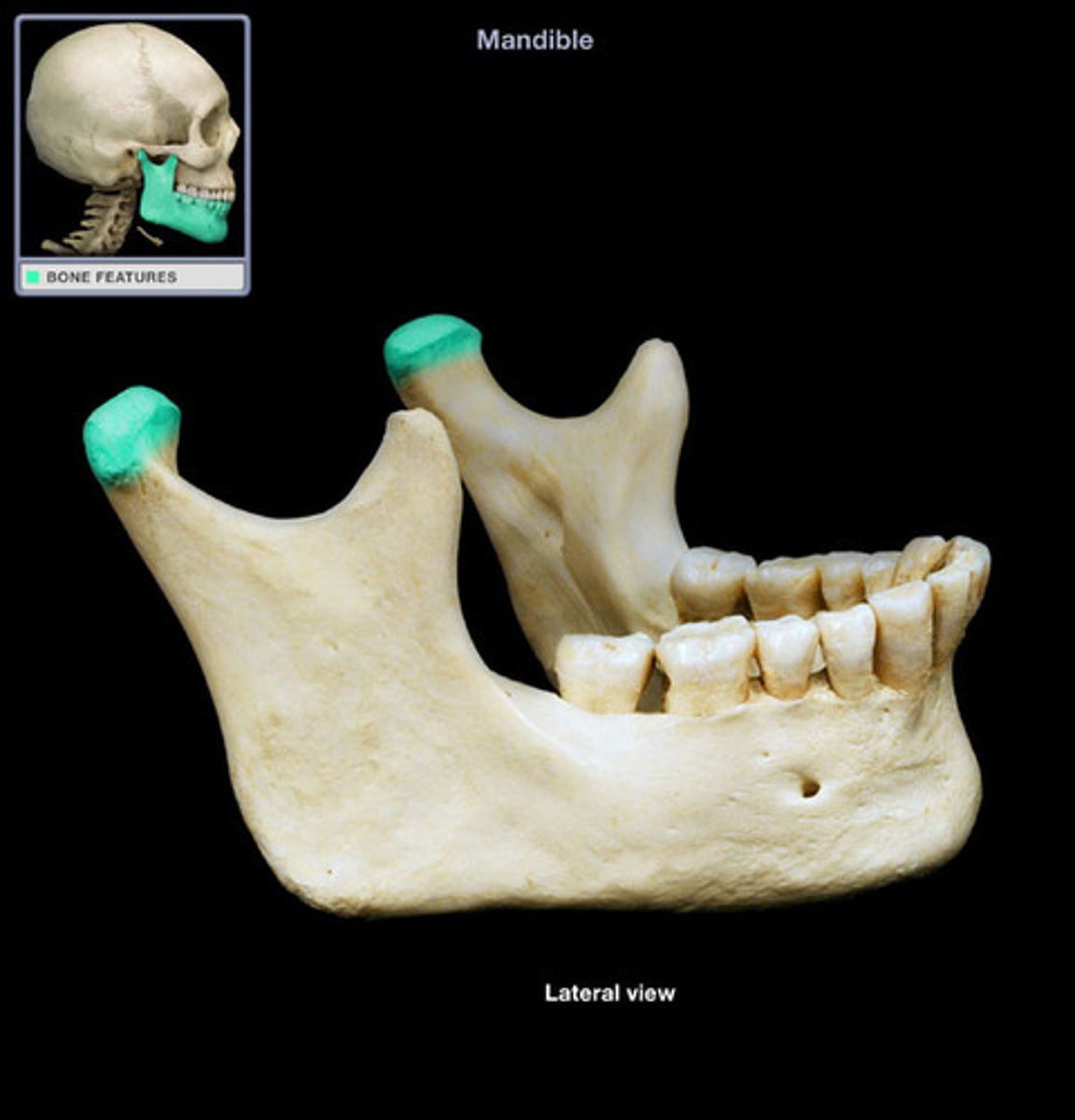
condylar process of the mandible bone
projection
back two projections of the mandible
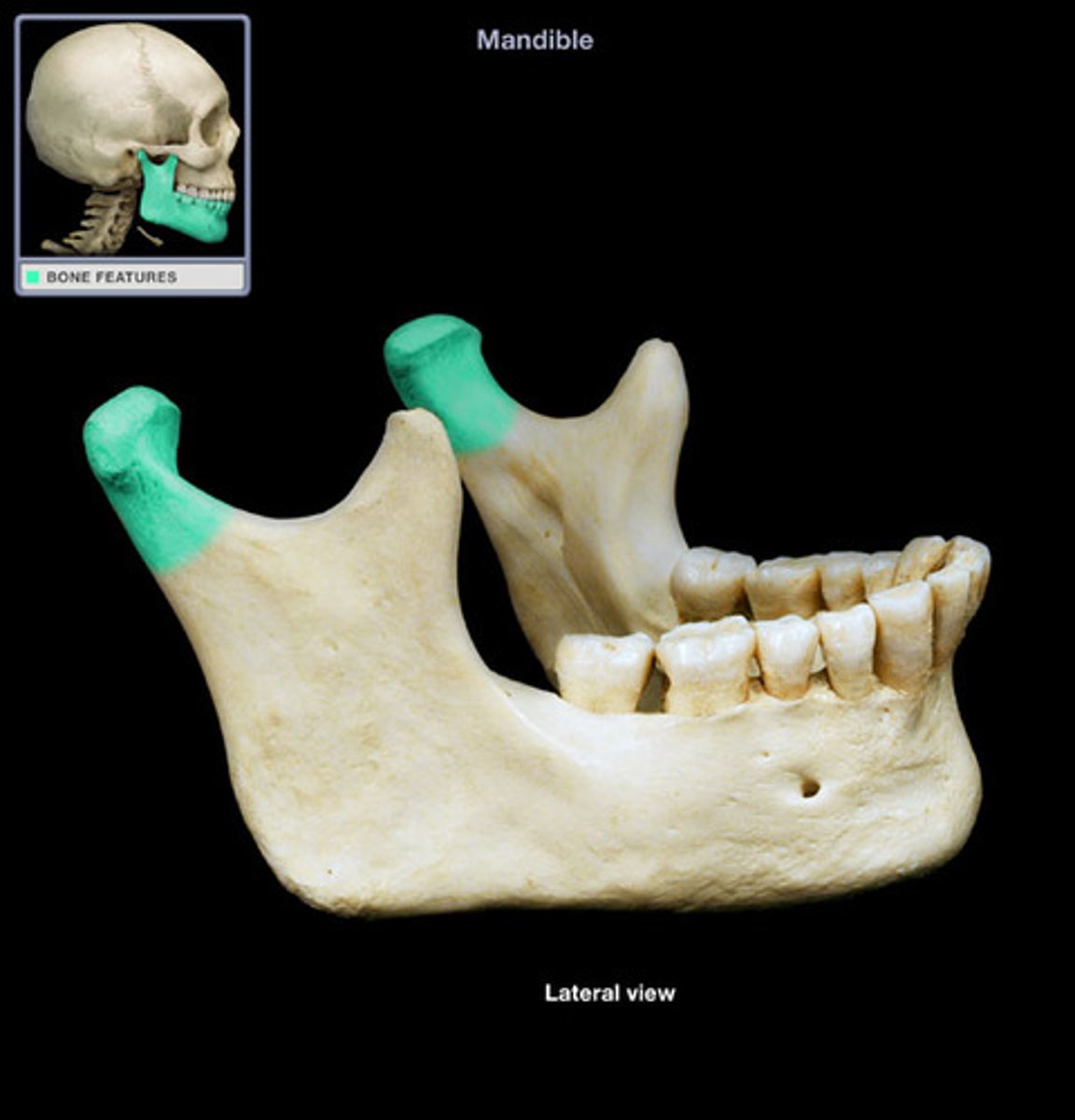
head of the mandible bone
tip of the condylar process
maxillae bone
bones that make up the upper jaw
body of maxilla bone
structure
c shape that makes up the top of the teeth
dental alveoli of the maxilla bone
depression
any upper teeth sockets
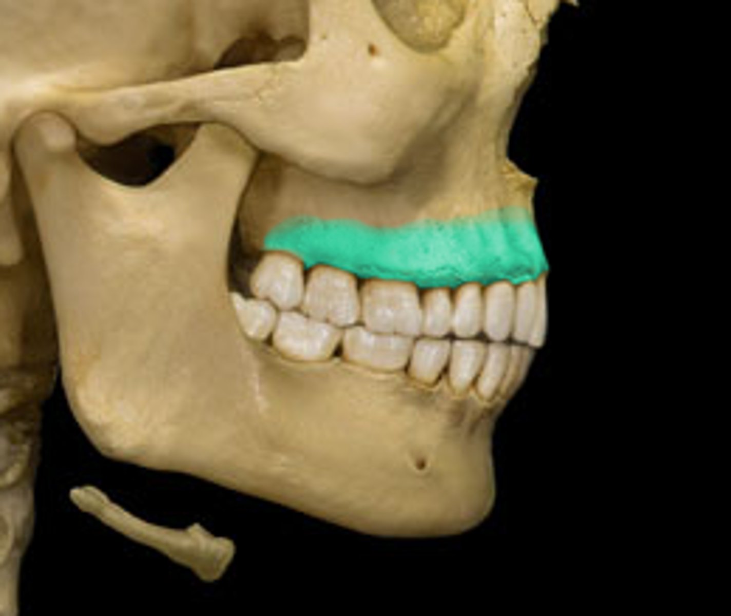
infraorbital margin of the maxilla bone
feature
thin line below eye sockets
palatine process of the maxilla bone
projection
just the purple part of the palate anterior to the other part
zygomatic bones
structure
cheek bones
temporal process of the zygomatic bones
projection
yellow part of the zygomatic arch makes up part of the eye socket
nasal bones
structure
two upper nasal bones
palatine bones
structure
bone that make up back part of the hard palate
horizontal plate of the palatine bones
structure
very top bar of the the palatine bone
perpendicular plate of the palatine bones
structure
twi legs of the palatine bone
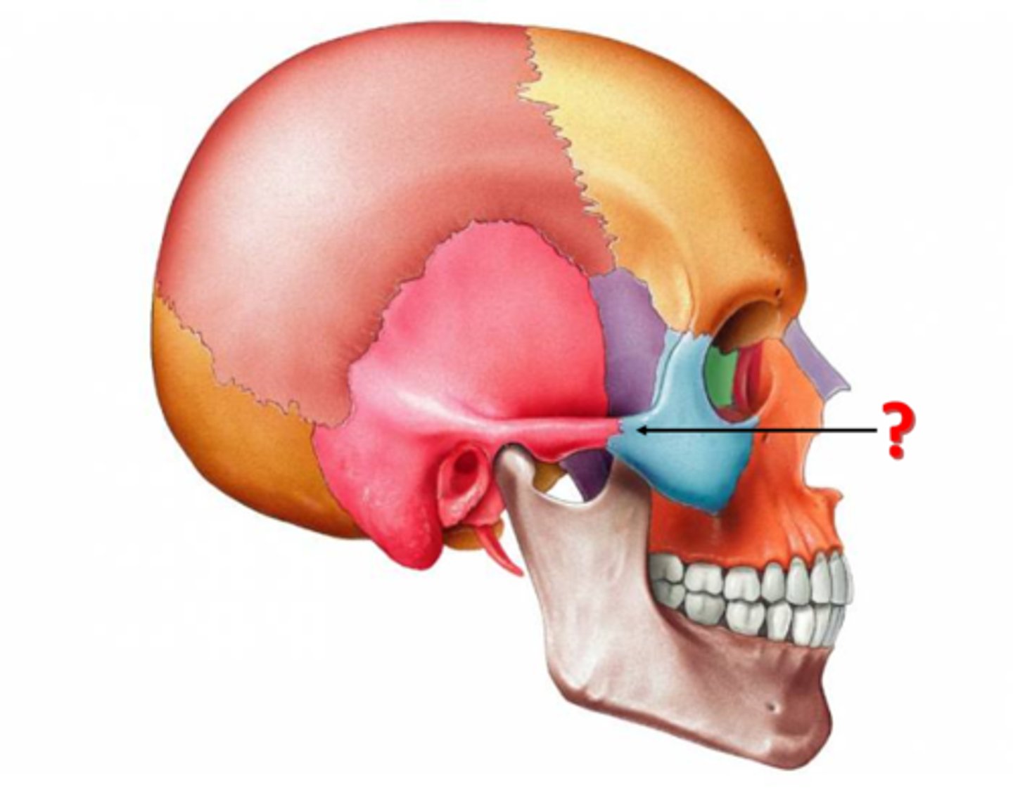
lacrimal bones
structure
inside the eye socket orange
opening for nasolacrimal duct of the lacrimal bones
2D space
where tears come from opening in eye socket small whole on side of nasal cavity
inferior nasal conchae
structure
lowest projections
yellow deep in eye

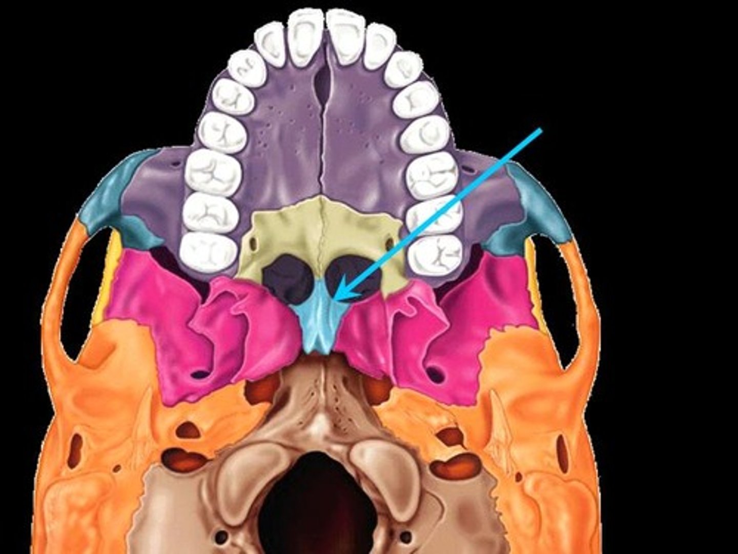
vomer
structure
yellow inside red on the underside of the skull. right below the lower part of the hard palate
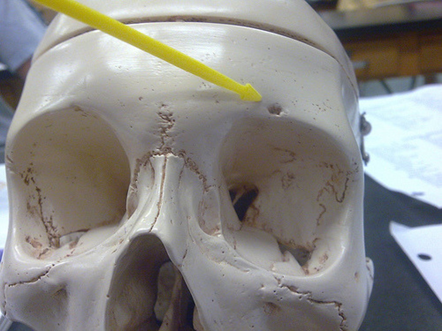
supraorbital foramen
hole
notch above the eye socket
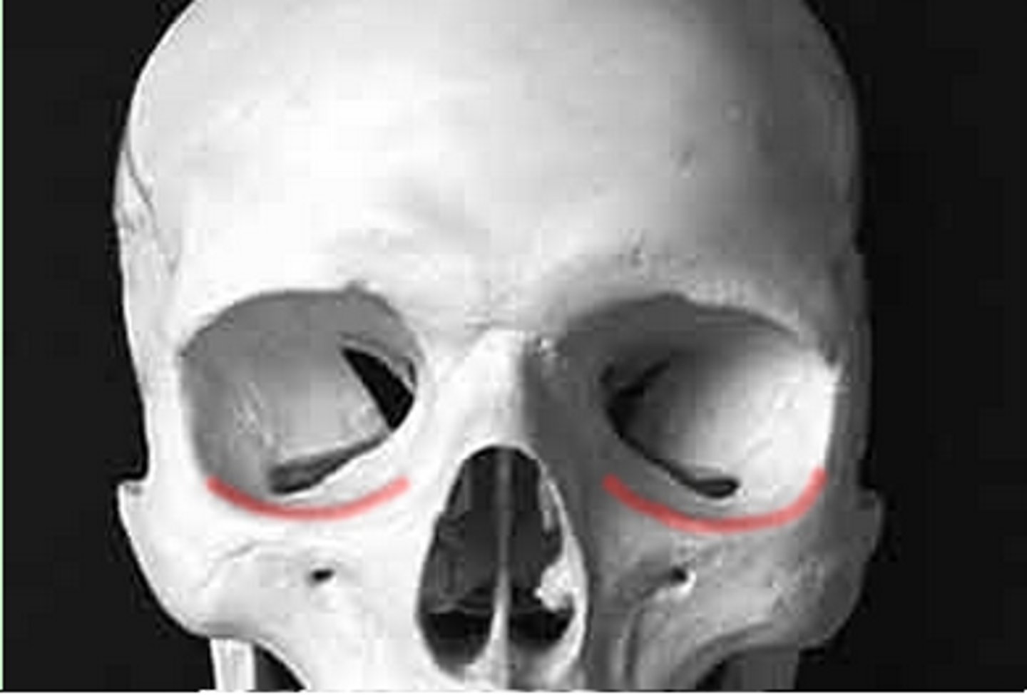
infraorbital foramen
hole
notch below the eye socket
mental foramen
hole
small hole on front of chin anterior
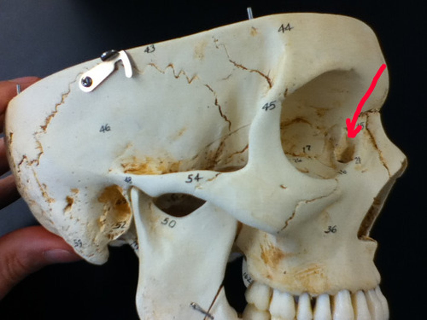
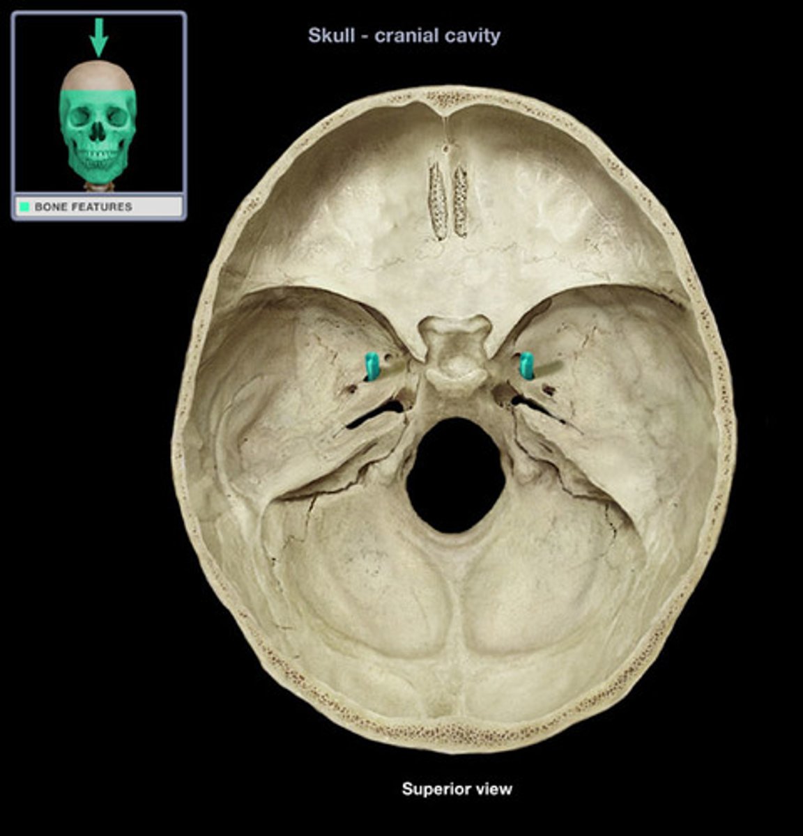
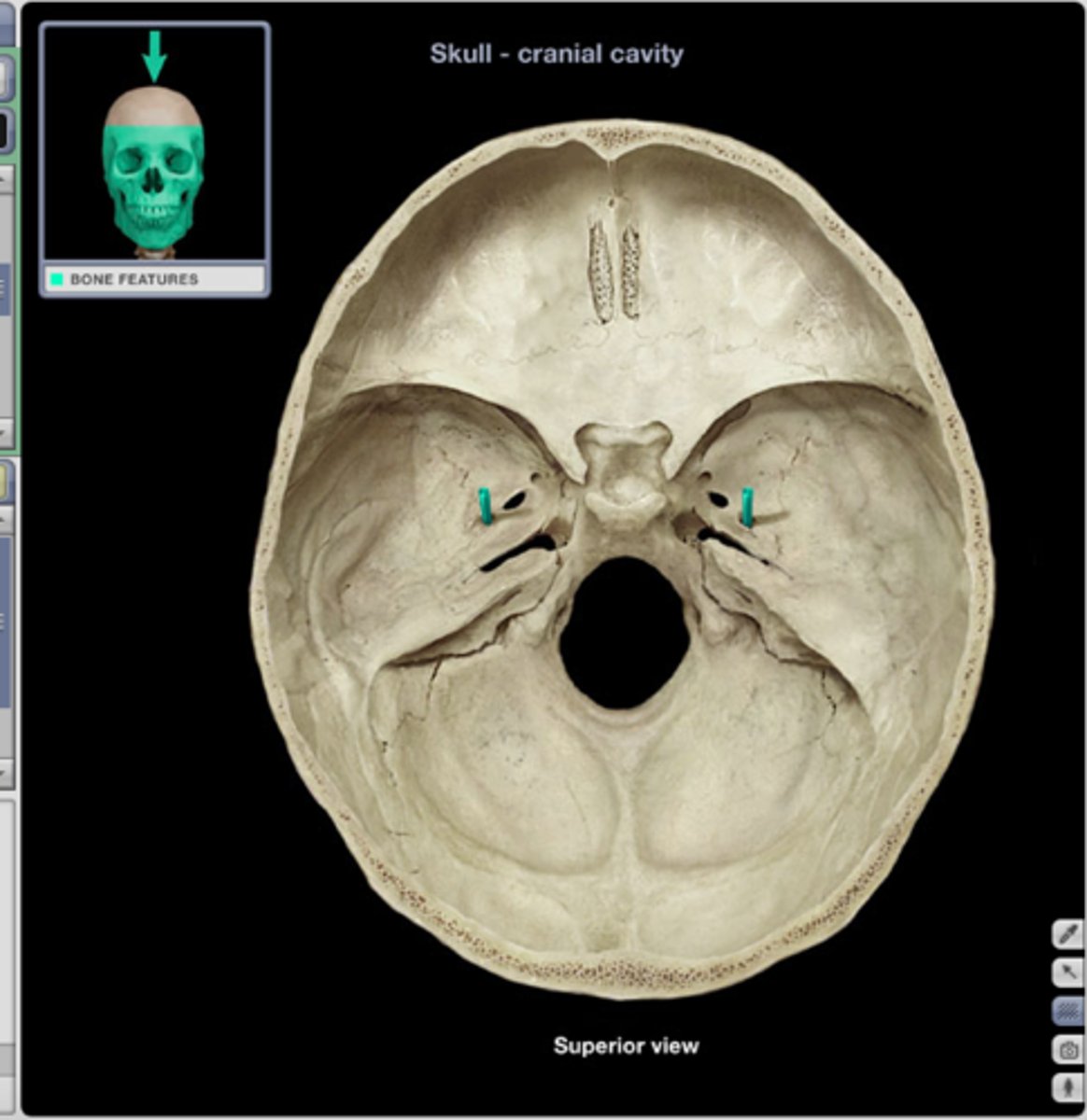
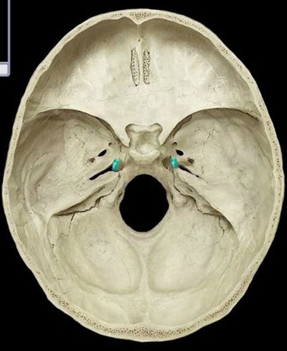
optic canal
hole behind the eye socket (circle) where the optic nerve goes through uperior part of the eyer socket

superior orbital fissure
space
A boomerang-type hole in the back of the eye socket (top part of the boomerang)
inferior orbital fissure
Bottom part of the boomerang shape of the orbital fissure.

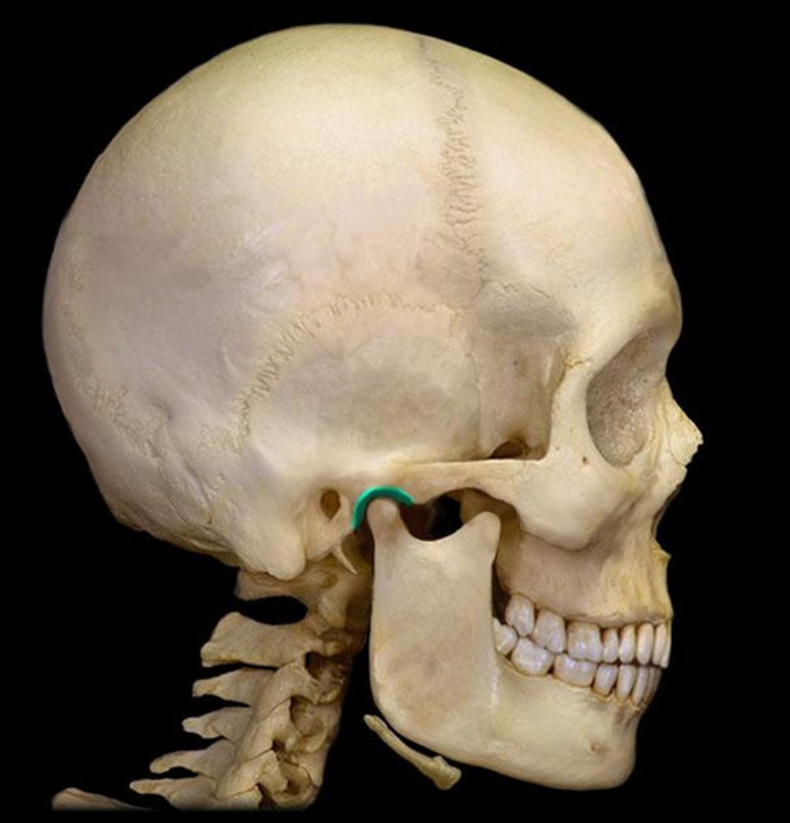
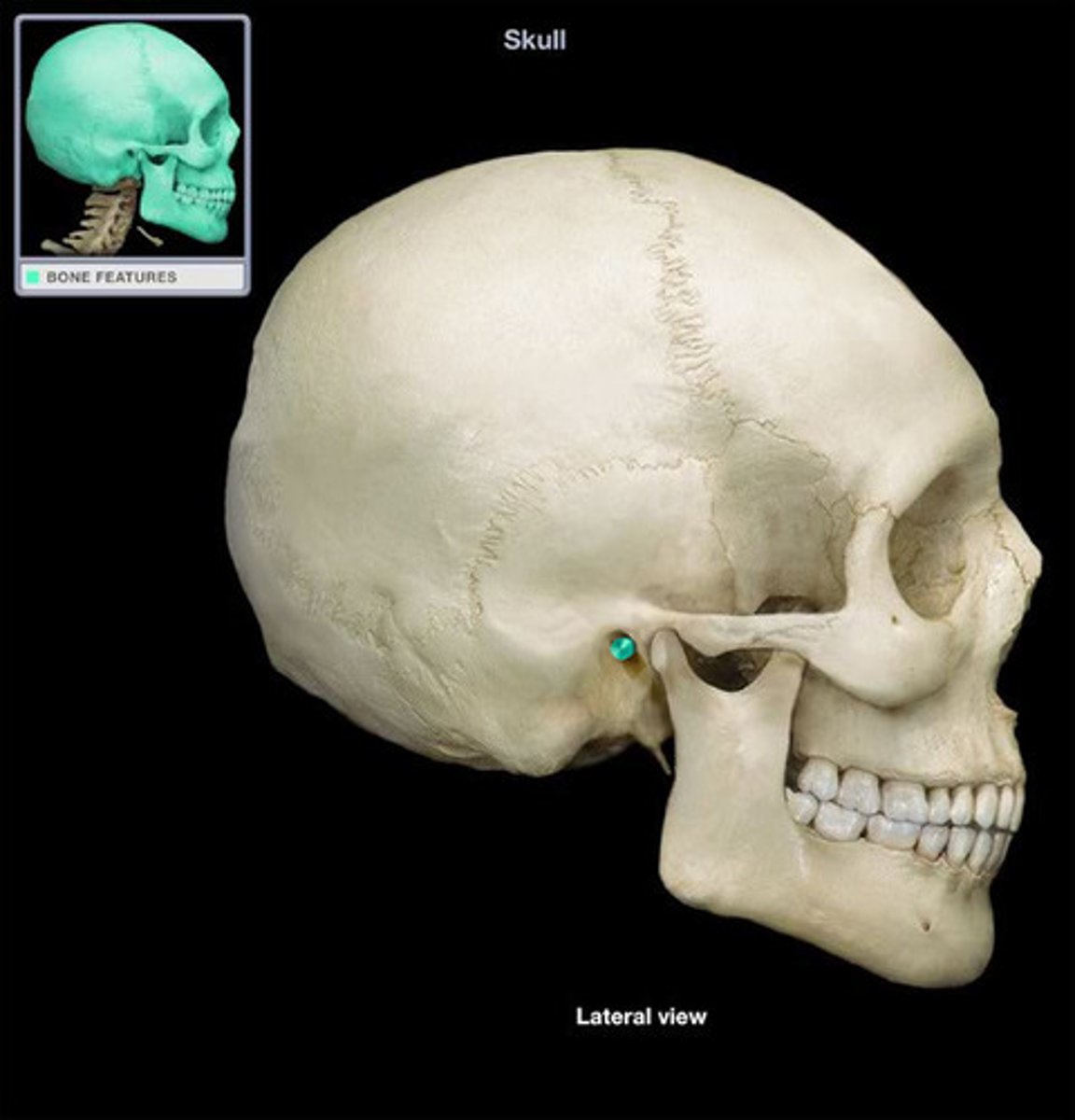
external acoustic meatus
structure
holes on either side of the head outside of the head where ear go
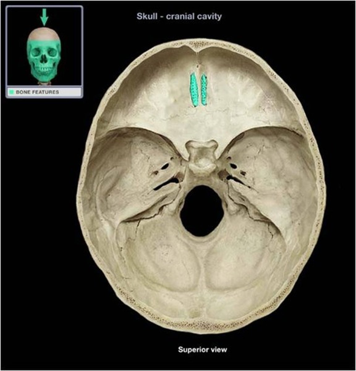
cribriform foramina
hole
the holes within the fribeform plate of the crista galli
optic canals
from the inside of the cranial base through the back of the eye socket

foramen rotundum
most anterior hole of the holes in the center of the cranial base

foramen ovale
hole
oval structure in the center portion of the cranial base
foramen spinosum
internal
pin hole of the cranial base most posterior whole of the middle of the cranial base
foramen lacerum
hole internal
having jagged edges the most inner wholes of the middle of the cranial base
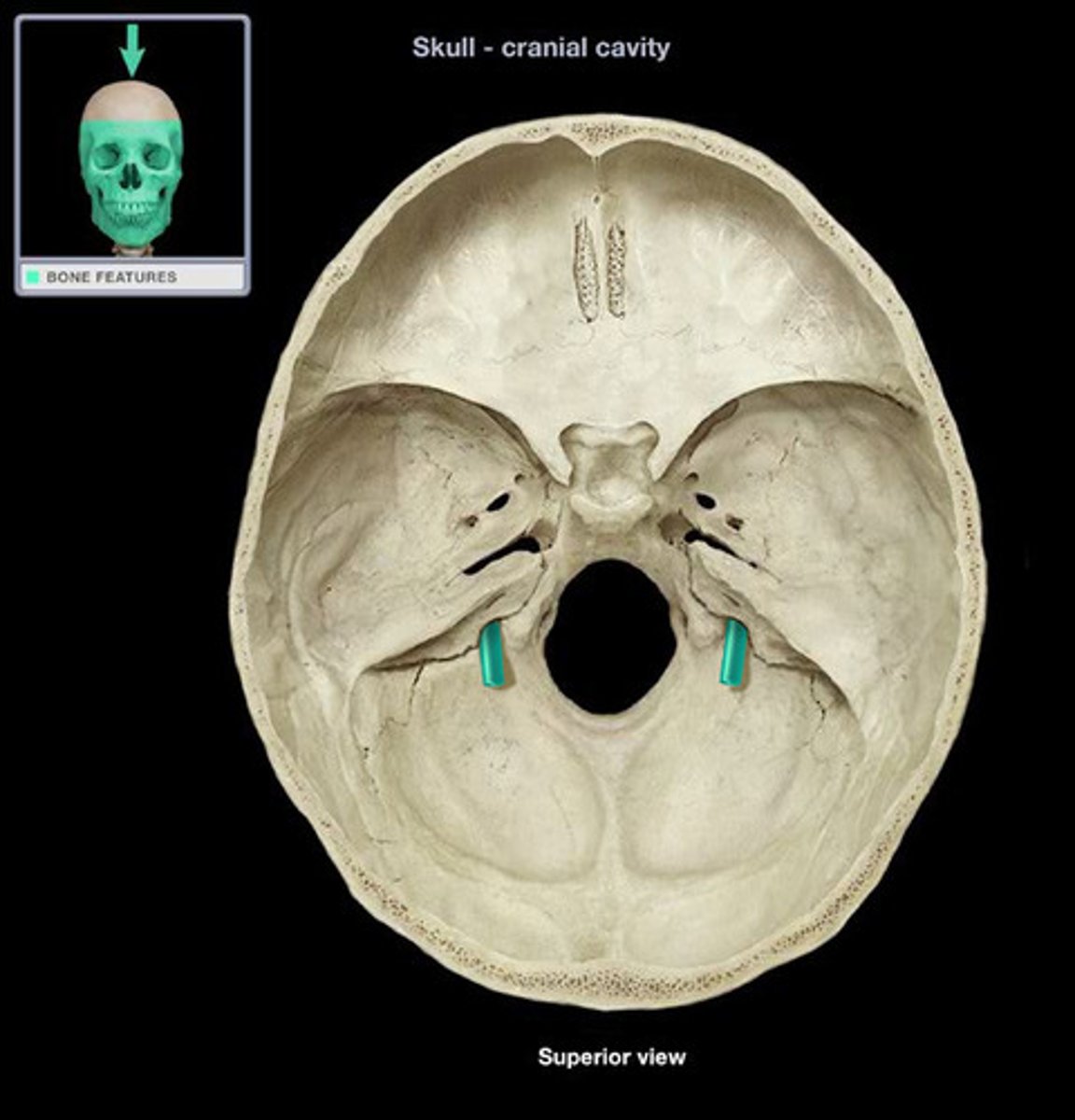
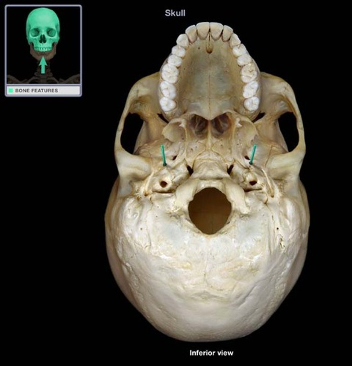
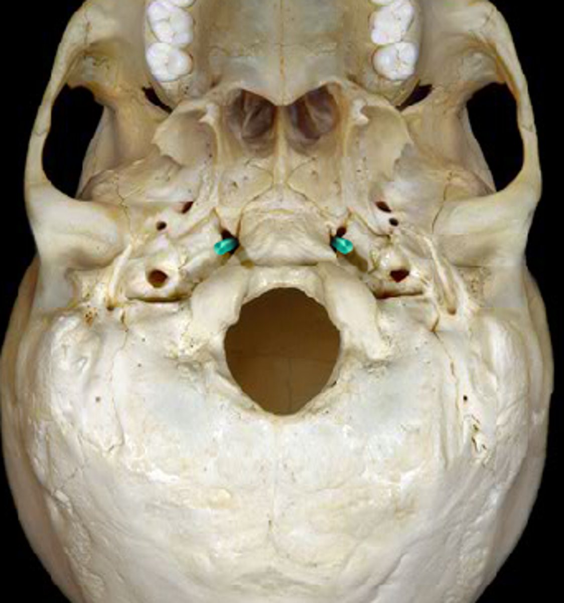
internal acoustic meatus
structure
internal wholes where the ears would go on either side of the skull

jugular foramen
hole
under the indents
posterior holes on the back of the skull
hypoglossal canal
hole
holes in the inside wall of the foramen magnum

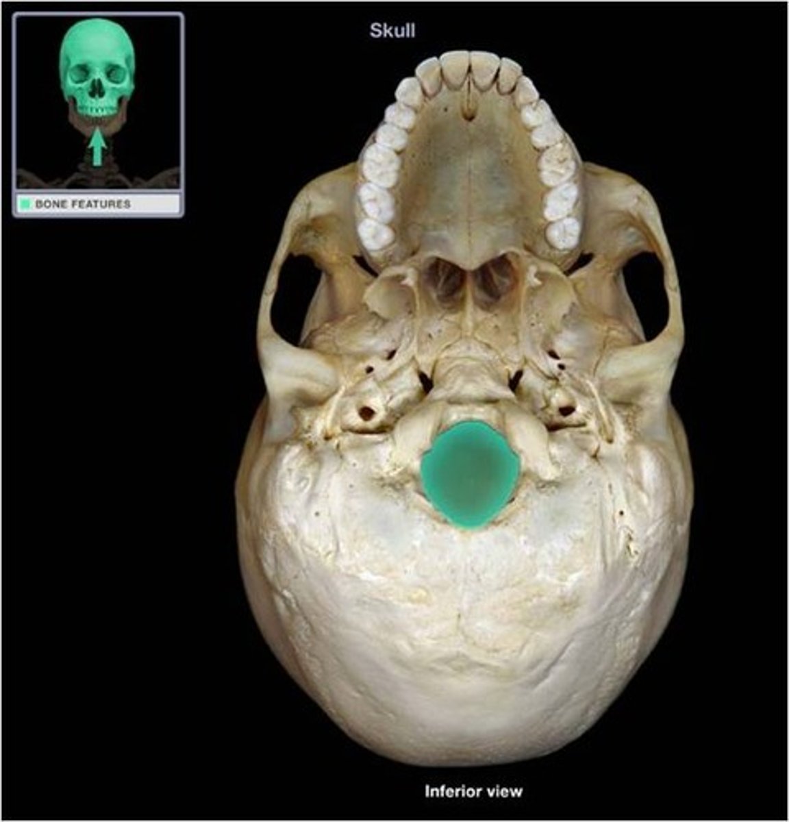
foramen magnum
hole interior
biggerst hole in the base of the skull
foramen ovale
hole exterior
ovals outside the jawbone

foramen spinosum
hole exterior
tiny holes
foramen lacerum
hole exterior
no structure passes completely through
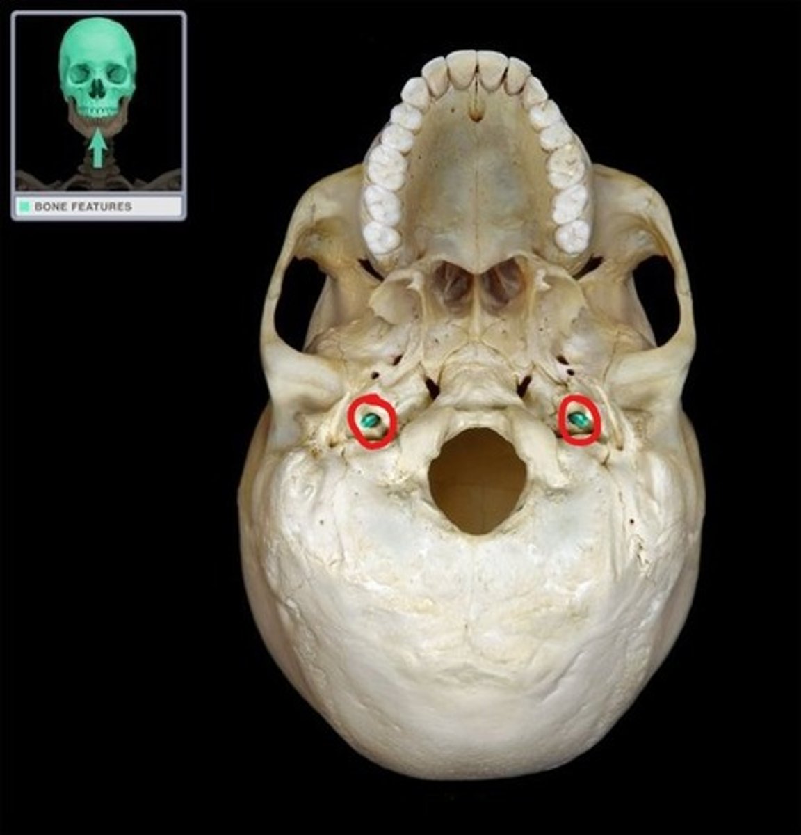
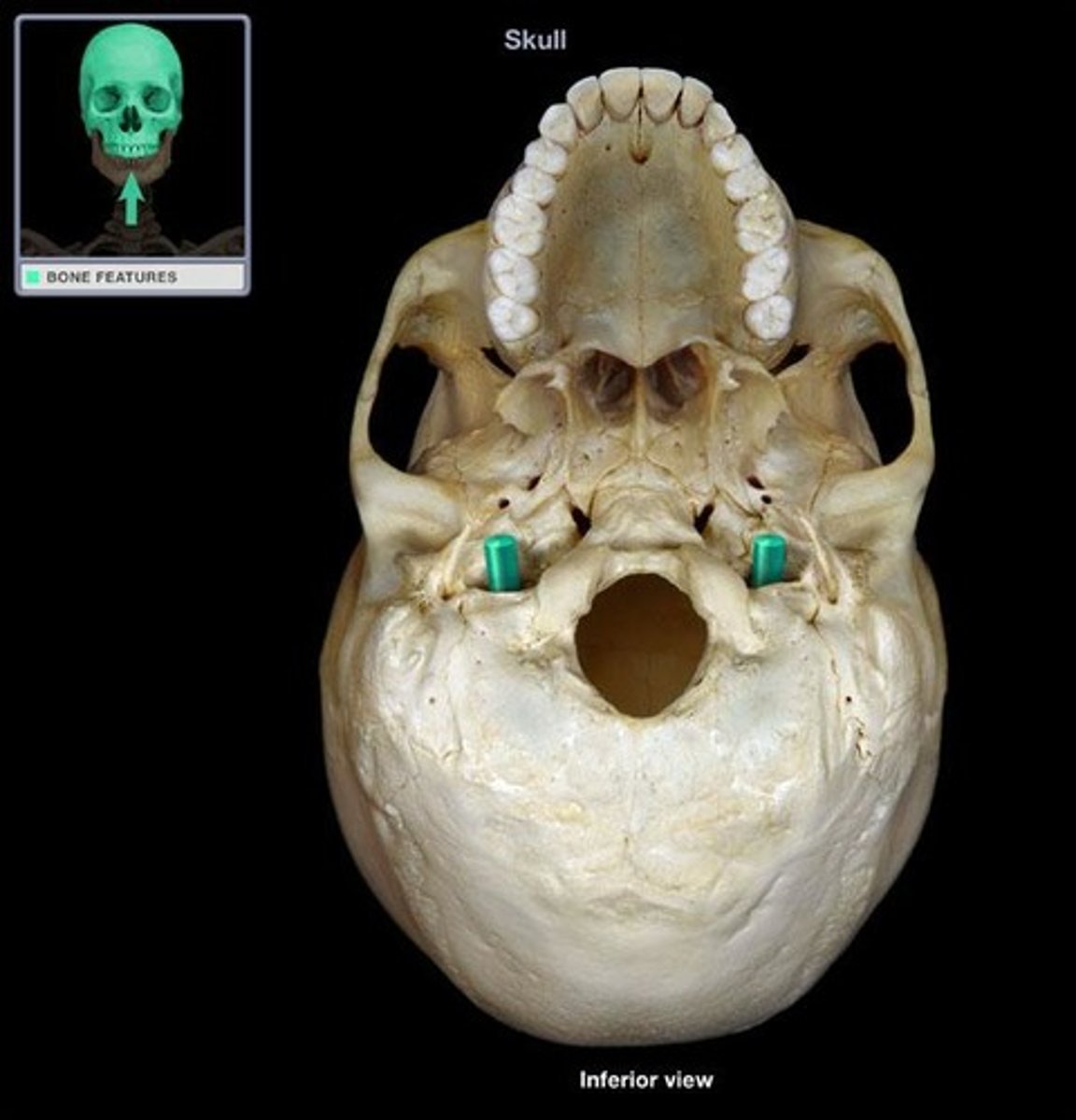
Carotid canal
whole posterior to the spinosum exterior
jugular foramen
hole exterior
jellybean shape
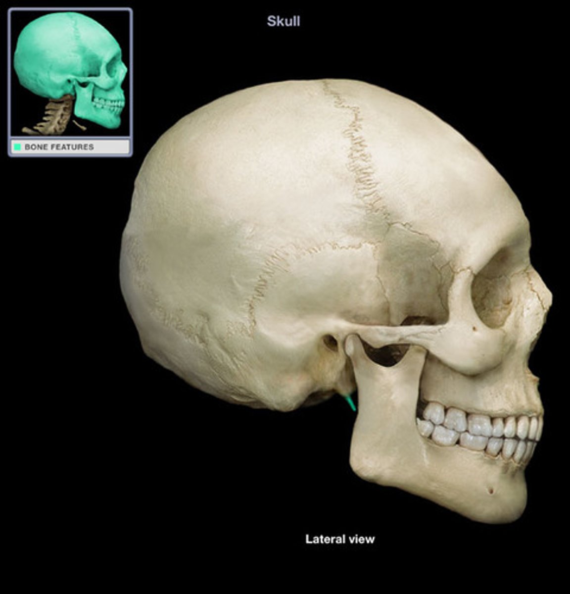
stylomastoid foramen
hole
hole between the styloid and mastoid process
hypoglossal canal
external
holes on the sides of the big hole

foramen magnum
external
large hole
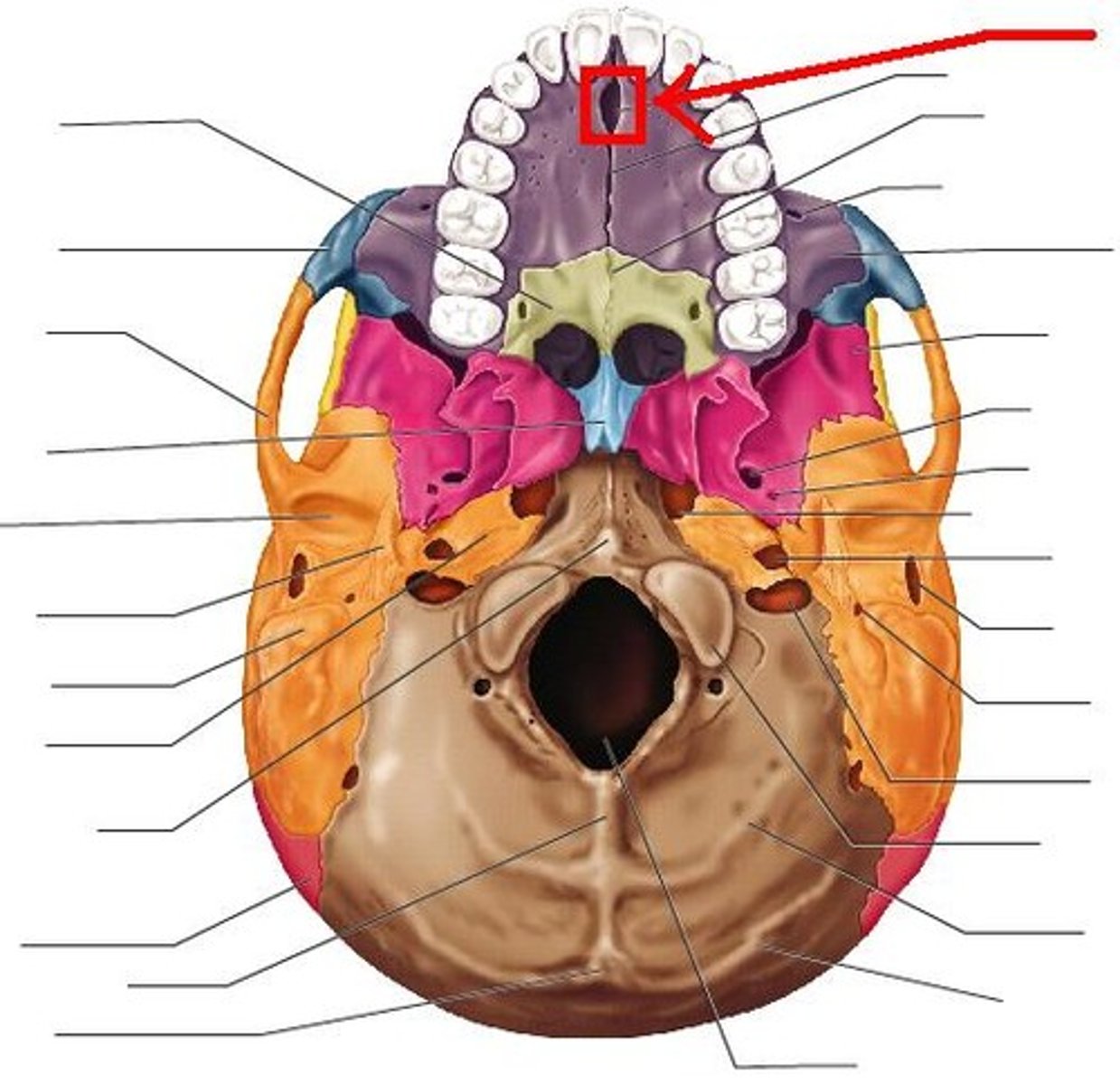
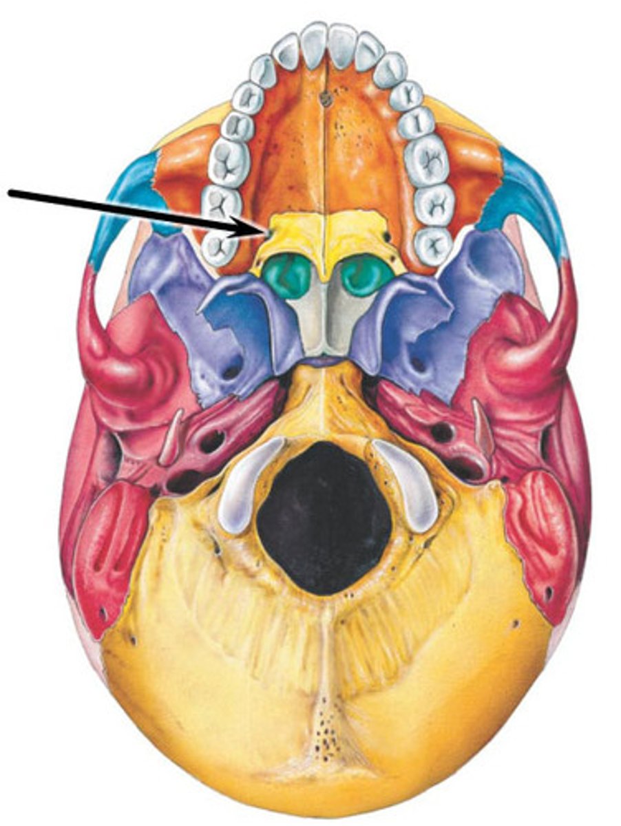
incisive fossa
depression
directly behind the two front teeth
greater palatine foramen
hole
holes back by the molars of the hard palate
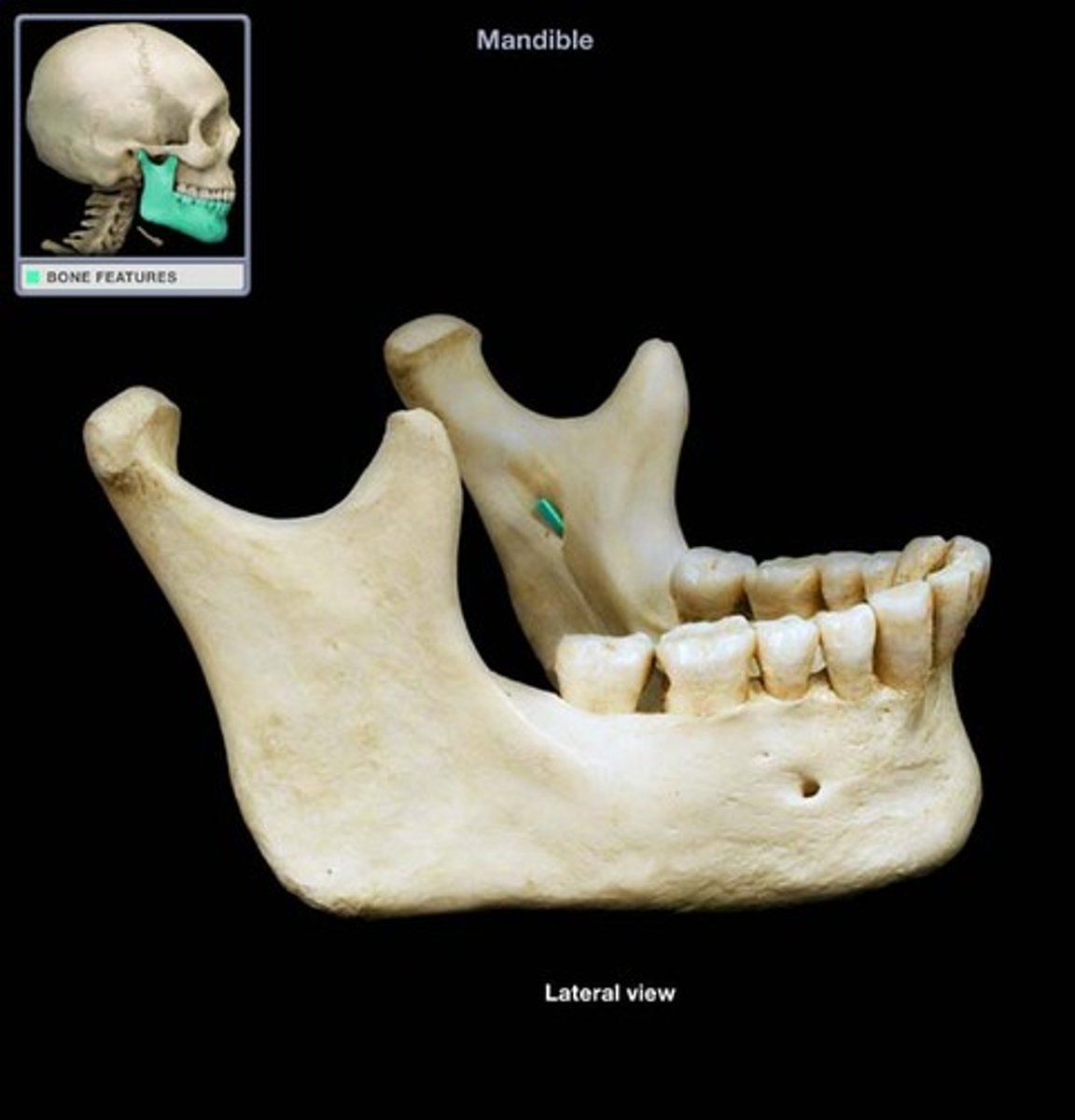
mandibular foramen
hole
internal surface of mandible
little holes on the back inside of the bottom jaw
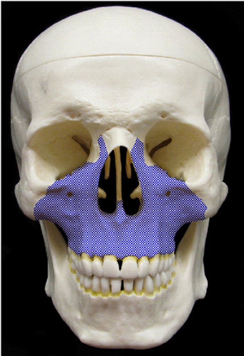
frontal sinuses
space behind the bones
right by/behind the eyebrows
paired, but rarely equal size
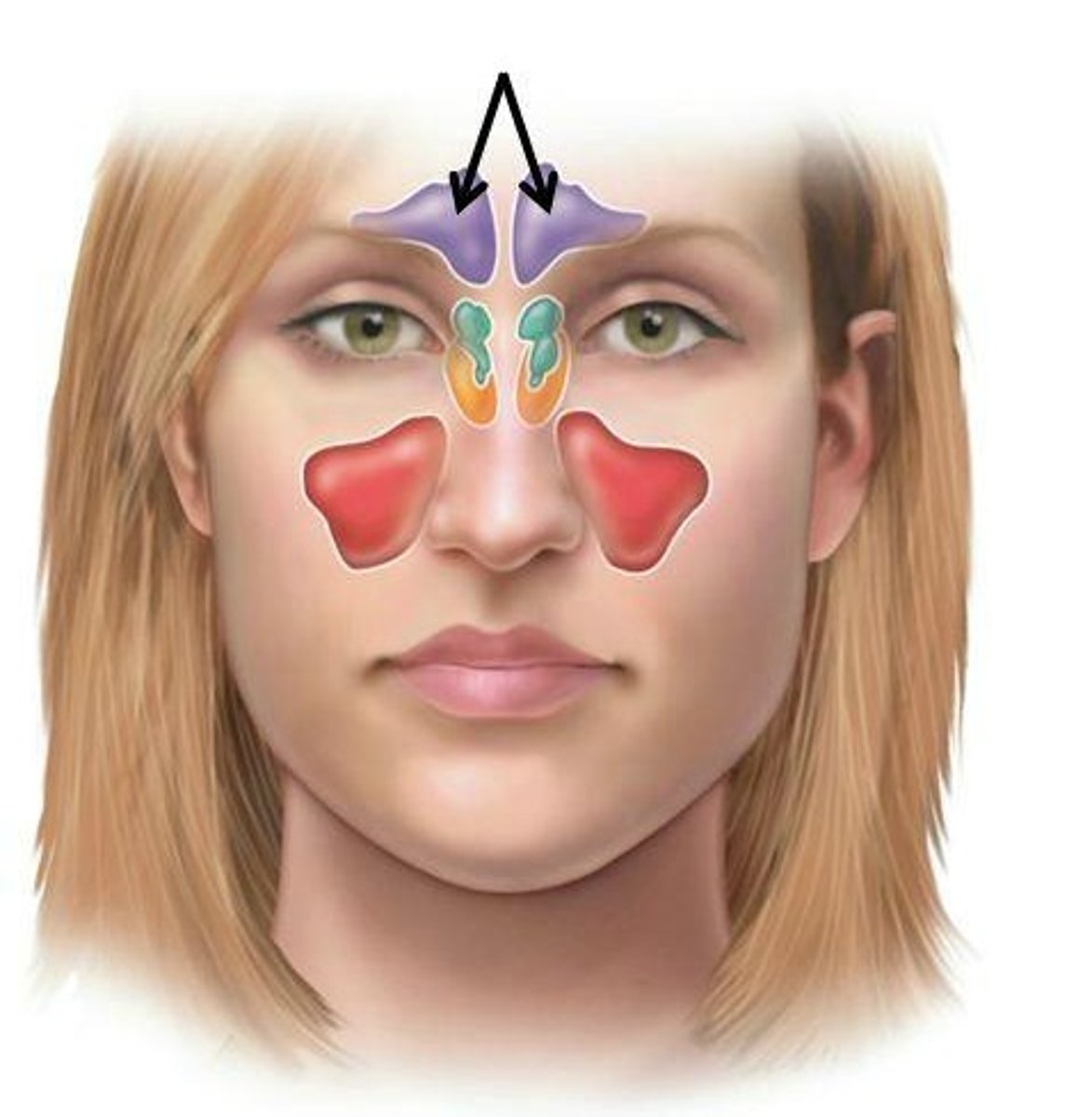
maxillary sinuses
space behind the bone of the maxillae bones that make up the top of the jaw
ethmoid air cells
space behind the bones
pinch the bridge of the nose
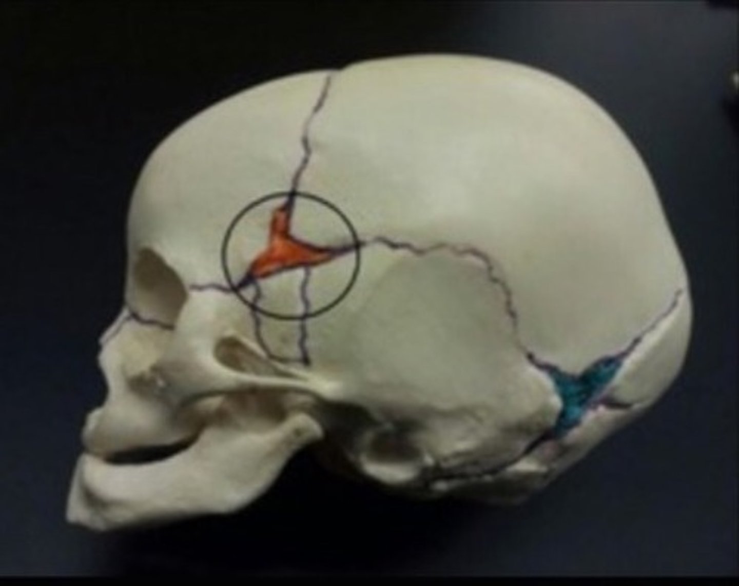
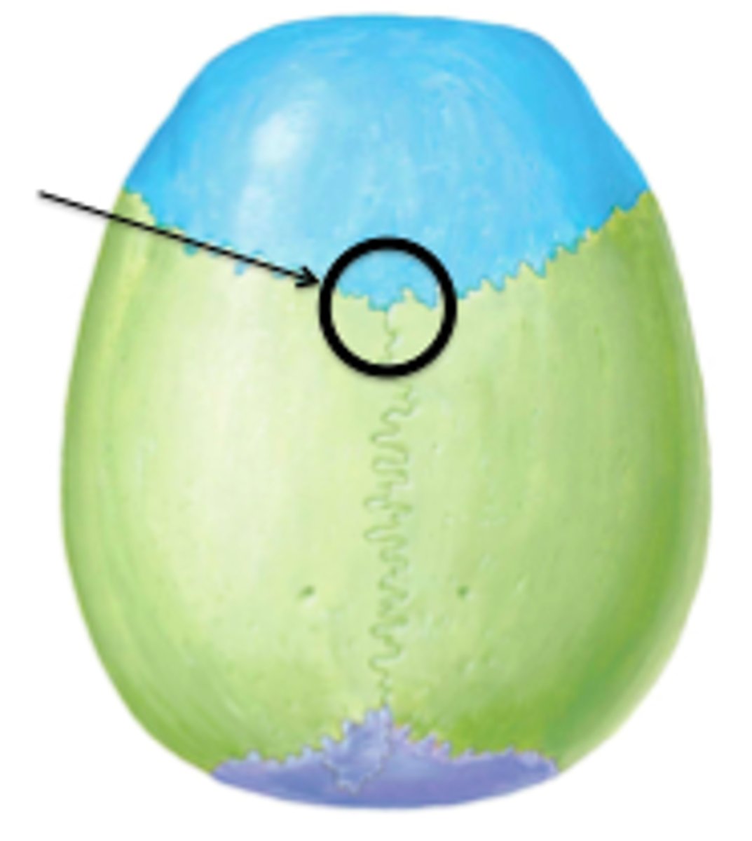
anterior fontanelle
junction of the skull sutures
tippy top, most anterior
where three sutures collide
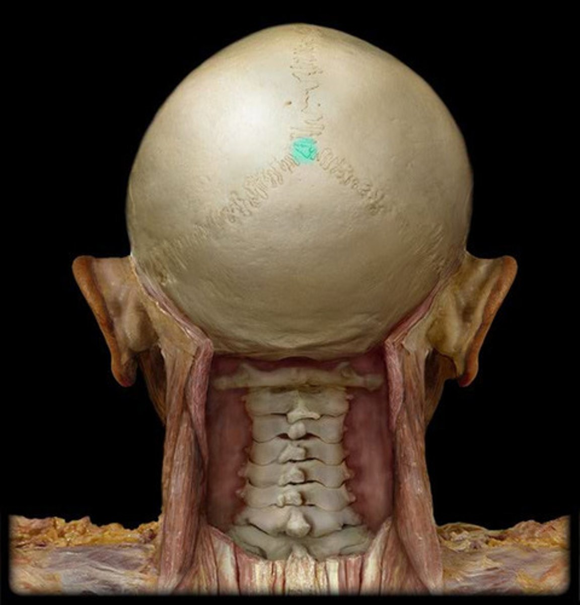
posterior fontanelle
junction
back of head where the three collide
sphenoid fontanelle
junction
around the temple of the head
on both sides of the skull