CH 12 BIO MO2A
1/76
Earn XP
Description and Tags
Name | Mastery | Learn | Test | Matching | Spaced | Call with Kai |
|---|
No analytics yet
Send a link to your students to track their progress
77 Terms
why cell division is needed for unicellular organisms
crucial for unicellular organisms because it is their primary method of reproduction, meaning each time a cell divides, it creates a new, identical organism, effectively allowing the population to increase and spread; essentially, cell division in a unicellular organism is equivalent to reproduction itself
how cell division contributes to growth and differentiation
creating new cells, which increases the overall number of cells in an organism, leading to an increase in size, while differentiation allows these new cells to specialize into different types of cells, forming tissues and organs with specific functions, thus enabling the development of a complex organism from a single fertilized egg
why cell division is needed to maintain tissue
allows for the continuous replacement of old or damaged cells within a tissue, ensuring its proper function and structure; without cell division, tissues would eventually deteriorate as cells die off and are not replenished.
genome
the complete set of DNA within a cell, meaning all the genetic information that is precisely replicated and distributed to each daughter cell during cell division, ensuring each new cell receives an identical copy of the original genome
all of the genes in a organism
Chromatin
the complex of DNA and proteins that makes up chromosomes, which becomes highly condensed during cell division, allowing for the organized separation of replicated DNA strands into two daughter cells
chromosome
a highly condensed structure made up of DNA, visible during cell division, that carries genetic information and is duplicated before mitosis to ensure each daughter cell receives an identical copy of the parent cell's genome
discrete piece of DNA and associated proteins
Somatic cell
any cell in a multicellular organism that makes up the body, excluding reproductive cells (gametes), and these somatic cells divide through the process called mitosis to create identical copies of themselves; essentially, they are the "body cells" that undergo mitosis for growth and repair
Germline
the lineage of cells within an organism that are destined to develop into gametes (sperm or egg cells)
gamate
a cell specifically designed for sexual reproduction, formed by a separate cell division process that halves the chromosome number to create haploid cells, unlike the diploid cells produced by mitosis
how many chromosomes are present in a human somatic cell
46
how many chromosomes are present in a human gamete
23
Sister chromatid
one of the two identical copies of a chromosome created during DNA replication, joined together at the centromere, which are eventually separated during cell division to ensure each new cell receives a complete set of chromosomes
centromere
a specialized region on a chromosome that acts as the attachment point for spindle fibers during cell division, essentially holding together the two sister chromatids and allowing them to be separated evenly during mitosis and meiosis
arm
one of the two sections of a chromosome, separated by the centromere
part of the chromosome on either side of the centromere
mitosis
the process where a cell duplicates its chromosomes and then separates them equally into two new nuclei, resulting in two genetically identical daughter cells, each with the same number of chromosomes as the parent cell; essentially, it's a type of cell division that ensures each new cell receives a complete set of replicated chromosomes
division of the nucleus
Cytokinesis
the final stage of cell division where the cytoplasm of a cell physically divides into two separate daughter cells, ensuring that each new cell receives a complete set of segregated chromosomes from the original cell during mitosis or meiosis; essentially, it is the process of "splitting the cytoplasm" after the chromosomes have been separated and pulled to opposite poles of the cell
cohesion
the process of holding sister chromatids together during cell division
the attraction of molecules for other molecules of the same kind
how and why sister chromatids are attached to one another
by a protein complex called "cohesin," which forms a ring-like structure that encircles the two identical DNA strands created during DNA replication, holding them together most tightly at the centromere region; this attachment is crucial to ensure proper chromosome segregation during cell division, allowing each new daughter cell to receive one copy of each chromosome.
when and why DNA is replicated
DNA replication occurs during the Synthesis (S) phase of interphase, which is the stage before mitosis, so that each new daughter cell created during mitosis receives a complete copy of the genetic material from the parent cell, ensuring identical genetic information in both daughter cells
G0
a cell enters a resting state, meaning it is not actively preparing to divide and is essentially paused outside of the normal cell cycle
G1
a cell primarily grows in size, synthesizes proteins and RNA, and accumulates the necessary building blocks to prepare for DNA replication
G2
the cell actively prepares for cell division by synthesizing proteins necessary for mitosis, replicating organelles, and essentially ensuring all the necessary components are ready to enter the M (mitotic) phase, where the actual division of the cell occurs
what happens during mitosis
a cell's duplicated chromosomes are separated into two identical sets, with each set moving to opposite poles of the cell, effectively dividing the genetic material into two identical daughter nuclei, which will eventually become two separate daughter cells
the sister chromatids of replicated chromosomes are separated
what happens during cytokinesis
final stage of cell division where the cytoplasm of a single cell physically splits into two separate daughter cells, effectively dividing the cell into two distinct entities, each with its own nucleus, by the formation of a cleavage furrow in animal cells or a cell plate in plant cells
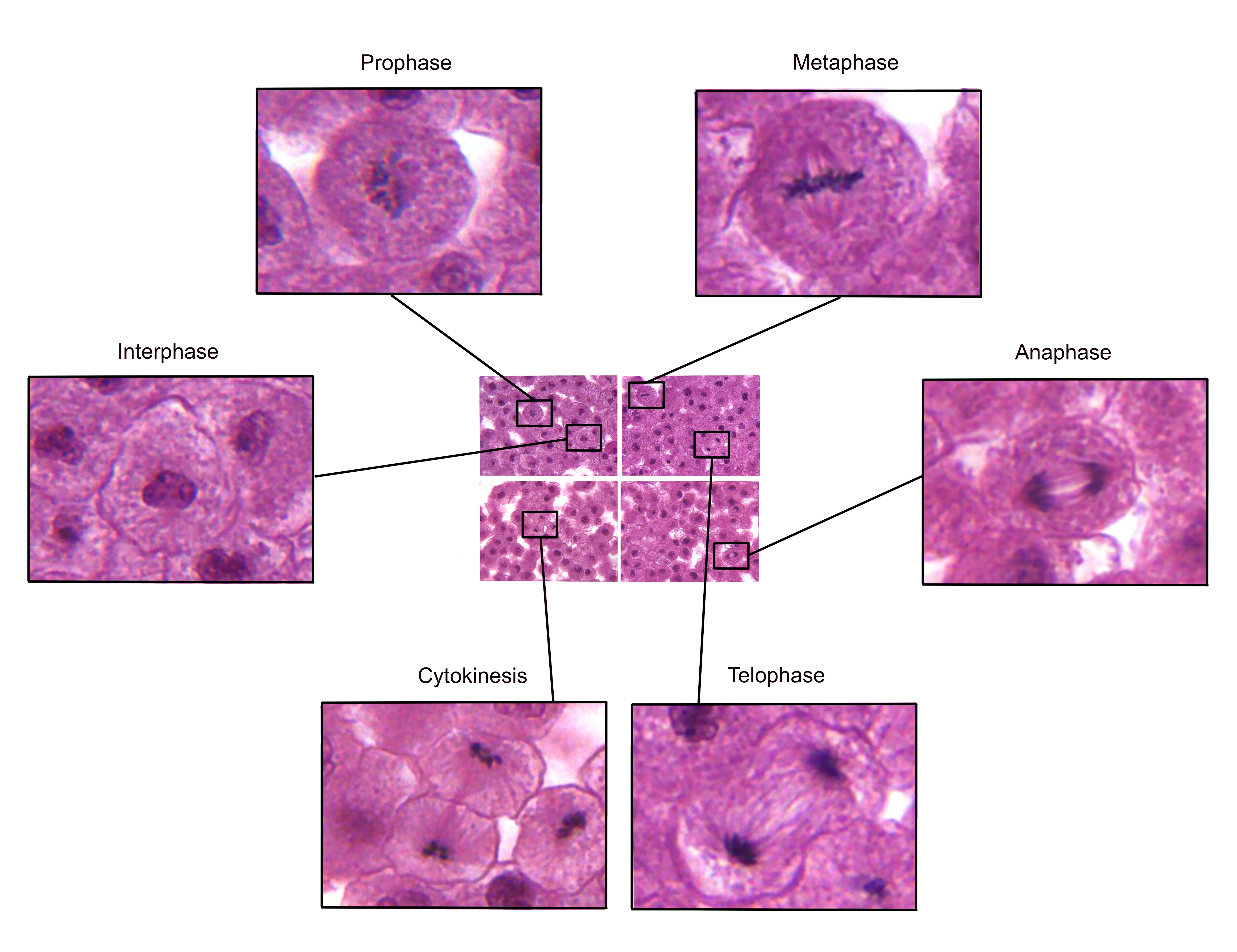
stages of mitosis
what happens to chromosomes in g1 phase
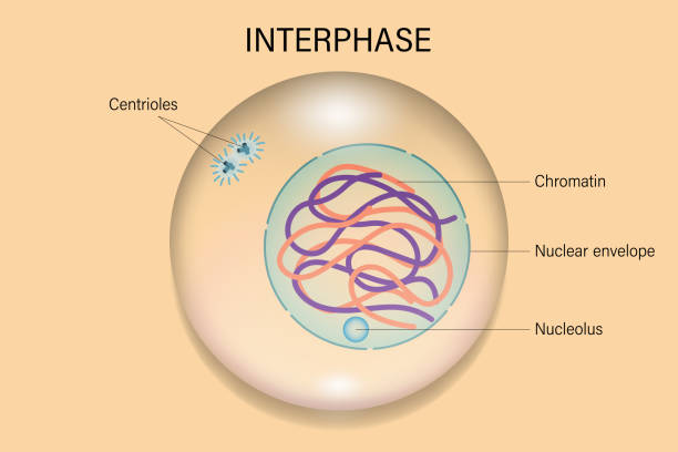
chromosomes remain in their normal, uncondensed state, meaning they are not actively being replicated and simply exist as single chromatids within the nucleus; the cell primarily focuses on growing and accumulating necessary materials for DNA replication which will occur in the subsequent S phase
what happens to Nuclear envelope in g1 phase
The nuclear envelope flattens at the end of the G1 phase. This flattening activates transcription factors that promote the transition from G1 to S phase
what happens to Centrosomes in g1 phase
a cell has one centrosome that is close to the nuclear envelope. During this phase, the cell grows, makes proteins and organelles, and synthesizes the centromere and other centrosome components
there is one centrosome per cell, consisting of two centrioles and the associated pericentriolar material
what happens to chromosomes in prophase
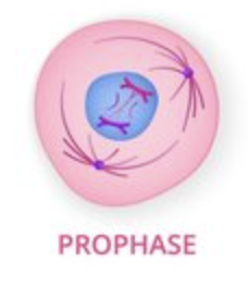
chromosomes condense and become visible under a microscope, appearing as distinct, X-shaped structures due to the presence of two identical sister chromatids joined at the centromere
what happens to nuclear envelope in prophase
still intact
what happens to centrosome in prophase
duplicated and separating
what happens to spindle in prophase
forming, not yet attach to chromosomes
what happens to chromosomes in Prometaphase
more condensed
what happens to nuclear envelope in Prometaphase
broken down
what happens to centrosome in Prometaphase
migrate to opposite poles of the cell, forming the bipolar spindle
what happens to kinetochore in prometaphase
becomes attached to microtubules from the spindle poles, allowing the chromosomes to be actively pulled and moved towards the center of the cell by the forces exerted by these microtubules
what happens to kinetochore microtubules in prometaphase
attach to the kinetochores (protein structures on the centromeres of chromosomes), pulling the chromosomes towards the center of the cell and setting the stage for proper alignment at the metaphase plate
what happens to non-kinetochore microtubules in prometaphase
extend between the poles of the cell without attaching to chromosomes, effectively pushing the centrosomes further apart and contributing to the elongation of the spindle apparatus
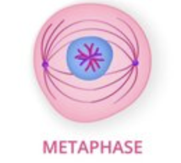
what happens to Chromosomes in metaphase
chromosomes line up in the middle of the cell, at the "metaphase plate," becoming highly condensed and visible under a microscope, with their centromeres precisely aligned, ready to be separated into individual sister chromatids
what happens to Nuclear envelope in metaphase
there is no longer a nuclear envelope
what happens to Centrosomes in metaphase
the centrosomes are positioned at opposite poles of the cell, having migrated there during prophase, and are actively involved in forming the spindle fibers that attach to the chromosomes
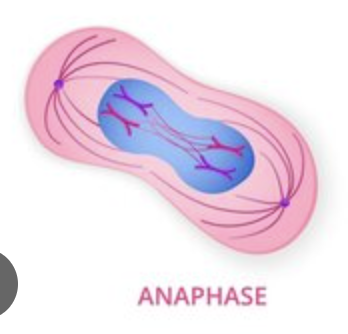
what happens to Chromosomes in anaphase
the sister chromatids of each chromosome separate at the centromere and are pulled apart by the spindle fibers, moving to opposite poles of the cell, effectively becoming individual chromosomes
what happens to Nuclear envelope in anaphase
the nuclear envelope is already broken down and remains disassembled
what happens to separase in anaphase
Separase is a protease that regulates chromatid cohesion during mitosis. Its activation leads to the cleavage of cohesin, which links replicated chromatids, and the subsequent separation of chromatids.
Separase is tightly regulated, and its malfunction can lead to genome instability and aneuploidy, which can cause cancers in higher vertebrates
what happens to Centrosomes in anaphase
centrosomes move further apart from each other as the spindle poles separate due to the elongation of interpolar microtubules, essentially "pushing" the centrosomes to opposite ends of the dividing cell
what happens to kinetochore microtubules in anaphase
shorten at the kinetochore end, effectively pulling the attached chromosomes towards the spindle poles by depolymerizing and releasing tubulin subunits, thus facilitating the separation of sister chromatids and their movement to opposite ends of the cell
what happens to non-kinetochore microtubules in anaphase
elongate, pushing the spindle poles further apart, contributing to the overall cell elongation as the chromosomes are pulled to opposite ends of the cell; essentially, they help "stretch" the cell during the separation of sister chromatids
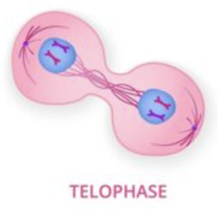
what happens to chromosomes in telophase and cytokinesis
chromosomes reach opposite poles of the cell, begin to decondense (uncoil) back into chromatin, and new nuclear membranes form around each set of chromosomes, essentially reversing the condensation process that occurred earlier in mitosis; in cytokinesis, which follows telophase, the cytoplasm physically divides, separating the two sets of chromosomes into distinct daughter cells, while the chromosomes themselves remain in their decondensed state and are not actively changing
what happens to Nuclear envelope in telophase and cytokinesis
During telophase, the nuclear envelope reforms around each set of separated chromosomes, effectively reappearing after breaking down in earlier stages of mitosis, while in cytokinesis, the nuclear envelope remains fully formed and undergoes no significant change as the cytoplasm divides to create two separate daughter cells
what happens to spindle in telophase and cytokinesis
Completely disassemble and break down, essentially disappearing as the chromosomes reach the poles of the cell and new nuclear membranes form around each set of daughter chromosomes
what happens to centrosome in telophase and cytokinesis
begin to disassemble as the mitotic spindle breaks down, and each daughter cell inherits one centrosome
how chromosomes move during anaphase
chromosomes move towards opposite poles of the cell by being pulled apart at their centromeres by spindle fibers, specifically the kinetochore microtubules
Contrast cytokinesis in animal cells and plant cells
In plants, cytokinesis takes place by the cell plate method, while in animals, it occurs by cell cleavage. The new cell membrane is usually derived from the Golgi Body in the case of plant cytokinesis, while in animals, it is derived from the endoplasmic reticulum
how cell division is different in bacteria, dinoflagellates, diatoms and yeast
in bacteria occurs primarily through binary fission, a simple splitting process, while dinoflagellates undergo a unique form of closed mitosis with a complex nuclear structure, diatoms divide with a specialized cell wall formation, and yeast typically reproduces by budding, where a smaller daughter cell grows from the parent cell
In humans, describe which cells continuously divide, which divide as needed, and which do not divide
skin cells (epidermis) and blood cells (produced in the bone marrow), which need constant replacement due to wear and tear; cells that divide as needed include liver cells, which can regenerate after injury; and cells that do not divide include most mature nerve cells (neurons) and cardiac muscle cells
Explain what happened in Johnson and Rao’s 1970 experiment when they fused mammalian cells from S phase with ones from G
the G1 nucleus began to replicate its DNA immediately, indicating that the cytoplasm of the S phase cell contained factors that could trigger DNA synthesis in a G1 nucleus, demonstrating that cell cycle progression is regulated by cytoplasmic signals and not solely controlled by the nucleus itself
Explain what happened in Johnson and Rao’s 1970 experiment when they fused mammalian cells from M phase with ones from G1
the G1 nucleus prematurely entered mitosis, indicating that the cytoplasm of the M phase cell contained factors that could trigger mitosis in a G1 nucleus, demonstrating that cell cycle progression is controlled by cytoplasmic signals that can influence the state of a nucleus even when it is in a different cell cycle phase
growth factor necessary for cell cycle progression
they act as extracellular signals that trigger a cell to enter the cell cycle from a resting state (G0 phase), essentially giving the "go-ahead" to proceed through the G1 phase and initiate DNA replication in the S phase
Attachment to proper surface/substrate
cells that are removed from a substrate being unable to proliferate
Absence of DNA damage necessary for cell cycle progression
damaged DNA can lead to mutations being passed on to daughter cells, compromising genomic integrity, and potentially causing cell death or cancer development
why is Attachment of kinetochore microtubule during transition from metaphase to anaphase necessary for cell cycle progression
it allows for the proper segregation of chromosomes to opposite poles of the dividing cell, ensuring that each daughter cell receives the correct number of chromosomes
what MPF is and how it was discovered
"Maturation Promoting Factor," is a protein complex that plays a crucial role in initiating the M phase (mitosis) of the cell cycle by activating various proteins necessary for cell division.
it was discovered by researchers Yoshio Masui and Clement Markert in the 1970s through experiments with frog eggs.
consist of cyclin-dependent protein kinase 1
Explain what the relation between cyclin level and MPF activity. How is cyclin-dependent kinase
regulated?
as cyclin levels rise, so does MPF activity; essentially, cyclin acts as the regulatory subunit that activates the catalytic activity of the cyclin-dependent kinase (CDK) to form the active MPF complex
CDK inhibitors regulate cyclin-dependent kinase
cancer
a disease that occurs when cells divide without control and spread into surrounding tissues. Cancer is caused by DNA changes, most often in genes, which are like a cell's instruction manual
Tumor formation in cancer progression
Tumor initiation; a genetic alteration causes a single cell to proliferate abnormally, which leads to the growth of a tumor.
Tumor progression; As clonal selection continues, the tumor becomes more malignant and grows faster
Invasion into neighboring tissue in cancer progression
Cancer cell invasion is the process by which cancer cells break through the basement membrane and extracellular matrix to penetrate surrounding tissues. This is an early step in the process of metastasis, which is when cancer cells spread to other parts of the body to form new tumors.
Spread via lymphatic and blood vessels in cancer progression
Cancer cells spread through the body via the lymphatic system and bloodstream in a process called metastasis
Metastasis in cancer progression
when cancer cells spread from the primary tumor to other parts of the body, forming new tumors
Radiation
to target and destroy cancer cells while minimizing damage to healthy tissues, primarily by disrupting their ability to replicate through mechanisms like DNA damage, which can be achieved through radiation therapy by delivering high-energy beams that directly damage the genetic material (DNA) of cancer cells, preventing them from dividing and causing them to die off
taxol
its ability to stabilize and prevent microtubules depolymerization, leading to cell cycle arrest at the G2/M phase and cell death
stopping cancer cells from separating into two new cells
Herceptin
by attaching to HER2 receptors on the surface of breast cancer cells and blocking signals that tell the cells to grow and divide
Tamoxifen
blocking estrogen receptors and preventing estrogen from telling cancer cells to grow
interphase
prophase
metaphase
anaphase
telephase
