Ocular Disorders
1/39
There's no tags or description
Looks like no tags are added yet.
Name | Mastery | Learn | Test | Matching | Spaced | Call with Kai |
|---|
No analytics yet
Send a link to your students to track their progress
40 Terms
Optometry vs Opthalmology vs Optician
optometry→healthcare professional
-complex optic cases
-advanced optic devices
opthalmology→MD
-mx care+surgery
-medical school+residency
optician→eyeglasses
-opticianry school/apprenticeship
Path of Light
light
cornea
lens
retina
optic nerve
optic tracts
occipital lobe
Cornea
transparent structure that refracts light
Iris
colored part
blocks excess light from coming in
Pupil
opening in the middle of the iris
controlled by muscles in the eye (opens+narrows iris)
-light→constriction
-dark→dilation
Lens
focuses light into retina
Retina
processes visual information (phototransduction)
components
cones
color
light
detail
-macula
-fovea
rods
motion
dim light
peripheral vision
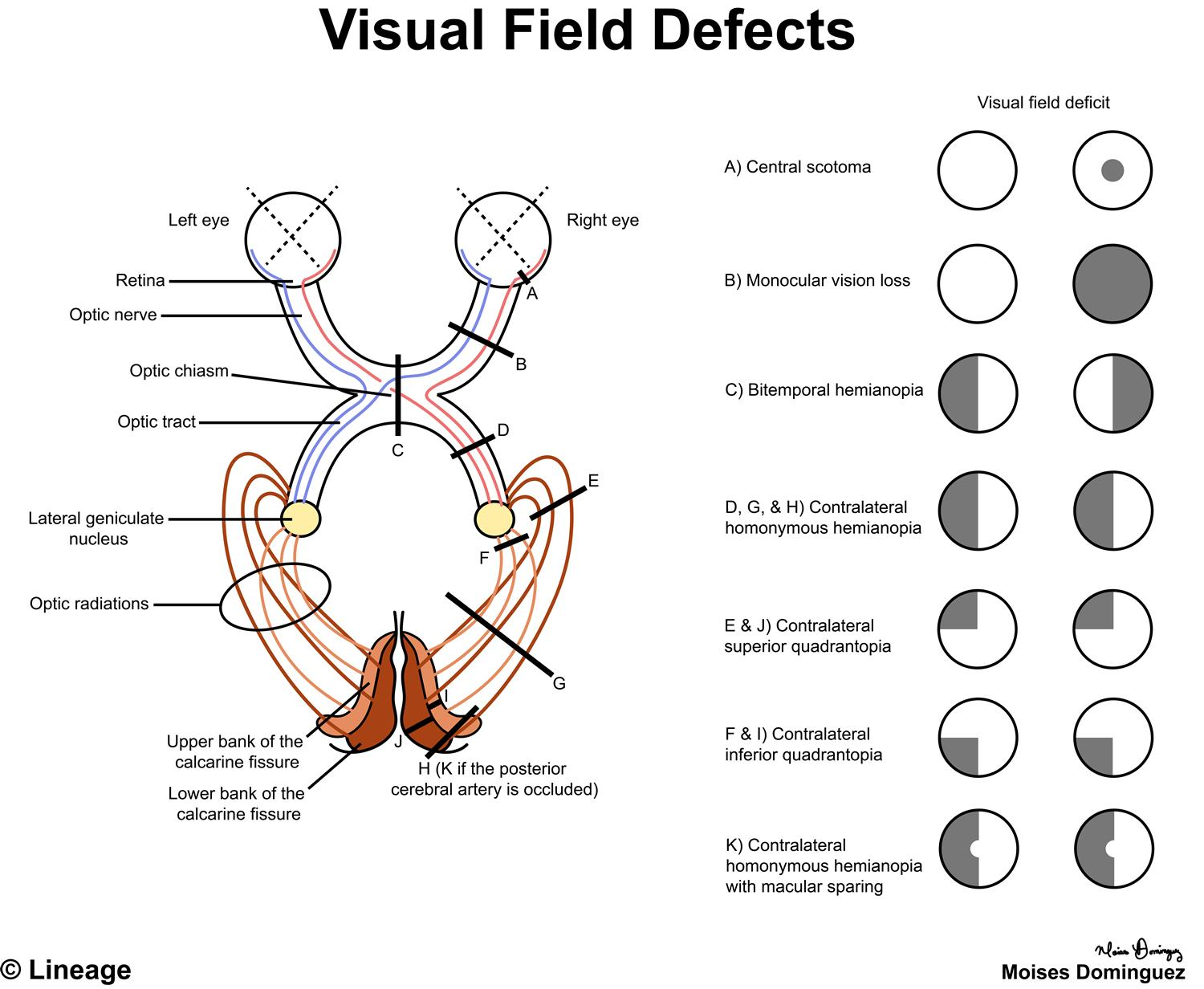
Optic Nerve
passes light+information from eye to the brain
Optic Nerve Tract
optic nerve→optic chiasm (crosses over to other lobe)→optic tract→occipital lobe
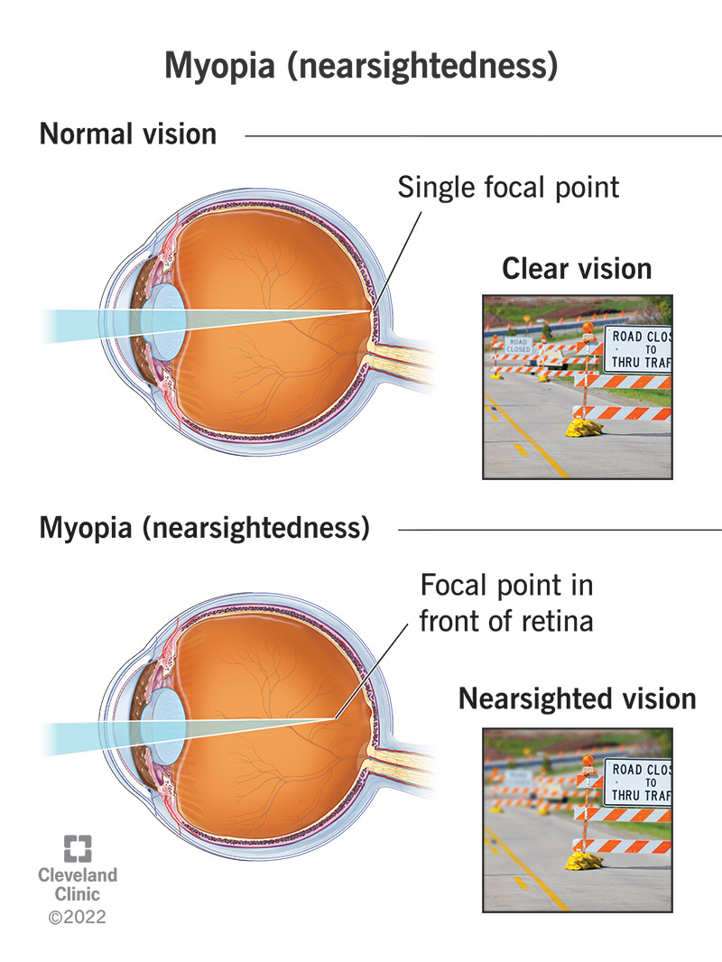
Myopia (Nearsightedness)
PP: eye grows too deep/far back into skull
RF: glaucoma
retina detachments
cataracts
macular degeneration
legal blindness
E: m/c→worldwide epidemic
CM: near→clear vision
far→blurry vision
TX: glasses(no lenses)
contact lenses
myopia management
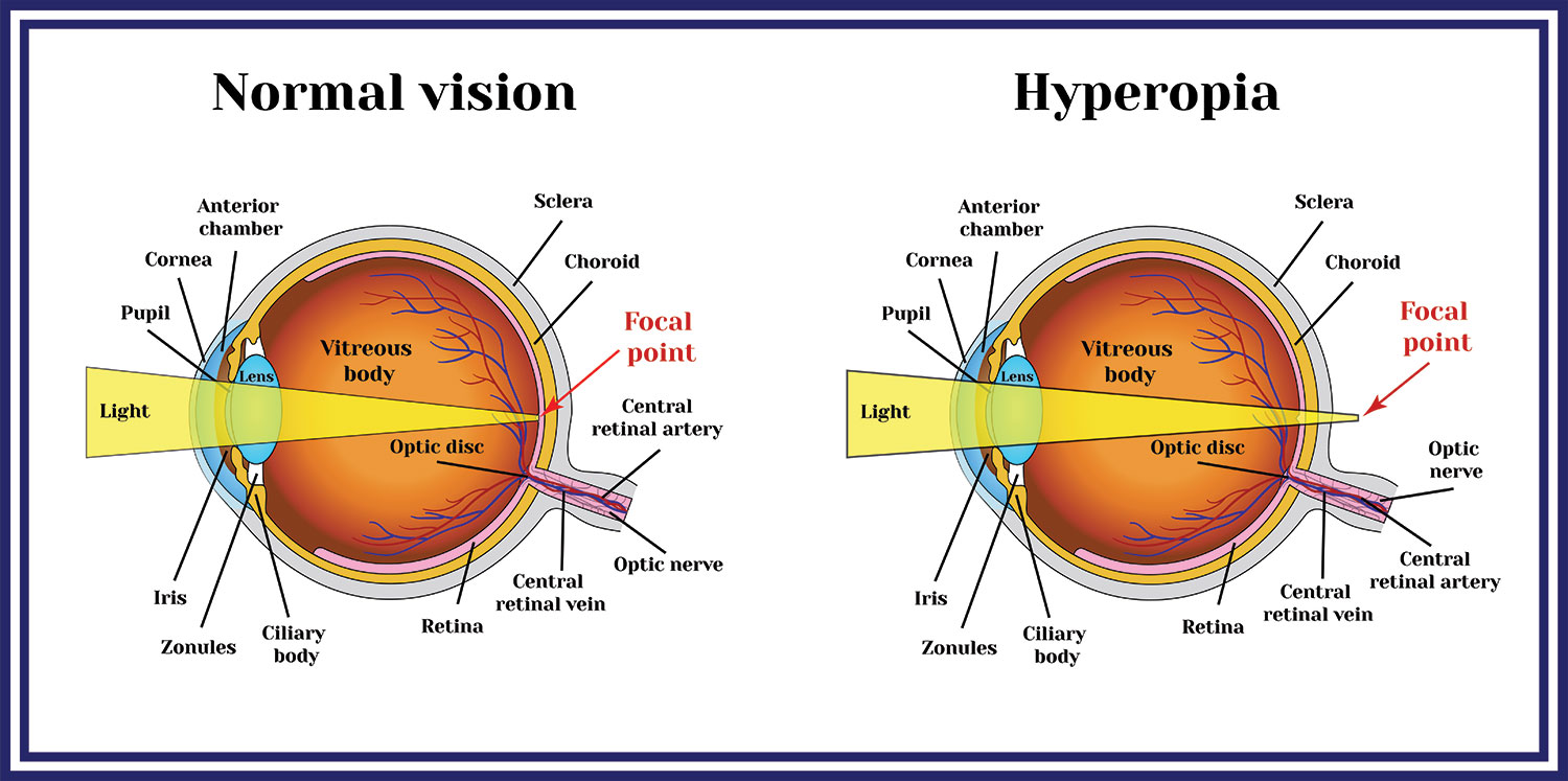
Hyperopia (Farsightedness)
PP: eye is too short→light passes past/through eye
RF: narrow angle glaucoma
CM: near→blurry vision
far→clear vision
TX: glasses+lenses
contact lenses
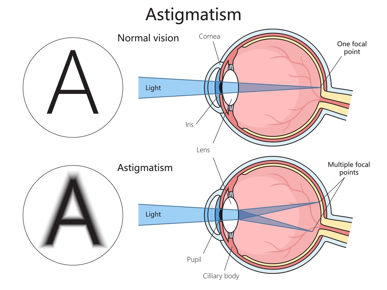
Astigmatism
PP: refractive lens error→problem with how eye focuses light
not a dx
E: misshapen cornea
misshapen lens
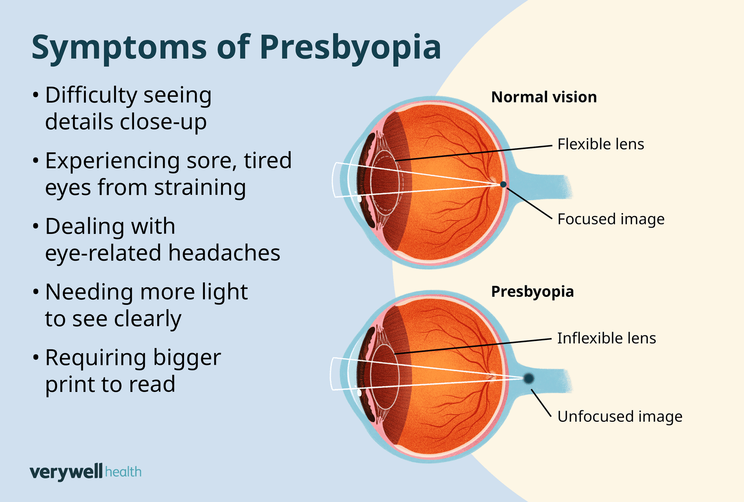
Presbyopia
PP: lens stiffens→decreased accommodation to light
E: ~40 y/o
age-related
CM: near→blurry vision
headaches
eye strain
tired eyes
Congenital vs Acquired Color Blindness
can’t see certain colors
congenital
x-linked recessive
males→7%
females→0.5%
bilateral
m/c→red-green
mother needs to be colorblind/carrier in order to have colorblind son
acquired
retina dx
optic nerve dx
Dark Adaptation
ability to adapt to different illuminations/brightness levels
modulated by rods+cones
-cones are faster than rods
poor dark adaptation etiology
aging
posterior segment ocular dx
-genetic
-acquired
poor diet→vitamin A deficiency
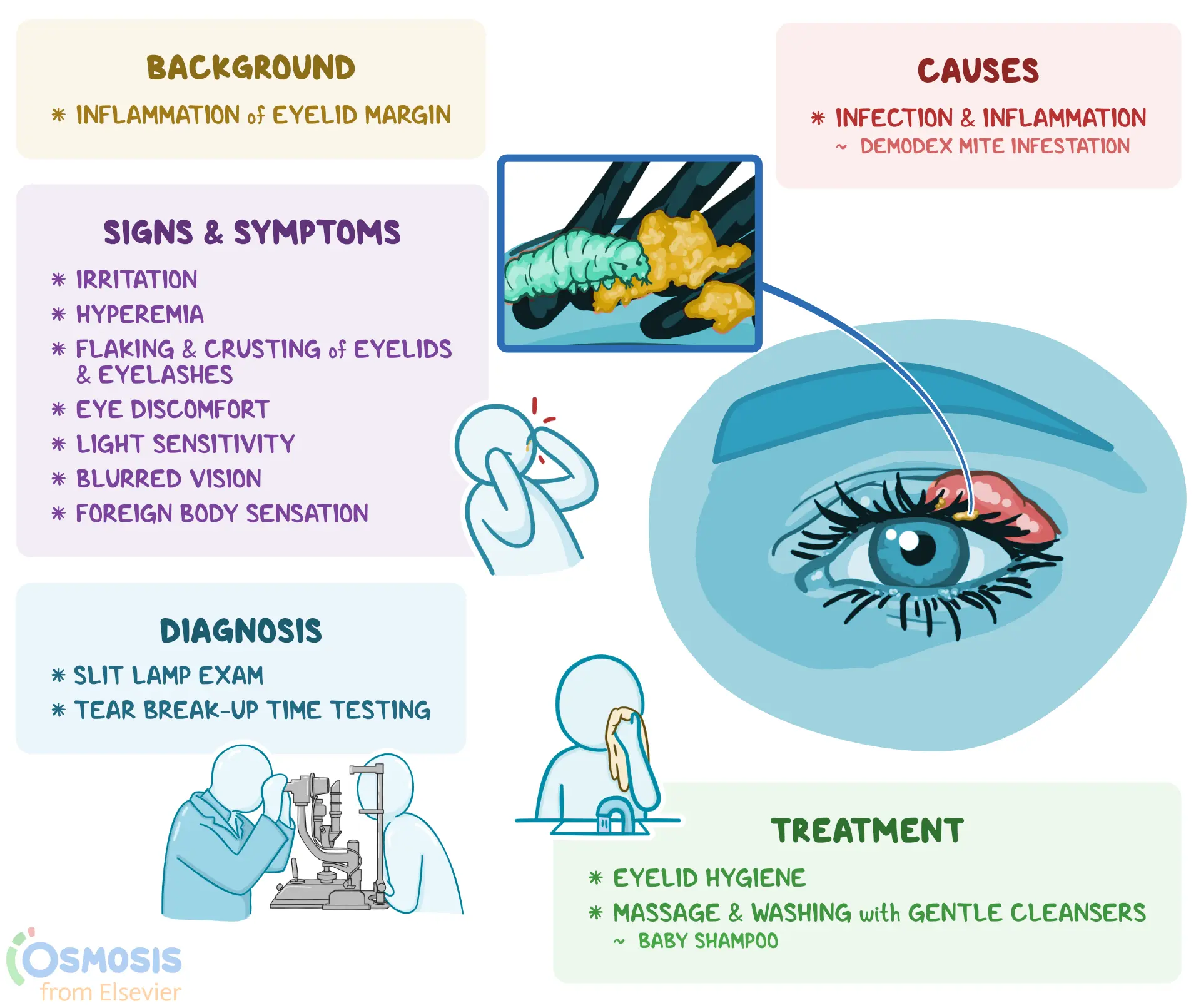
Blepharitis
E: demodex mites in hair follicles
s aureus
epidermidis
CM: erythema
irritation
gritty feeling
TX:
mites→lotilaner eye drop
bx+mites:
tea tree oil
hypochlorous acid spray cleaner
Entropion
PP: eye lid turns inward→eyelashes irritate eye
E: m/c→involutional (skin shrinking with age)
trauma
congenital
tumor
infection
-lower eyelid (m/c)
CM: can’t see eyelid
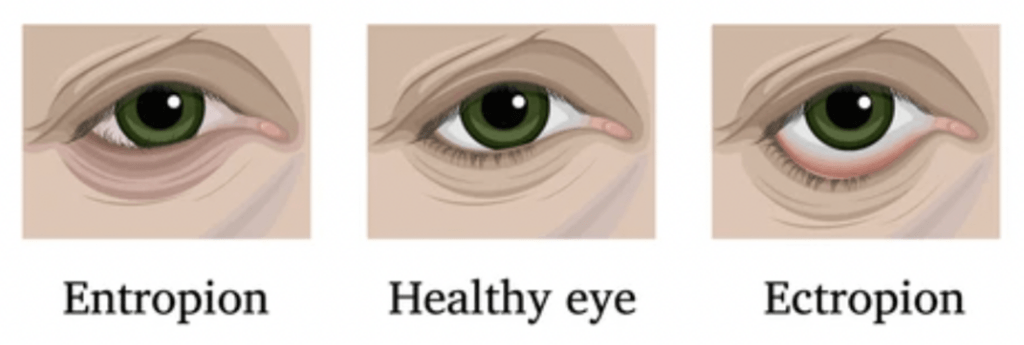
Ectropion
PP: eyelid turns outward→eyelid+eye exposed
E: m/c→involutional (skin shrinking with age)
tumors
trauma
7th nerve paralysis
congenital
-lower eyelid (m/c)
CM: red portion of lower eye

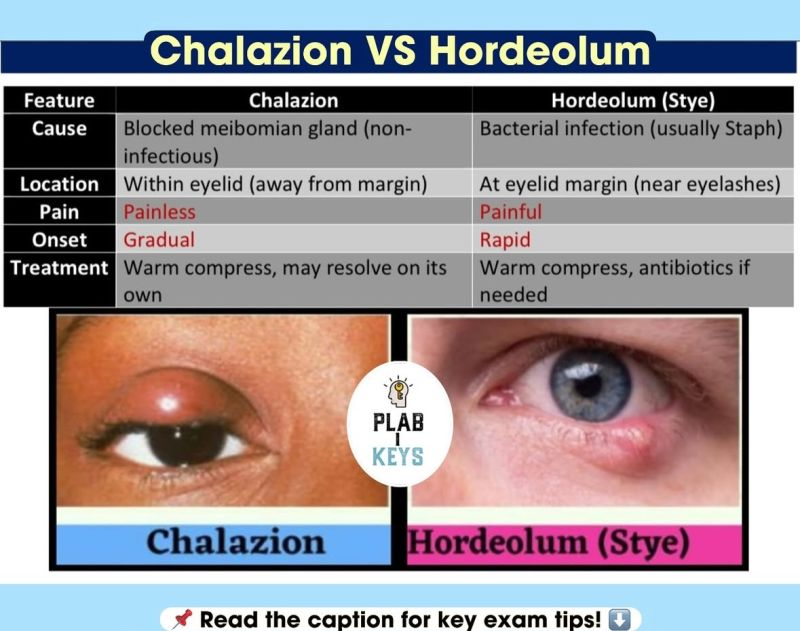
Hordeolum vs Chalazoin
hordeolum
active lesion
new
painful
lower eyelid
“stye”
TX: warm compress
PO abx
chalazoin
inactive lesion
long standing
no pain
upper eyelid
TX: warm compress
intense pulsed light therapy (IPL)
surgical excision
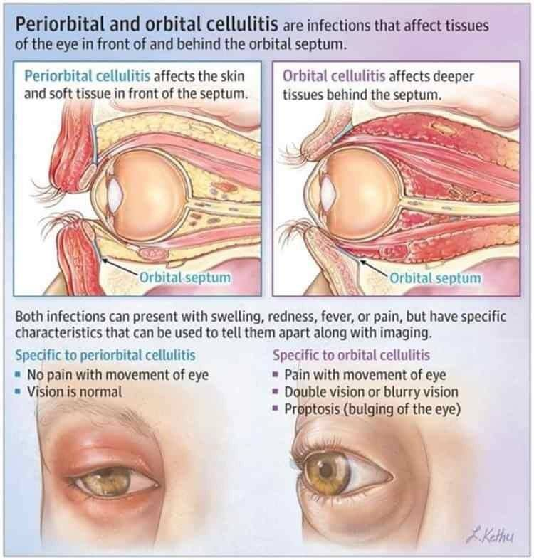
Preseptal vs Orbital Cellulitis
preseptal
erythema limited to the eyelid
TX: PO abx
orbital
erythema surrounds whole eye
limited ocular movement
decreased vision
chemosis (swollen conjunctiva)
proptosis (eyes pop out)
TX: IV abx
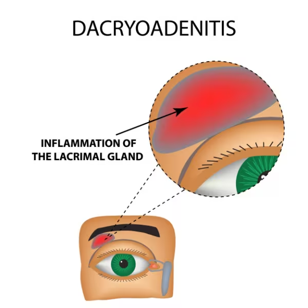
Dacryoadenitis
PP: inflammation of lacrimal gland
E: m/c→inflammation
mumps
mono
influenza
bx (rare)
CM: erythematous tender swelling of outer 1/3 of upper eyelid
TX:
NP: cool compress
calm inflammation
MX: abx
steroids
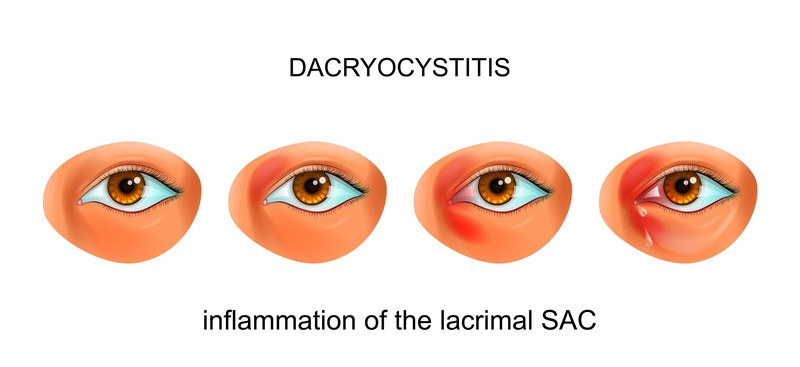
Dacryocystitis
PP: inflammation of lacrimal sac
E: bx infxn
nasolacrimal duct obstruction
tumor
TX: warm compress
abx
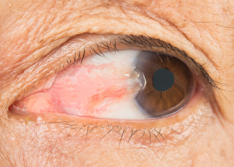
Ptergyium
PP: fibrovascular tissue from conjunctiva→cornea→dry eye/astigmatism
E: UV exposure
chronic irritation
CM: wing-shaped fold on eye
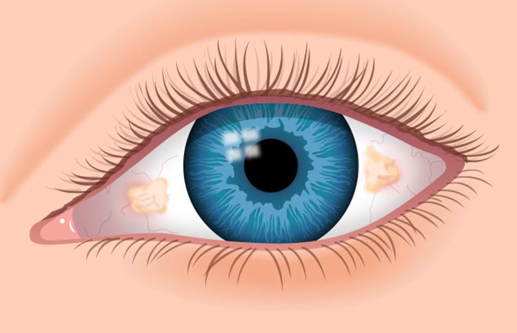
Pinguecula
PP: noncancerous growth of the conjunctiva→dry eye
E: UV exposure
chronic irritation
CM: white-yellow growth on conjunctiva of eye
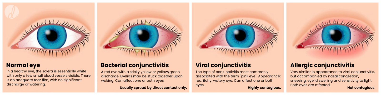
Bacterial Conjunctivitis
E: s aureus
s pneumonia
h influenzae
gonorrhea
chlamydia
CM: unilateral/bilateral
mucopurulent discharge
open eyes→“glued shut”
discomfort
TX: topical abx eye drops (gtts)
Viral Conjunctivitis
E: adenovirus
URI virus
CM: foreign body sensation
discomfort
red eye+watery discharge
UL→BL
TX: no eyedrops (gtts)
referral→betadine rinse
preservative free artificial tears
steroid eye drops
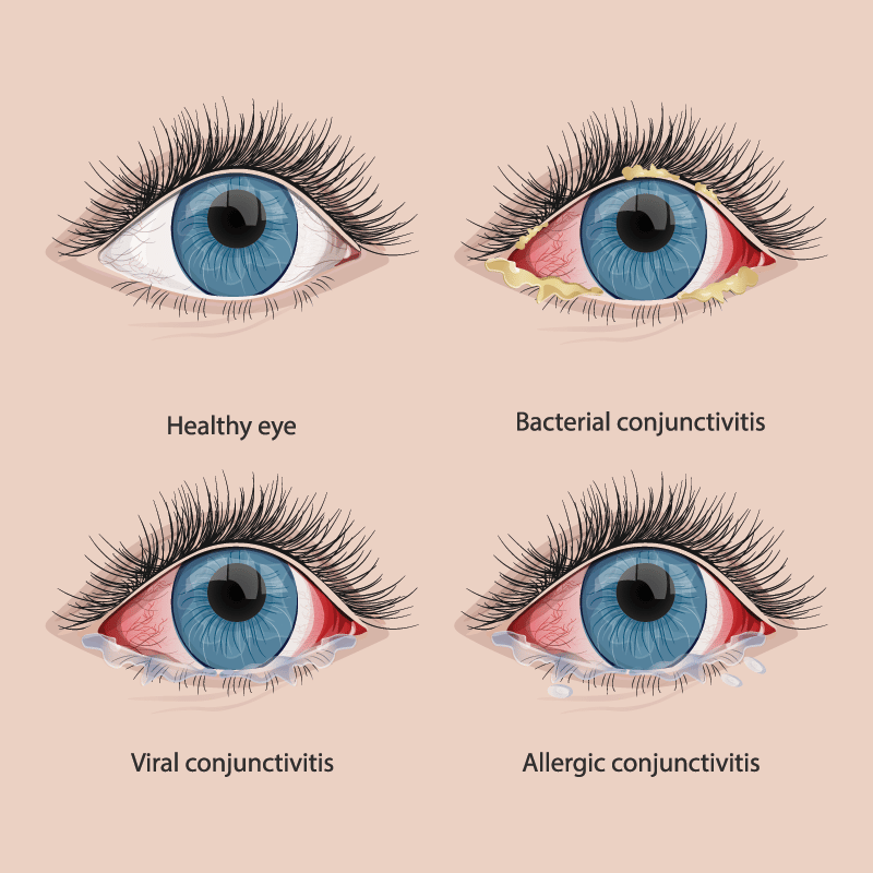
Allergic Conjunctivitis
E: type 1 hypersensitivity rxn
type 4 hypersensitivity rxn
CM: bilateral at same time
itchy
discomfort
swelling+puffy-eyed appearance
TX: pataday
alaway
zaditor
lastacraft
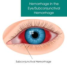
Subconjunctival Hemorrhage
PP: rupture of conjunctival vessels
E: trauma
sneezing
coughing
vomiting
straining
TX: self limiting
stop NSAIDS
stop anticoagulants
chronic:
coagulation DX
HTN
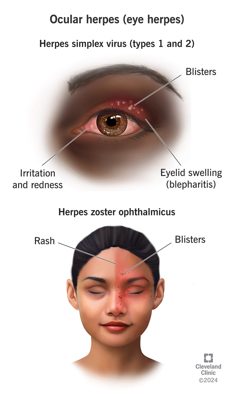
Herpes Zoster Opthalmicus
PP: dendritic/pseudodendritic lesions on cornea
E: varicella zoster virus
DX:
slit lamp exam+NaFL stain+lissamine green/rose bengal stain
TX:
PO acyclovir x within 72 hrs
PO valacyclovir x within 72 hrs
2nd infxn prophx→topical abx eye drops (gtts)
immunocompromised→admit+IV acyclovir
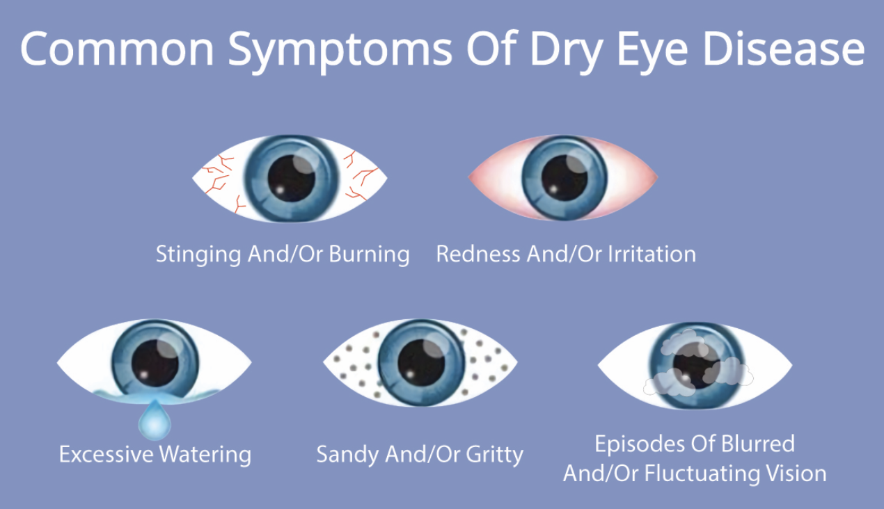
Dry Eye Disease
PP/E: mimic for eye conditions
CM: bruning
itching
redness
tearing
foreign body sensation
gritty feeling
blur→clears with blinking
TX: depends on type of dry eye
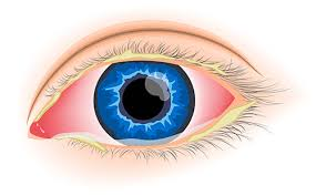
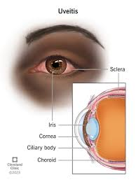
Uveitis/Iritis
PP: inflammation of choroid+ciliary body+iris
E: idiopathic
autoimmune
inflammatory
CM: UL or BL
red
painful
light sensitive
TX: topical steroid
pain→cycloplegic
referral
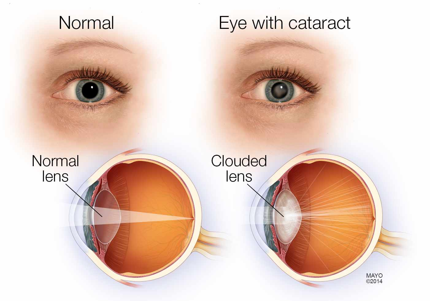
Cataracts
PP: clouding of the lens (behind iris+pupil)→vision loss
E: m/c vision loss over 40+ y/o
#1 cause of blindness worldwide
age
UV light
DM
smoking
injury
steroids
CM: blurry vision (even with glasses on)
glare
decreased color brightness
TX: cataract extraction surgery
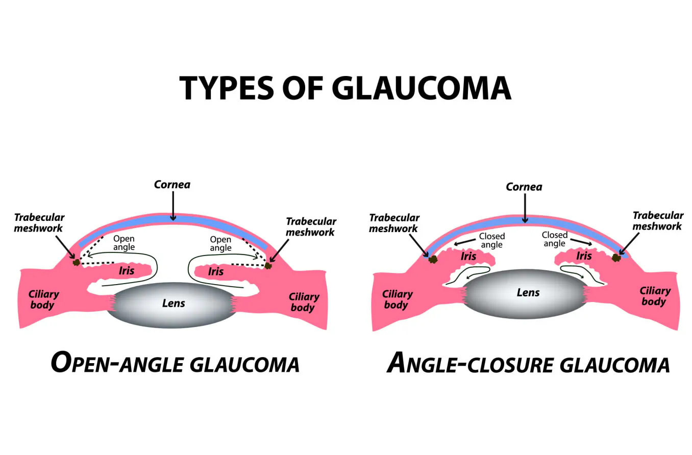
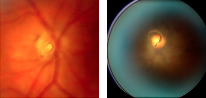
Primary Open-Angle Glaucoma
PP: increased eye pressure→optic nerve cell death
RF: 65+ y/o
black
DM
myopia
ocular HTN (IOP 21+ mmHg)
larger cup→higher risk
E: leading cause of visual impairment/blindness in US
2.5 million
50% unaware
CM: gradual loss of peripheral vision
no pain
DX: optic cup enlargement
high eye pressure
TX:
beta blocker drops
prostaglandin drops
carbonic acid inhibitor drops
alpha agonist drops
surgery

Primary Closed-Angle Glaucoma
PP/E: iris blocks exit of aqueous humor from anterior chamber→increased intraocular pressure→optic nerve cell death
RF: larger cup→higher risk
CM: intense ocular pain
blurred vision
halos around lights
nausea
vomiting
DX: diffuse conjunctival injection
dilated+fixed pupil
clouding of cornea
Posterior Vitreous Detachment
PP: gel surface that attaches on to retina (vitreous)→detaches from retina
CM: asx
or
floaters
flashes of light
DX: discrete translucent/broken circle shaped opacity near optic disc
retinal break (8-10%)→dilated eye exam
Vitreous Hemorrhage
E: vitreous detachment
DM
retina break/detachment
sickle cell dx
retinal vein occlusion
macular degeneration
trauma
tumor
etc.
CM: sudden+painless vision loss
TX: stop blood thinners
no heavy lifting
vitrectomy
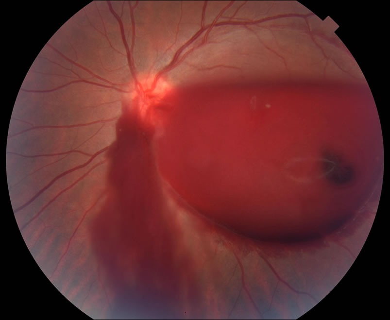
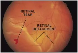
Retinal Hole/Tear/Detachment
RF/E: high myopia (nearsightedness)
retinoschisis (split retina)
neoplasms
vascular dxs
trauma
etc.
CM: flashes
floaters
curtain/veil over vision
vision loss
TX: urgent retinal specialist referral→surgery
P: macula on→better vision prognosis→more of an emergency
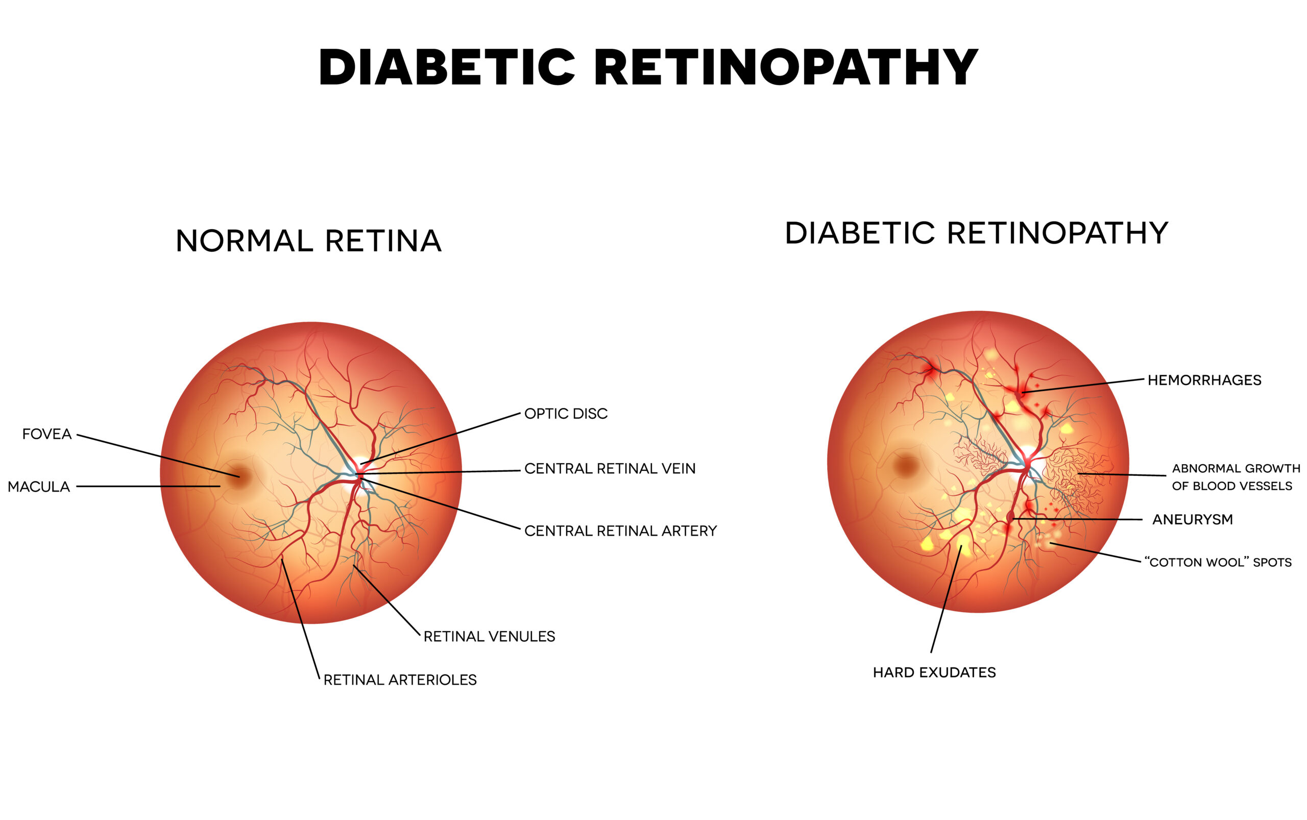
Diabetic Retinopathy
RF: high blood sugar
DM length
E: leading cause of blindness in US
proliferative vs non-proliferative
-mild
-moderate
-severe
CM: vision fluctuations
floaters
vision spots
shadow in vision
distorted/blurry vision
slow healing eye lesions
diplopia
macular edema
TX: depends on severity
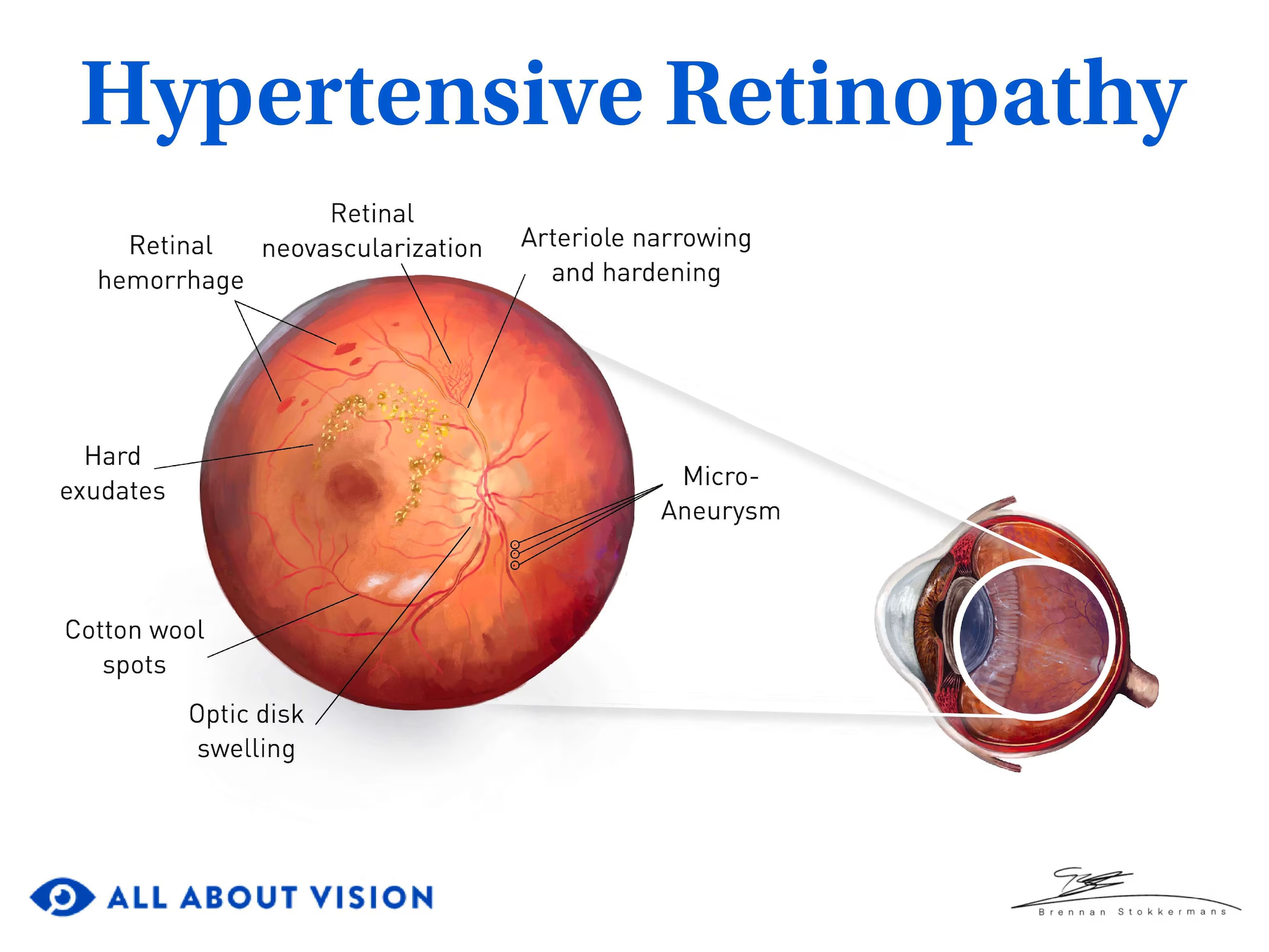
Hypertensive Retinopathy
CM: asx
or
decreased vision
artery narrowing
severe:
hard exudates
flame-shaped hemorrhages
optic nerve edema
TX: control BP
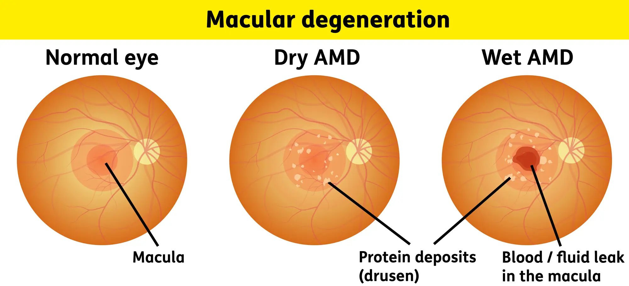
Macular Degeneration
PP: part of retina deteriorates→can’t remove waste
RF: age
FHx
cigarette smoking
E:
dry→gradual breakdown of cells in macula
wet→new abnormal blood vessels grow under central retina→leaking+bleeding+scarring
-exudative
-neovascular
CM: decreased/blurry central vision
slow dark adaptation
DX:
dry:
macula→drusen
wet:
retinal exudates
neovascularization
TX:
wet→retina specialist referral→anti-vegf injections
vision loss→low vision services