Differentiation of the Ectoderm
1/65
There's no tags or description
Looks like no tags are added yet.
Name | Mastery | Learn | Test | Matching | Spaced |
|---|
No study sessions yet.
66 Terms
Parts of the Central Nervous System
Brain and Spinal Cord
Parts of the brain
cerebrum, cerebellum, and brainstem
Parts of the brainstem
midbrain, pons, and medulla
Formation of the Neural Tube
Neurulation
Primary Neurulation – the cells surrounding the neural plate direct the cells to (blank), (blank), & (blank) from the surface to form a hollow tube
proliferate, invaginate, pinch off
Secondary Neurulation – the neural tube arises from a (blank) that sinks into the embryo & subsequently hollows out (cavitates) to form a hollow tube
solid cord of cells
Steps in Primary Neurulation
Initial epithelium, columnarization, rolling/folding, closure, neural tube complete
Steps in Secondary Neurulation
dispersed mesenchyme, mesenchymal condensation, medullary cord/neural rod, epithelial transition/cavitation, neural tube complete
Neurulation in fish
exclusively secondary
Neurulation in Birds
the anterior portions of the neural tube are constructed by primary neurulation, while the neural tube caudal to the twenty-seventh somite pair (i.e., everything posterior to the hindlimbs) is made by secondary neurulation
Neurulation of amphibians
mostly, the neural tube is made by primary neurulation but the tail neural tube is derived from secondary neurulation.
Neurulation in mammals
both but secondary neurulation (begins at or around the level of somite 35)
Differentiation of the neural tube
gross anatomical level, tissue level, cellular level
what happens in gross anatomical level
the neural tube and its lumen bulge and constrict to form the chambers of the brain and the spinal cord
what happens in tissue level
the cell populations within the wall of the neural tube rearrange themselves to form the different functional regions of the brain and the spinal cord
what happens in cellular level
the neuroepithelial cells differentiate into the numerous types of nerve cells (neurons) and supportive cells (glia) present in the body
what forms in the gross-anatomical level?
lateral ventricles, third ventricle, cerebral aqueduct, fourth ventricle, and central canal of the spinal cord
what forms in the tissue level
lateral sulcus, frontal lobe, precentral Gyrus, central sulcus, postcentral Gyrus, parietal lobe, occipital lobe, temporal love, cerebellum, medulla oblongata, and pons
what forms in the cellular level?
neuron, oligodendrocyte, microglia, astrocyte
early mammalian neural tube is in what structure?
straight structure
before the posterior portion of the tube has formed, what starts undergoing drastic changes?
anterior portion
the neural tube balloons into three primary vesicles, what are those?
forebrain (prosencephalon), midbrain (mesencephalon), and hindbrain (rhombencephalon)
3 primary vesicles
forebrain, midbrain, hindbrain
5 secondary vesicles
telencephalon, diencephalon, mesencephalon, metencephalon, and myelencephalon
the forebrain forms what secondary vesicles?
telencephalon and diencephalon
the midbrain forms what secondary vesicle?
mesencephalon
the hindbrain forms what secondary vesicles?
metencephalon and myelencephalon
the telencephalon has 3 adult derivatives, what are these and their function?
olfactory lobes - smell
hippocampus - memory storage
cerebrum - association (“intelligence”)
the diencephalon has 4 adult derivatives, what are these and their function?
optic vesicle - vision (retina)
epithalamus - pineal gland
thalamus - relay center for optic and auditory neurons
hypothalamus - temperature, sleep, and breathing regulation
the mesencephalon has 1 adult derivative, what is this and its function?
midbrain - fiber tracts between anterior and posterior brain, optic lobes, and tectum
the metencephalon has 2 adult derivatives, what are these and their function?
cerebellum - coordination of complex muscular movements
pons - fiber tracts between cerebrum and cerebellum (mammals only)
the myelencephalon has 1 adult derivative, what is this and its function?
medulla - reflex center of involuntary activities
what are the parts of the vertebrate eye?
anterior chamber aqueous humor, choroid, cornea, fovea, iris, lens, optic nerve, retina, sclera, suspensory ligaments, vitreous humor, and vitreous chamber
The major sensory organs of the head develop from the interactions of the neural tube with a series of epidermal thickenings called the (blank)
cranial ectodermal placodes
the most anterior of the cranial ectodermal placodes are the
two olfactory placodes
what does the two olfactory placodes do?
form the ganglia for the olfactory nerves, which are responsible for the sense of smell
what cranial ectodermal placodes invaginate to form the inner ear labyrinth?
auditory placodes
the neurons of auditory placodes form the (blank), which enables us to hear
acoustic ganglion
what is in direct contact with the optic vesicle?
presumptive lens placode
what does the optic vesicle acocmplish?
the activation of the head ectoderm's latent lens-forming ability and the positioning of the lens in relation to the retina
what does the optic vesicle turn into?
two-walled optic cup, whose two layers differentiate in different directions
the cells of the outer layer of the optic cup produce (blank)
melanin pigment
the melanin pigment produced becomes the?
pigmented retina
one of the few tissues other than the neural crest cells that can form this melanin pigment
outer layer of the optic cup
The cells of the inner layer proliferate rapidly and generate a variety of (blanks - 4)
glia, ganglion cells, interneurons, and light-sensitive photoreceptor neurons
the glia, ganglion cells, interneurons, and light-sensitive photoreceptor neurons collectively constitute what?
neural retina
these are neurons whose axons send electrical impulses to the brain
retinal ganglion cells
the axons of the retinal ganglion cells meet at the base of the eye and travel down the optic stalk called?
optic nerve
other term for goosebumps
piloerection, cutis anserina, and horripilation
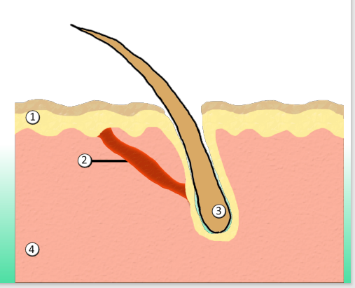
define the parts numbered
epidermis
arrector pili muscle
hair follicle (root)
dermis
in most vertebrates it shortly becomes a two-layered structure
presumptive epidermis
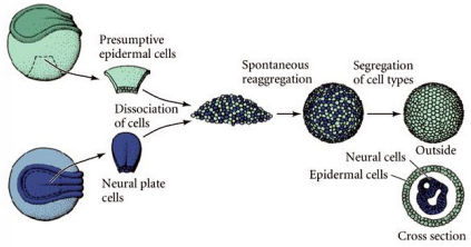
explain the image
This image illustrates an experiment showing how embryonic cells can self-organize based on their identities. Scientists took two types of cells from an early embryo—presumptive epidermal cells (which will form skin) and neural plate cells (which will form the nervous system)—and separated them. After mixing these dissociated cells together, the cells were allowed to reaggregate on their own. Remarkably, the cells sorted themselves out: the neural cells moved to the inside, and the epidermal cells moved to the outside, forming a layered structure similar to how tissues naturally organize during development. This demonstrates that cells have intrinsic properties and recognition mechanisms that guide them to assemble into the correct structures, even without external instructions.
the outer layer of the epidermis gives rise to the?
periderm
a temporary covering that is shed once the inner layer differentiates to form a true epidermis
periderm
the periderm is a temporary covering that is shed once the inner layer differentiates to form a (blank)
true epidermis
the inner layer of epidermis is called?
basal layer or stratum germinativum
the basal layer is a (blank) that gives rise to (blank) of the cells
a germinal epithelium that gives rise to all the cells of the epidermis
the basal layer divides to form what?
spinous layer
what are the two epidermal layers of the Malpighian layer?
basal layer and spinous layer
the cells of the Malpighian layer divides to produce what?
granular layer
granules of the protein keratin
granular layer
the cells of the granular do not divide, but differentiates into epidermal skin cells called?
keratinocytes
the keratin granules become more prominent as the keratinocytes of the granular layer (blank) and (blank) to form the what?
age and migrate outward; cornified layer or stratum corneum
the cornified layer becomes what of keratin protein?
flattened sacs of keratin protein
what happens to the nuclei of the cornified layer?
nuclei are pushed to one edge of the cell
what are the parts of the dermis?
keratohyaline granule, desmosome, desquamating cell, stratum corneum, granular layer, spinous layer, and basal layer