Nematodes intro small intestine nematodes , large intestine nematodes , heart worm, lungworms , eye worms
1/42
There's no tags or description
Looks like no tags are added yet.
Name | Mastery | Learn | Test | Matching | Spaced | Call with Kai |
|---|
No analytics yet
Send a link to your students to track their progress
43 Terms
What is the other name for a nematode
Roundworm fyi, about 80% of animals on earth are nematodes
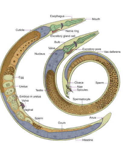
Nematode morphology, body cavity? digestive tract? rest and circulatory system? Dioecious or monoecious?
In addition, to their digestive cavity, roundworms have a body cavity called pseudocoelom consisting of a fluid-filled space between the body wall and digestive tract.
Nematodes have a complete digestive tract with a mouth and an anus
Nematodes lack respiratory and circulatory systems
Excretory cells empty waste from the pseudocoelom into two lateral excretory canals.
Nematodes are covered by a thick, non-cellular cuticle that is produced by a thin, multinucleate epidermis.
Nematodes have only longitudinal muscles and these are divided into 4 bundles or quadrants.
Most nematode species are diecious, with separate male and female individuals
Males are usually smaller than females
During copulation, one or more chitinized rods (spicules) are inserted into the genital pore of the female
Picture female is left and male is right
What is a bursa in nematodes
lobular modification of the male posterior end, which is highly elaborated in some nematodes, e.g; those in order stronglyida. It is a distinctive clasping organ with finger-like projections (“birsal rays”) that have sensory function

What is the purpose of the four larval mounts in nematodes
They must undergo it for larval development. Sexually mature adults form from the final mould. The adults mate and produce eggs and in most cases nematodes leave the host as eggs in the feces. Eggs can leave embyonated or unembyonated
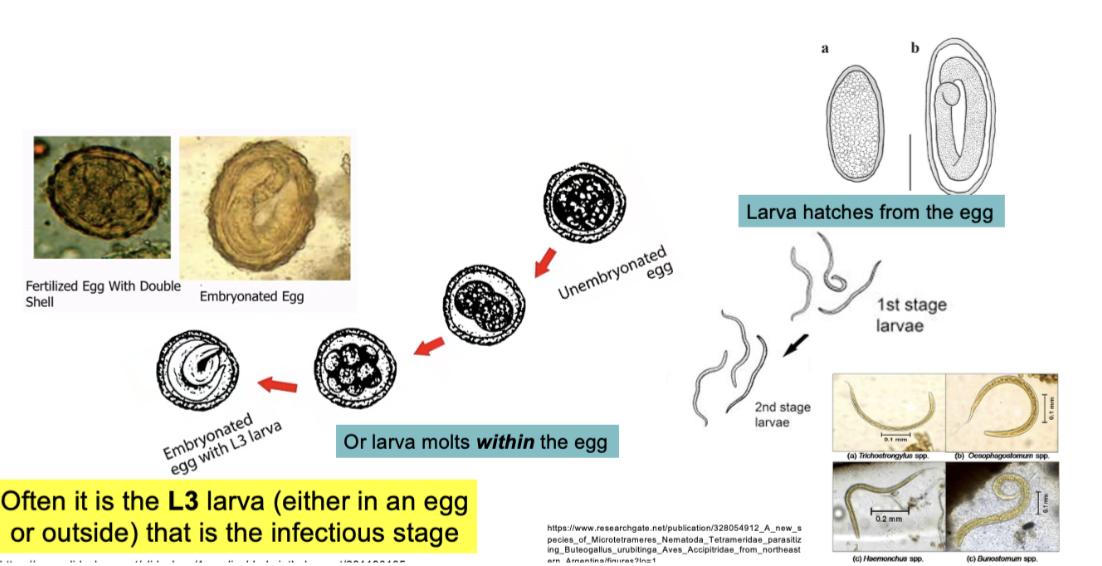
What is the infectious stage in nematodes
It is often the L3 larva (either in a egg or outside)
Family Ascarididae, common name? what do adults feed on? tell me about the females? give me some examples of ascarididae and who they infect?
Common name→ ascarids
feed on→ gut contents in SI
Females→ lay relatively thick-walled, unembyonated eggs
Examples→
Ascaris – infecting pigs, people
Parascaris – infecting horses
Toxocara – infecting dogs, cats
Toxocara leonina - infecting dogs and cats
Baylisascaris procyonis - infecting racoons
For ascaris suum, tell me the host? Where adults are found? importance of this nematode? Clinical signs? Main route of parasite?
Host: Pigs
Adults found→ SI
Importance→ is the most important swine nematode due to economic loss
Clinical signs→
unthriftiness
weight lloss
coughing: rapid shallow breathing
colic
Main route of parasite→ Mouth to SI to bloodstream to lungs to SI
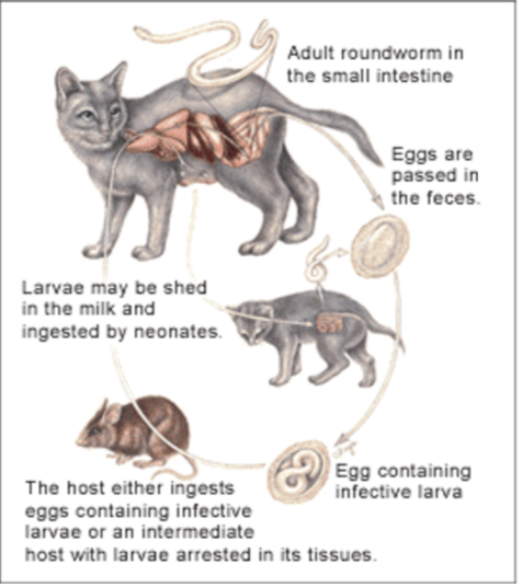
Life cycle and type of life cycle of Acaris suum
Direct life cycle:
Infection occurs on ingestion of infective eggs (#1).
The eggs hatch in the small intestine and the L3 larvae penetrate the intestinal wall, enter the blood stream,
and then migrate intravascularly to the liver, heart and lungs (#4).
Here they molt and burrow out of the blood vessels to enter the bronchioles.
From here they migrate up through the air passages of the lungs, to the trachea. They then enter the throat and are swallowed (#2),
finally ending up in the small intestine where they mature and mate (#3).
The adult parasite lives in the lumen of the small intestine. The female parasite lays unembryonated eggs (#5) which embryonate/larvate outside the host yielding eggs containing infective L3s (#6).
Pathophysiology of Ascaris suum
Due to hepatopulmonary migration of larvae
•Liver
–Interstitial hepatitis
–Localized fibrotic areas; reaction to larvae - “milk spots”
•Lungs
–Hemorrhage, bronchitis, edema
–Pneumonia in young pigs
Due to L4/adults in the small intestine
- Catarrhal enteritis (small intestine inflammation)
- Obstruction or perforation (rare)
Parascaris equorum (horse roundworm), Where is it found, where does infection mainly manifest? some symptoms? life cycle?
Found→ in SI
Manifestation→ mainly in nursing and weaned foals less than a year old
Symptoms→ due to slow growth get diarrhea and GI blockage
Life cycle same as ascaris suum just with equine
What is the name of the human roundworm
Ascaris lumbrivoides
Tell me about the Dog roundworm, main name?DH? Pathogenesis? clinical signs? Diagnosis? Treatment and prevention? Zoonoses?
Name→ Toxocara Canis
DH→ Dogs, worms live in SI
Pathogenesis→ majority of dogs affected show no signs of disease
Clinical signs→ Shown more in puppies, noisy breathing, cough, vomiting, diarrhea, stunted growth, distended abdomen. Abdominal discomfort
Diagnosis→ Confirmed when you see eggs in feces. Adults dogs harder to detect.
Prevention→ anthelmintics
Zoonoses→ YES, humans can get it by ingesting parasite eggs. Affected more children due to putting things in there mouths. Migrates to liver, lung.
Life cycle of Toxocara Canis
Females lay unembyonated eggs→ passed in feces
Embyonate in soil and become infective
Eggs ingested by dog, hatch and release larvae
Penetrate the intestinal wall
In younger dogs, the larvae migrate through the lungs and bronchial tree; larvae are coughed up, swallowed, and returned to the small intestine, where they mature. Adult female worms deposit eggs in the small intestine. Although older dogs may be infected in the same way, larvae are more likely to encyst in tissues.
Encysted stages are reactivated in female dogs during late pregnancy and infect the puppies via the transplacental or transmammary route. As a result, adult worms become established in the small intestine of the puppies (a major source of environmental contamination).
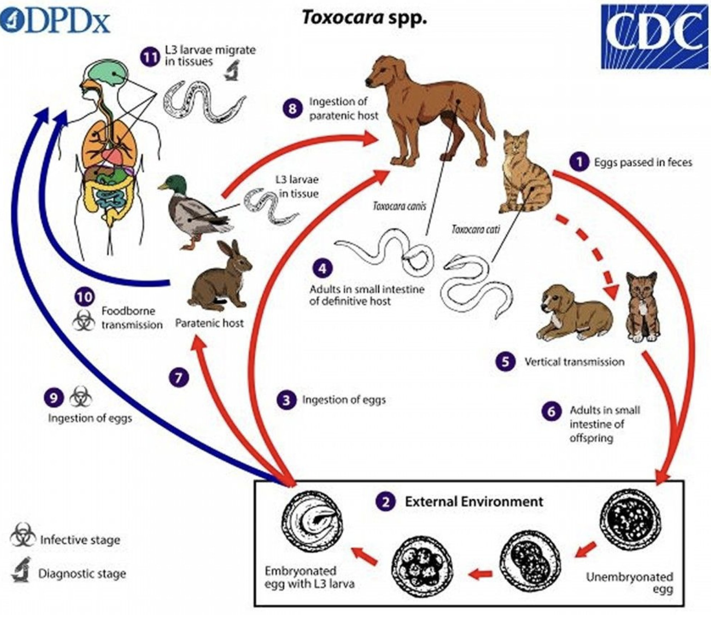
How T.canis effects humans, explain Visceral larva migrant and ocular larva migrant
Migrating larvae can damage internal tissues. The migration of helminth larvae through tissue in suboptimal hosts is termed larval migrans and may affect the viscera (visceral larval migrans), the eye (ocular larval migrans), or the nervous system (neural larval migrans).
VLM→ Consumed by human and emerges and migrates to liver, lung
OLM→ Migrate to eyes. Granulomatous mass may form in posterior chamber of eye
Toxocara Cati ( cat roundworm) , 3 ways of infection? pathogenesis and clinical signs? Diagnosis? treatment and prevention? Zoonoses?
3 ways of infection→
Ingesting infective eggs
Via paratenic host (rodents, birds)
Transmammary route
Pathogenesis and clinical signs→
Same for T.canis
Diagnosis→ seeing eggs in feces
Treatment and prevention→ Regular deworming, kittens assumed to be infected and automatically dewormed.
Zoonoeses→ YES
Life cycle of Toxocara Cati
Is the same as T.canis except that transplacental transmission is not known to occur in cats
What is a cervical alae?
Clear cuticular flanges running along the anterior lateral margins of the worms
Cervical alae of T.canis
Cervical Alae in T.cati
Is more broad and end abruptly. Gives a more “arrow-head appearance
Toxascaris leonina, HOST? LIFE CYCLE, what’s different compared to other toxocara? how are dogs infected? and cats infected? pathogenesis and clinical signs? diagnosis?
Host→ Dogs and cats
Life cycle→ These parasites do not migrate through the body in the way that, e.g., T. canis does. In addition, no prenatal or transmammary infection is seen.
Dogs are more commonly infected by ingestion of eggs with infective L2 larvae.
Cats are most commonly infected by ingestion of paratenic hosts,usually rodents.
Subsequent development of T. leonina larvae to adults takes place in the intestines without a migratory phase.
Pathogenesis and clinical signs→ Heavy infection can cause diarrhea, dehydration, poor health, rarely death
Diagnosis→ Found in fecal exam. Has a thick, smooth outer shell.
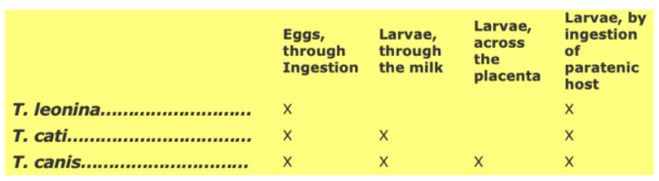
Review of common cat and dog GI roundworm transmission modes (the toxocaras)
Baylisascaris Procyonis, host? where adult worms live in DH? Zoonoses? Pathogenesis and clinical signs(cause of what in humans, what disease can it cause)? Diagnosis and treatment?
Host
Racoons
Adult worms → SI
Zoonoses
YES, widespread geographically
Pathogenesis and signs
Nonpathogenic in racoons
The larvae (not adults) occur in humans (and other mammals and birds) where they are one of several causative agents of "larval migrans".
Outbreaks of fatal central nervous system disease (NLM) caused by B. procyonis have occurred on farms and in zoos and research animal colonies and natural infections have also been recognized in several species.
Diagnosis and treatment
Identification of parasite eggs in feces
No effective therapy for incidental hosts, anthelmintics slow down or kill exposed larvae as they migrate. Prevention important!!!
Eggs has thick shell that protects from most disinfectants. Best way to rid of contaminated soil or area is flaming it.
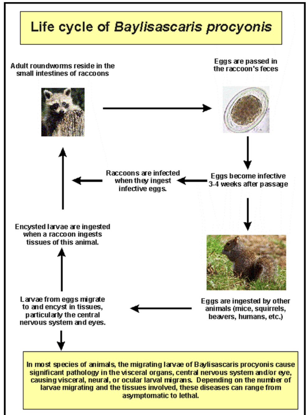
Life cycle of Baylisascaris procyonis
When raccoons ingest infective eggs, larvae hatch, enter wall of SI, and develop to adult worms in the small intestine
What are the two general hookworms (nematodes)
Ancylostoma and Unicinaria spp.
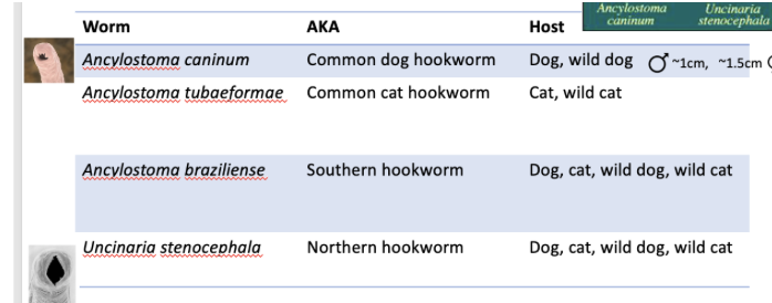
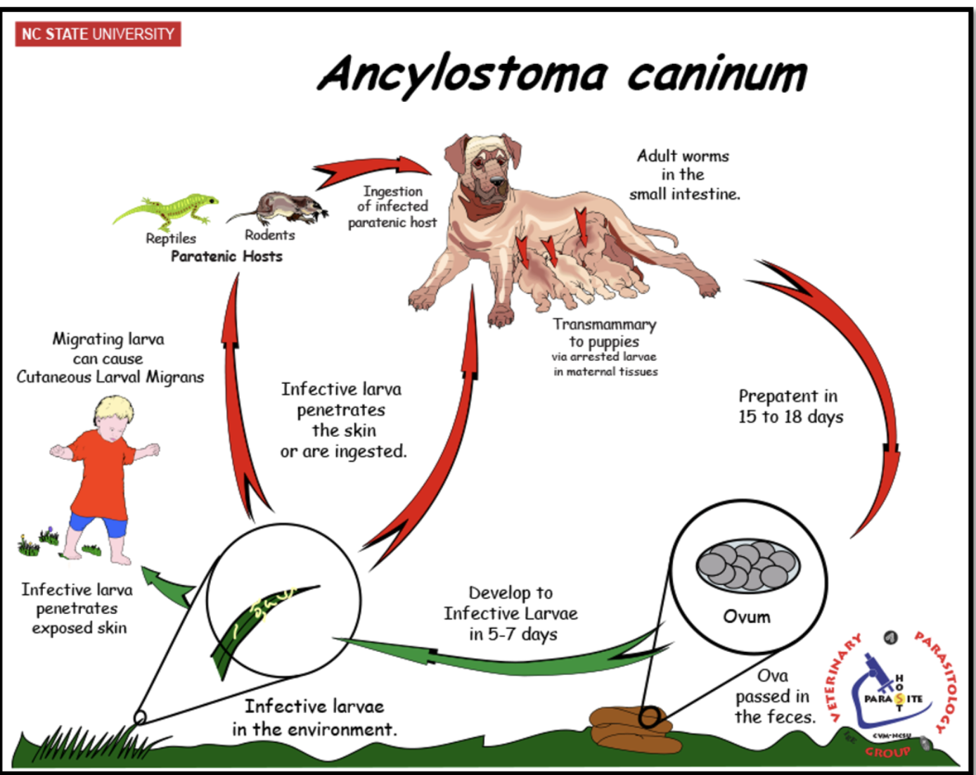
Ancylostoma caninum (common dog hookworm) , Life cycle? PPP? What are the 3 ways of transmission?
Life cycle:
Eggs passed in feces
Hatch in soil, L3 is infective stage
Dogs get infected by: skin penetration, oral ingestion, transmmary transmission
Larva migrate (skin → blood→ lungs→ SI)
PPP
2-3 weeks, less in puppies
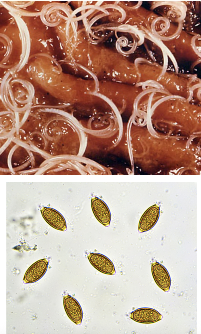
Hookworm spp. Pathology, clinical signs, diagnosis , treatment, prevention, public health concerns?
Pathology: Hookworms are bloodsuckers
Clinical signs: Anemia, black tarry feces, death dermatities; esp. Interdigital
Diagnosis: Fecal exam to look for eggs
Treatment: fenbendazole, pyrantel and others
Prevention: sanitation - larva killed by sunlight (larva more susceptible to harsh conditions)
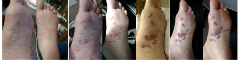
Public health concerns: cutaneous larva migrans (CLM)
Explain to me Cutaneous larva migraines
•Humans exposed to L3 stage larvae
•L3 penetrate the skins and cause a creeping eruption where localized migration occurs
•Extremely pruritic
•Self-limiting – larvae die in 5-6 weeks
•Larvae cannot reach maturity in the human host
Most commonly occurs with A.braziliense (don’t have to know for exam)
•Also known as “plumber’s itch”, which, as the name implies, affects plumbers who crawled in areas contaminated with larvae
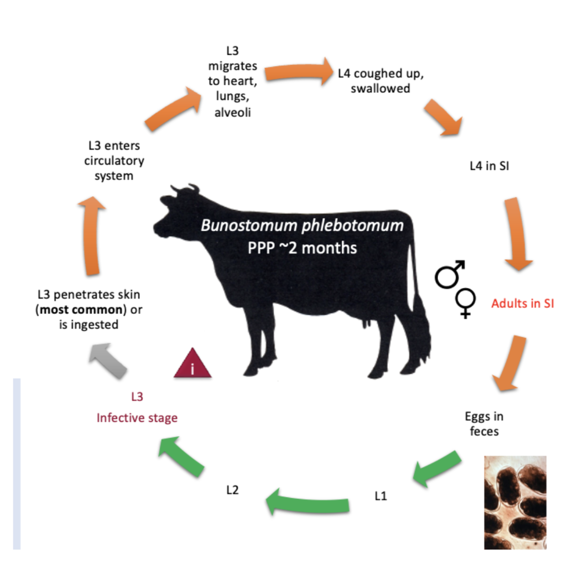
Hookworm of ruminants: Bunostomum phlebotomum, pathogenesis and signs? What can they cause in humans? Life cycle?
Pathogenesis and signs:
Hematophagous(blood feeders) though generally nonpathogenic; but, heavy infection can produce clinical signs including anemia, intermittent diarrhea, and rapid weight loss
What can they cause in humans→
Can cause short-lived CLM(cutaneous larval migrans) in humans
Life cycle→
Larvae penetrates skin of cow→ go into blood→ lungs→ alveoli→ small intestine→ produce eggs and is where larval stage emerges which is infectious stage.
What is the large intestine nematode of importance
Trichuris Spp→ whipworms
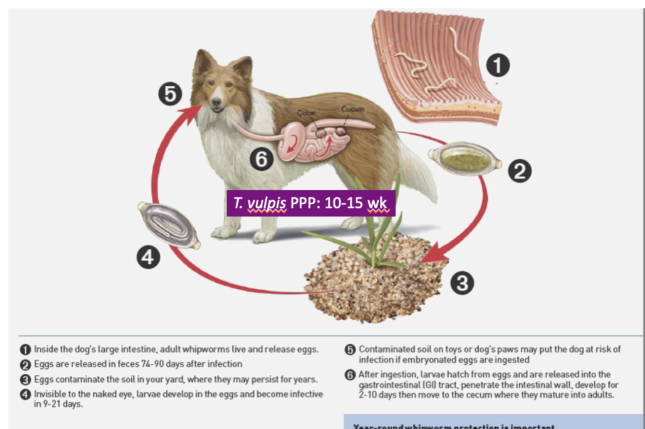
Trichuris spp→ Type of life cycle? type of egg? What larval stage is infective? Pathology? Clinical signs? diagnosis? where do adults live?
Type of life cycle→ Direct life cycle
Egg→ single celled bipolar egg passed
Adults→ live in colon and cecum
Larval stage→ L1, egg protects larvae and allows it to survive in the environment year-round
Pathology→ Typhlitis (inflammation of the cecum) with heavy worm burden
Clinical signs→ Mucoid diarrhea, mushy stools, rarely blood involved
Diagnosis→ fecal float
Trichuris life cycle
Adults in large intestine and imbed into intestinal mucosa
Females lay embryonated eggs
Eggs passed in feces
In soil they embryonate and develop into infective L1 larvae
Host digest embryonated eggs via contaminated food, water, soil
Eggs hatch in SI and molt locally
Immature worms migrate to cecum and colon and mature into adults and embed into mucosa
What is some prevention you could let a client know for parasites
•Pick up dog feces. If parasite eggs/larvae are not in the environment, other animals cannot become infected. Follow this practice at your clinic and convince your clients to do so at home.
•Cover sandboxes. Covered sandboxes cannot become contaminated by dogs and cats.
•Deworm dogs regularly. Using an approved anthelminthic, deworm puppies at 2, 4, 6, and 8 weeks of age and then monthly, if possible.
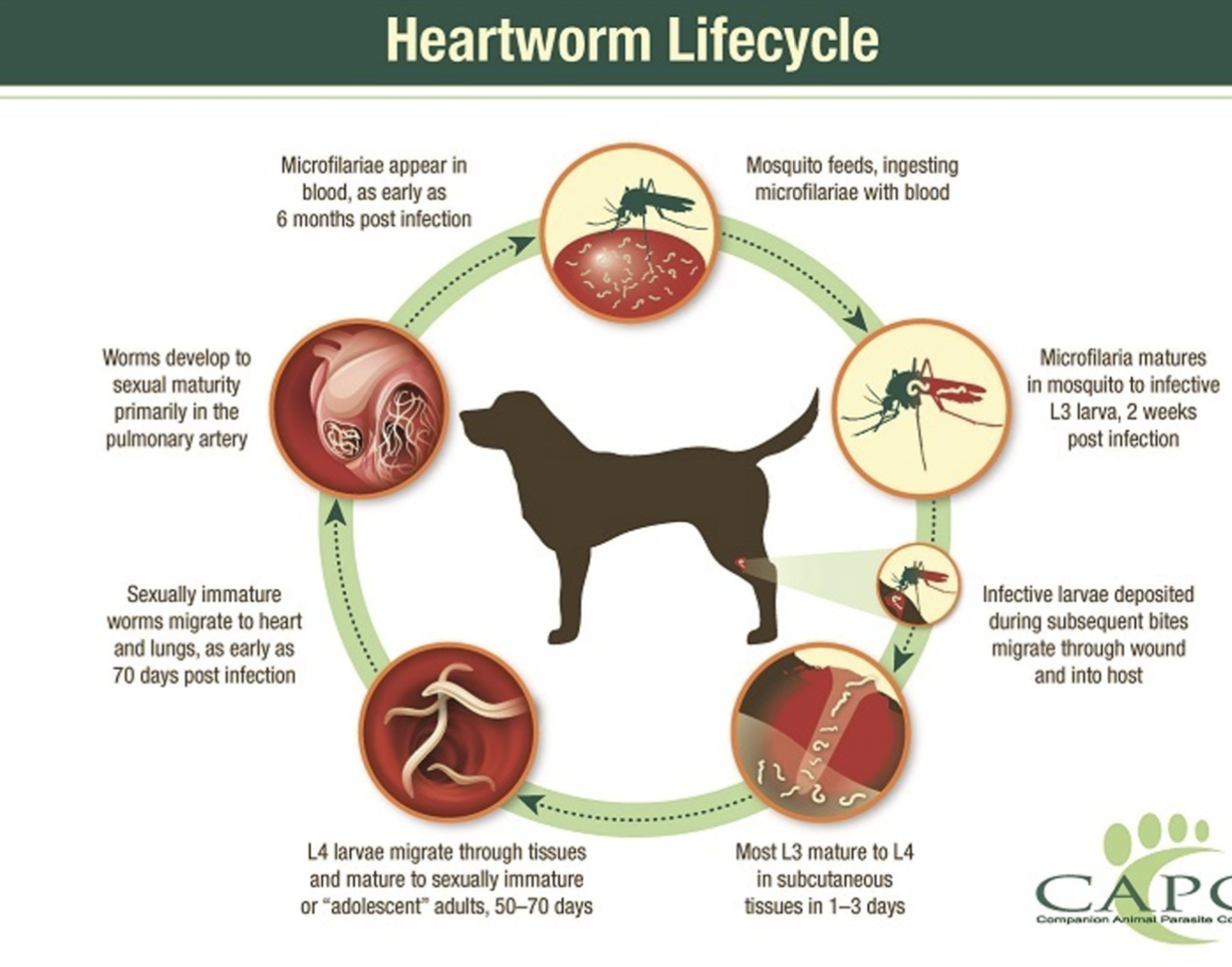
Heartworm (Dirofilaria immitis), DH? Type of life cycle? What are larvae called? what is the infective stage? Pathology? Clinical signs? Diagnosis? Mosquitos associated?
DH→
Dogs, ferrets, sea lions, rarely humans (cats considered “atypical” hosts)
Type of life cycle→
Indirect life cycle
Larvae called →
Microfilariae
Infective stage→
L3 is infective
Pathology→
Due to adult worms (Adult heartworms can live for 5-7 years in dogs and up to 2-3 years in cats)
•Obstruction of pulmonary arteries
•Progressive pulmonary endarteritis (inflammation of the inner lining of an artery) & fibrosis (thickening & scarring of connective tissue). Thickened fibrotic arterial lining causes turbulence & decreased blood flow
•Chronic pulmonary hypertension causes right heart failure
•Vena cava syndrome – large number of worms obstruct venous return to vena cava & right atrium
Clinical Signs→
chronic progressive cough, Exercise intolerance/ fatigue, weight loss, ascites, Death
Diagnosis→
ID microfilariae in blood
Antigen or antibody detection
Radiography (enlarged R heart and enlarged pulmonary arterial trunk)
ECG
Necropsy
Mosquitoes associated
Aedes sp, culex sp, anopheles sp
Heartworm life cycle
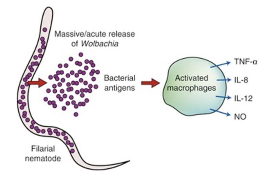
Wolbachia? What is it? How it contributes to Heartworm disease? What is it susceptible to?
What is it→
Gram negative intracellular bacteria
How it contributes to heart worm dz→
Symbiotically colonizes dirofilaria immitis.
Antigen stimulation of mammalian host by released bacterial antigens causes inflammation when the parasite dies.
Contributor to pathology and clinical signs of D.immits
Susceptible to→
Tetracycline
What gets confused with D.immitis? What is its DH? IH? Pathology? Clinical signs? Diagnosis? Treatment?
Acanthochellonema reconditum
life cycle: Indirect
DH→ Dog
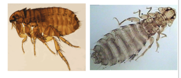
IH→ ctenocephalides fels flea and heterodoxies sponger lice
Pathology→ none
Clinical signs → none
Diagnosis: Microfilaria in blood - can be differentiated from D. immitis by using modified knotts technique
Treatment: Ivermectin treats the microfilaria
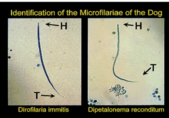
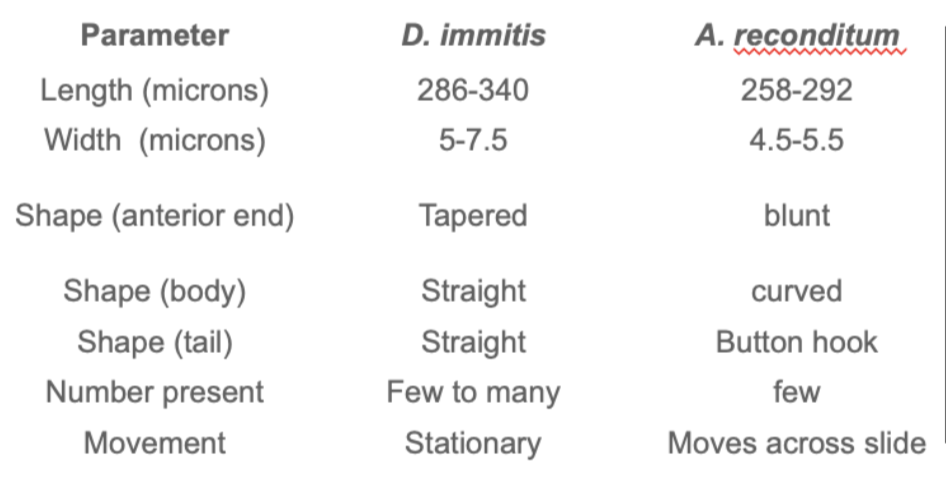
Differences between morphology of D.immitis and A.reconditum
What are some examples of some nematode lungworms and what is the one that mainly infects cats?
Capillaria aerophilia
Eucoleus aerophilus
Filaroides firthi
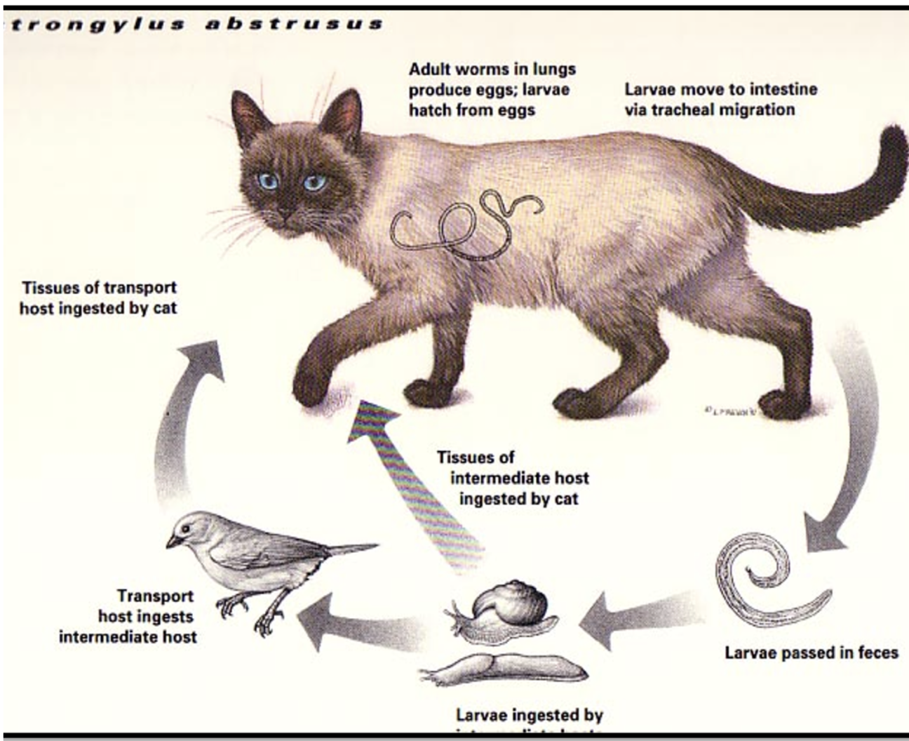
One that infects cats→ Aelorostrongylus abstrusus
Aelurostrongylus abstrusus life cycle and type of life cycle? IH? PH? Where are adults found?
Indirect life cycle→
Eggs passed by adult female worms hatch in the lungs, and larvae pass up the trachea, then down the intestinal tract, and out in feces.
IH→ Snail and slugs are the intermediate hosts
paratenic hosts→ e.g., rodents, birds, amphibia, and reptiles.
Ingested larvae are liberated in the intestine, penetrate the mucosa, and migrate in the blood to the lungs.
Adult worms are found→in the alveolar ducts and terminal bronchioles 8 to 9 days after infection.
Aelurostrongylus abstrusus? Life cycle (what stage passed in feces and what stage is infective)? DH? IH? PH? Pathology? Clinical signs? Diagnosis? Treatment? control?
Life Cycle: L1 larvae passed in feces, L3 infective
Indirect
Definitive Host: cat - passes L1
Intermediate Host: snails, slugs - 3rd stage
Paratenic host: birds, rodents, amphibians, reptiles - 3rd stage
Prepatent Period: ;1 in feces at about 6 weeks
Pathology:
•Eggs induce granulomatous reactions leading to the formation of subpleural nodules within the lung parenchyma
Clinical signs:
•Usually none, but could have a chronic cough & dyspnea (labored breathing)
Diagnosis:
•Can try Baermann but worm is not very motile – struggle to leave feces
•Direct fecal smear – low concentration may prevent ID
•Radiographic evidence
•TTW (Transtracheal wash) or BAL (bronchiolar lavage) is the best option
Treatment:
•Fenbendazole or ivermectin
Control:
•Prevent interaction w/ snails & slugs, and paratenic hosts
What is an eye worm nematode?
Thelazia app.
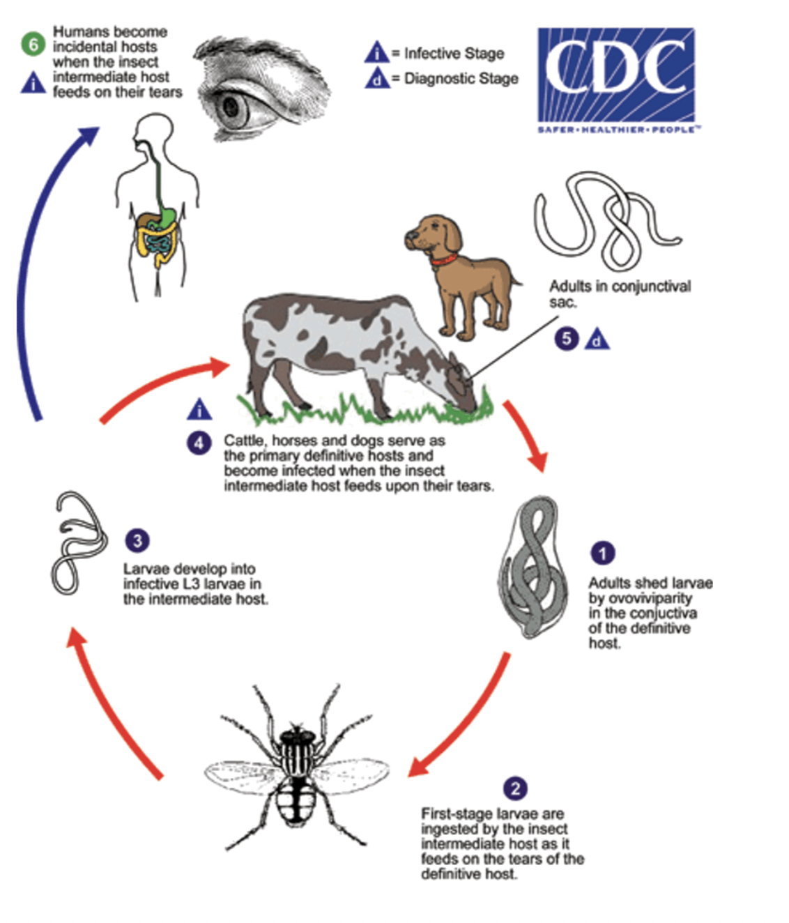
Thelazia SPP. Life cycle type? PPP? Zoonoses? DH? IH? Where are adults found? 3 Thelazia spp important for vet med?
Life cycle → Indirect
Female worm produces larvae (L1) in conjunctival sac
Adult flies consume L1
L1 molts to L3 within fly
L3 crawls to mouthparts of fly and escapes into the orbit of the DH when the fly feeds
L3 develops into an adult without migration
PPP→ 3-6 weeks
DH→
Cattle, horse, dog, cat, sheep, human
IH→
Diptera
Adults found→
Conjunctiva and lacrimal ducts
Zoonoses→
YESSSS
T.californiensis and T.callipaeda→ dogs and cats
T.lacrymalis→ horses
Thelazia californiensis
Both the adult parasites and larval stages can cause pain and discomfort leading e.g., to keratitis (inflammation of the cornea) or conjunctivitis (inflammation of the conjunctiva – the mucous membrane covering the front of the eye).