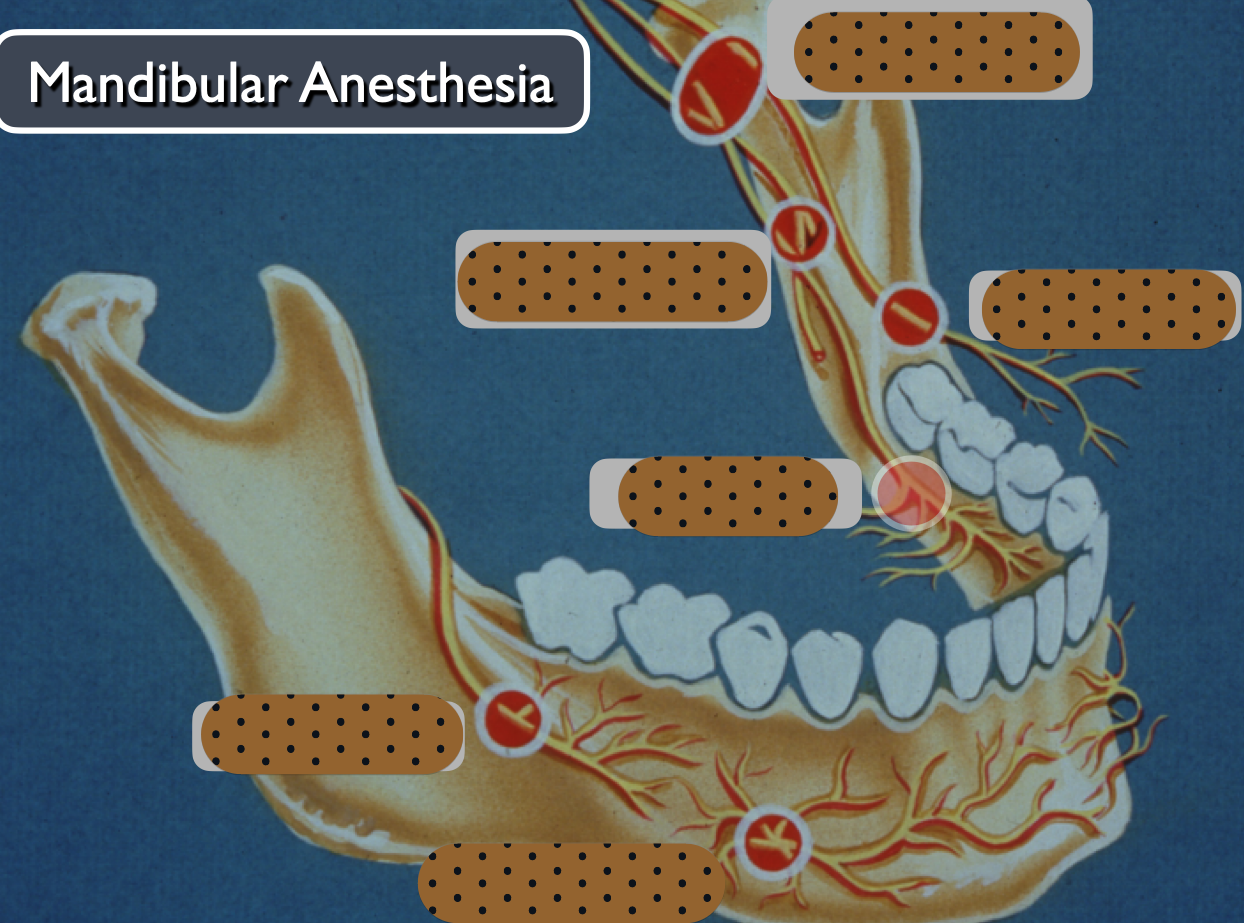
4. mandibular anesthesia part 2
1/44
There's no tags or description
Looks like no tags are added yet.
Name | Mastery | Learn | Test | Matching | Spaced | Call with Kai |
|---|
No analytics yet
Send a link to your students to track their progress
45 Terms
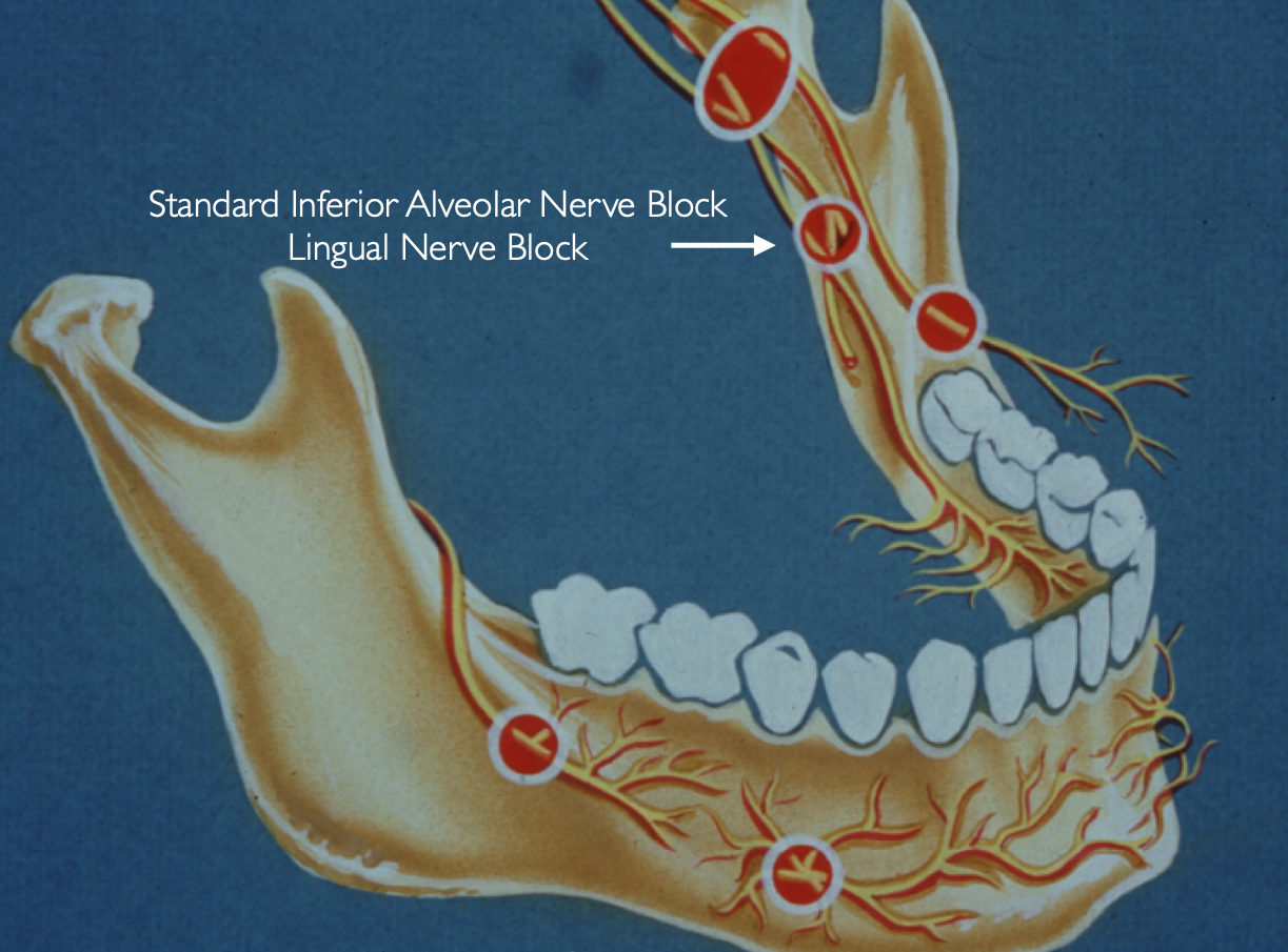
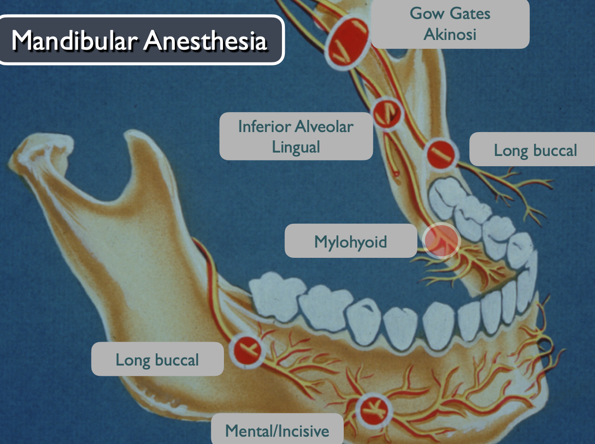
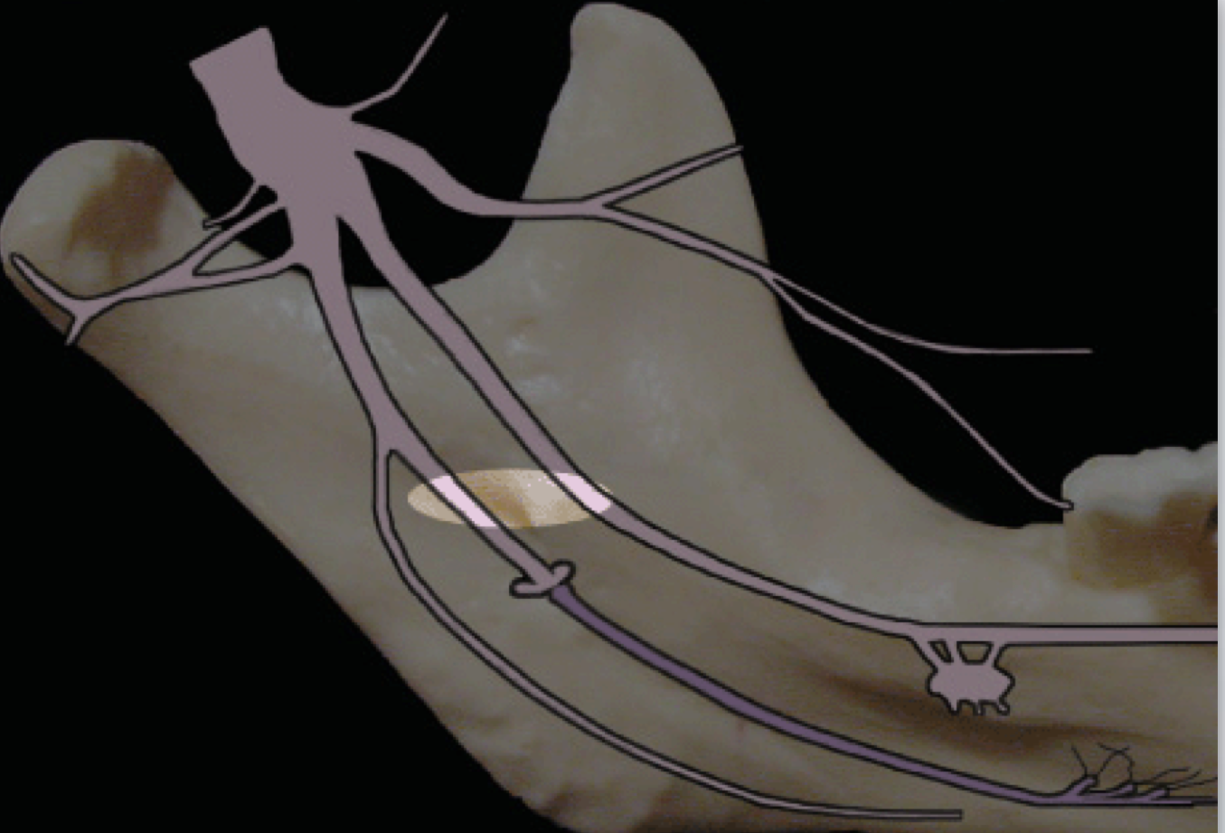
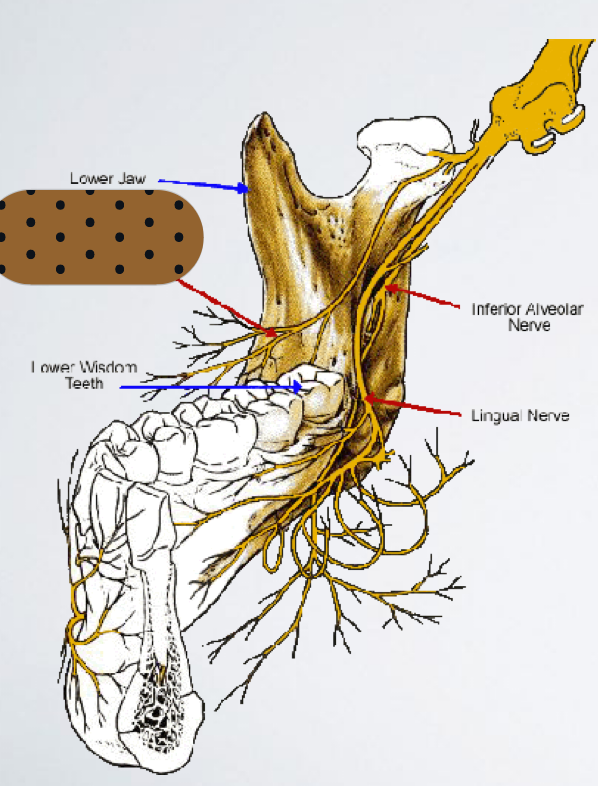
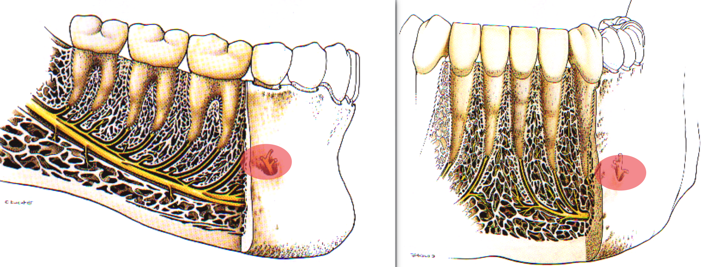
standard IAN block, lingual nerve
failures of IAN block
Deposit the anesthetic too low (below the mandibular foramen)
To correct: reinject 5-10 mm above the previous site
Deposit the anesthetic too anteriorly on the ramus
Deposit the anesthetic too posteriorly on the ramus
Accessory innervation (Mylohyoid nerve)
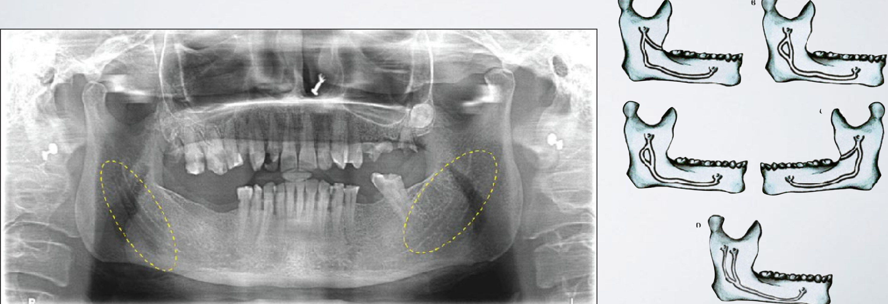
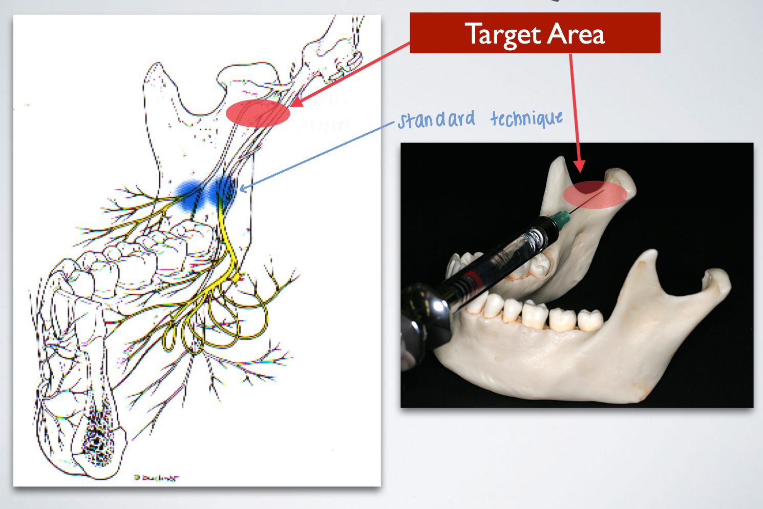
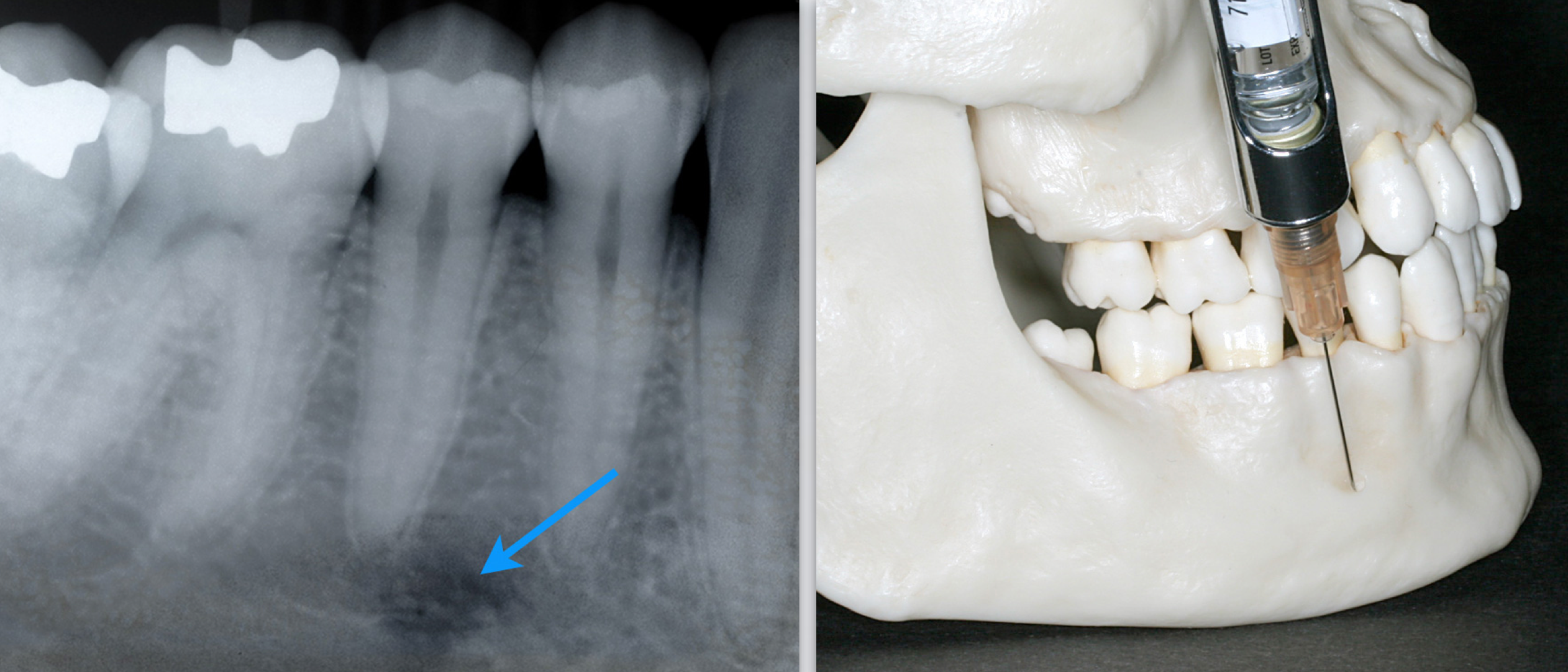
describe failure of IAN block and relationship to bifid inferior alveolar nerve/canal
A second mandibular foramen may exist
To correct: second injection inferior to the normal anatomical landmark
Indication: When lingual soft tissue anesthesia is required
Contraindication: Infection/acute inflammation in the area of injection
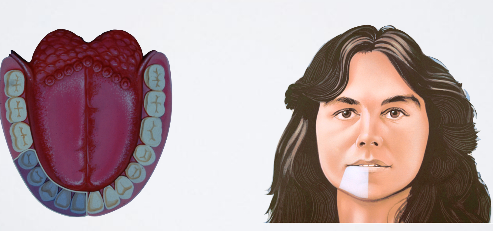
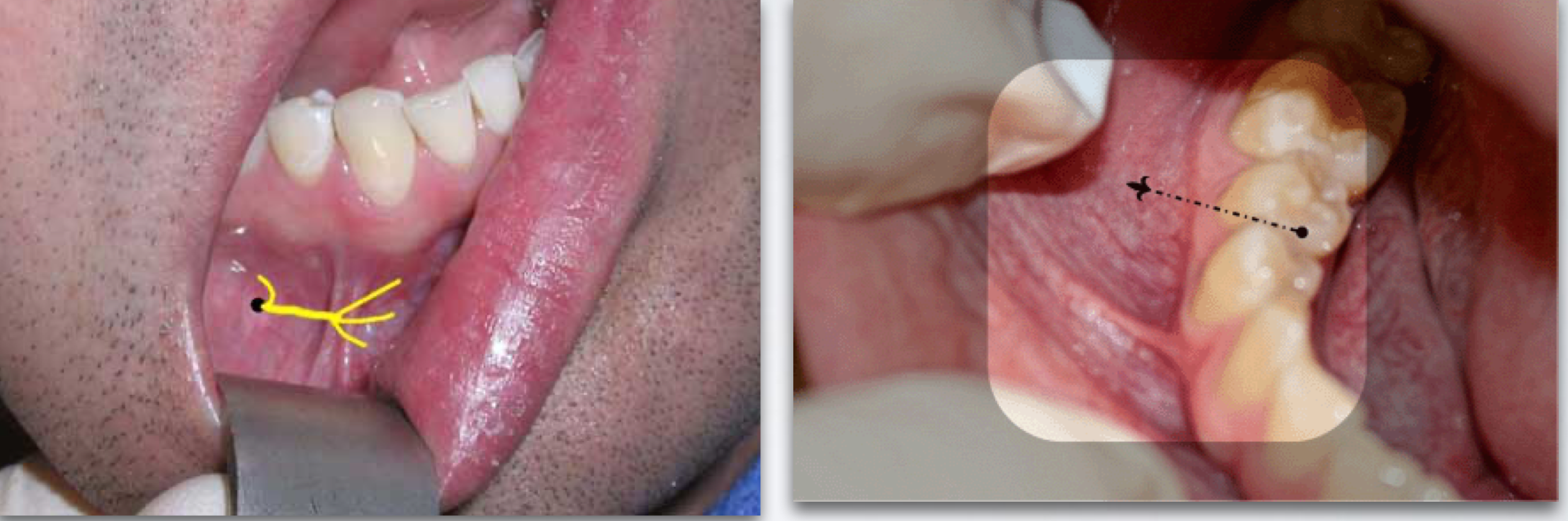
lingual nerve block
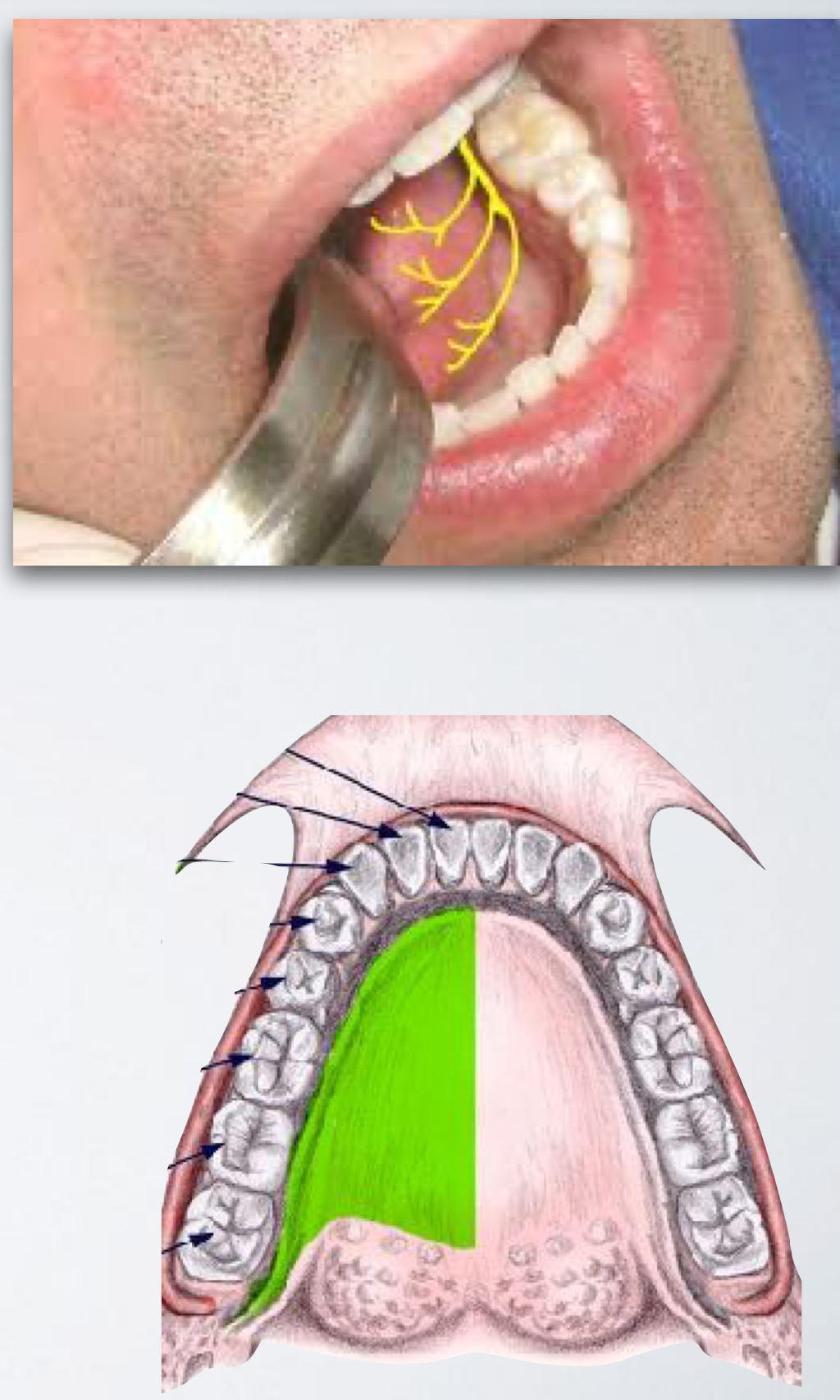
Area Anesthetized:
Lingual soft tissue
Floor of the mouth
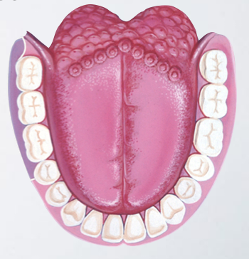
Anterior two thirds of the tongue to midline
lingual nerve block
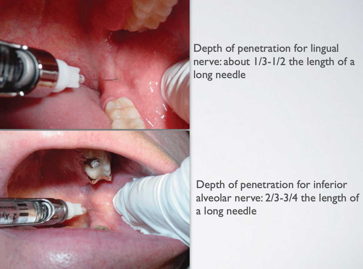
separate injection usually necessary for lingual nerve block
false
Technique Factor
A separate injection is usually not necessary (part of inferior alveolar nerve block)
If administered as a separate injection, may use a long needle but only need to advance halfway
lingual nerve block
Injection/Needle Target Site
Same for inferior alveolar nerve block
Withdraw needle halfway after inferior alveolar nerve block and deposit local anesthetic solution
lingual nerve block
lingual nerve block
lingual nerve block
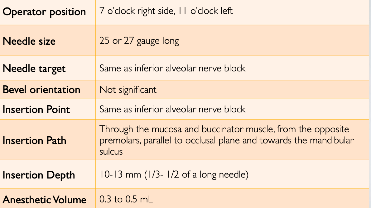
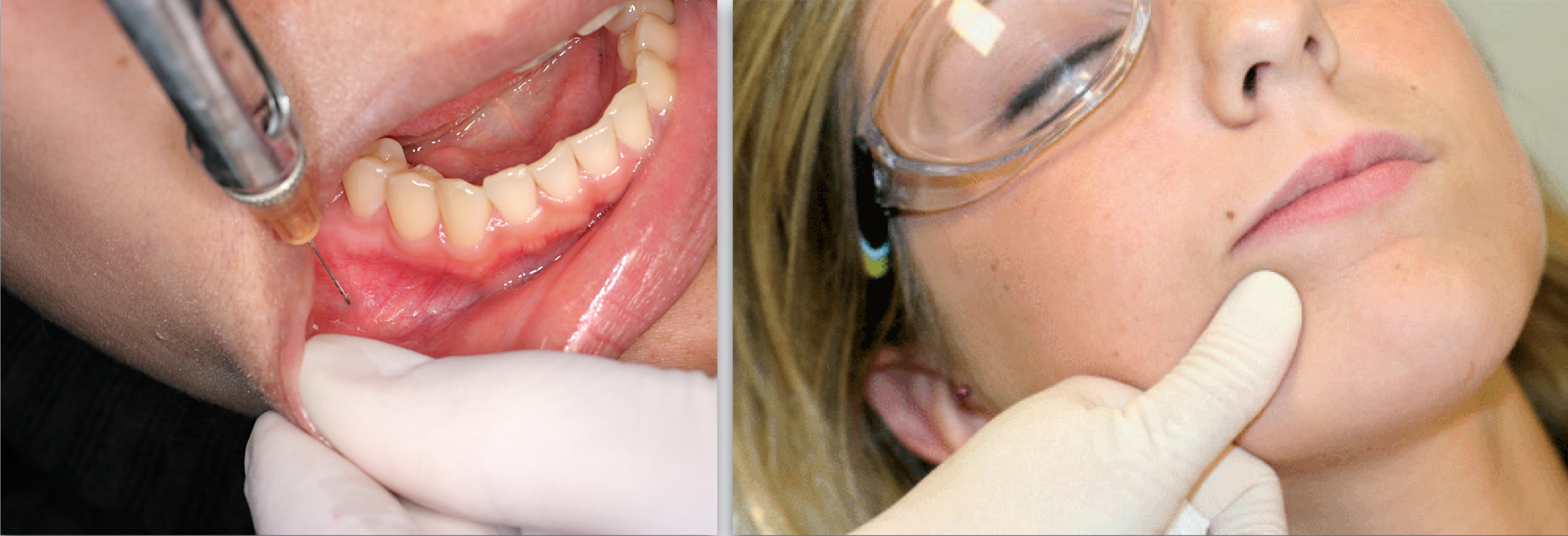
Indication: When mandibular posterior buccal soft tissue is required for dental procedure
Contraindication: Infection/acute inflammation
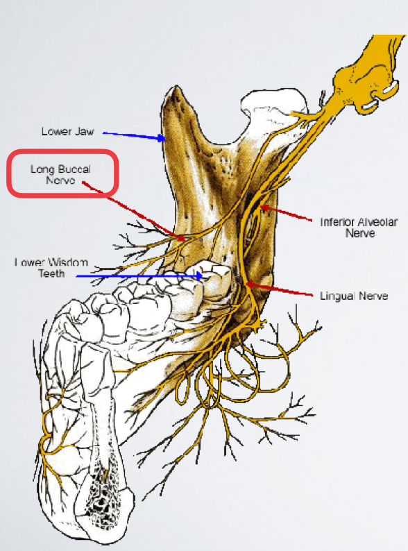
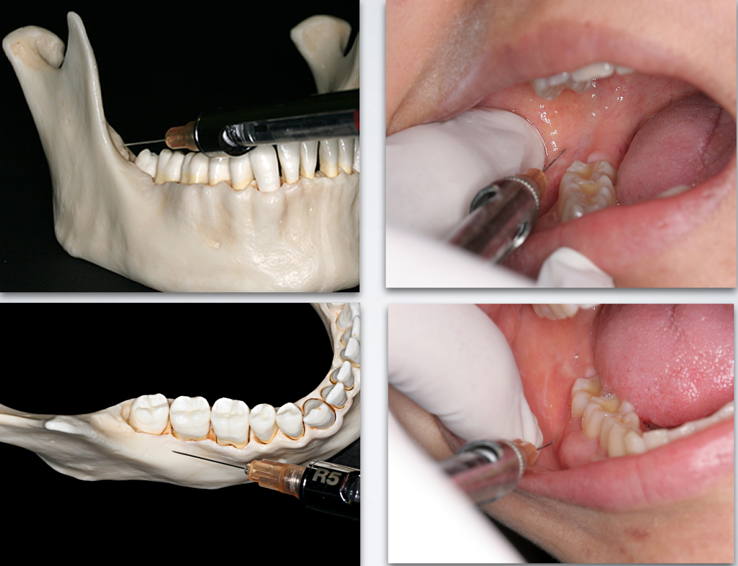
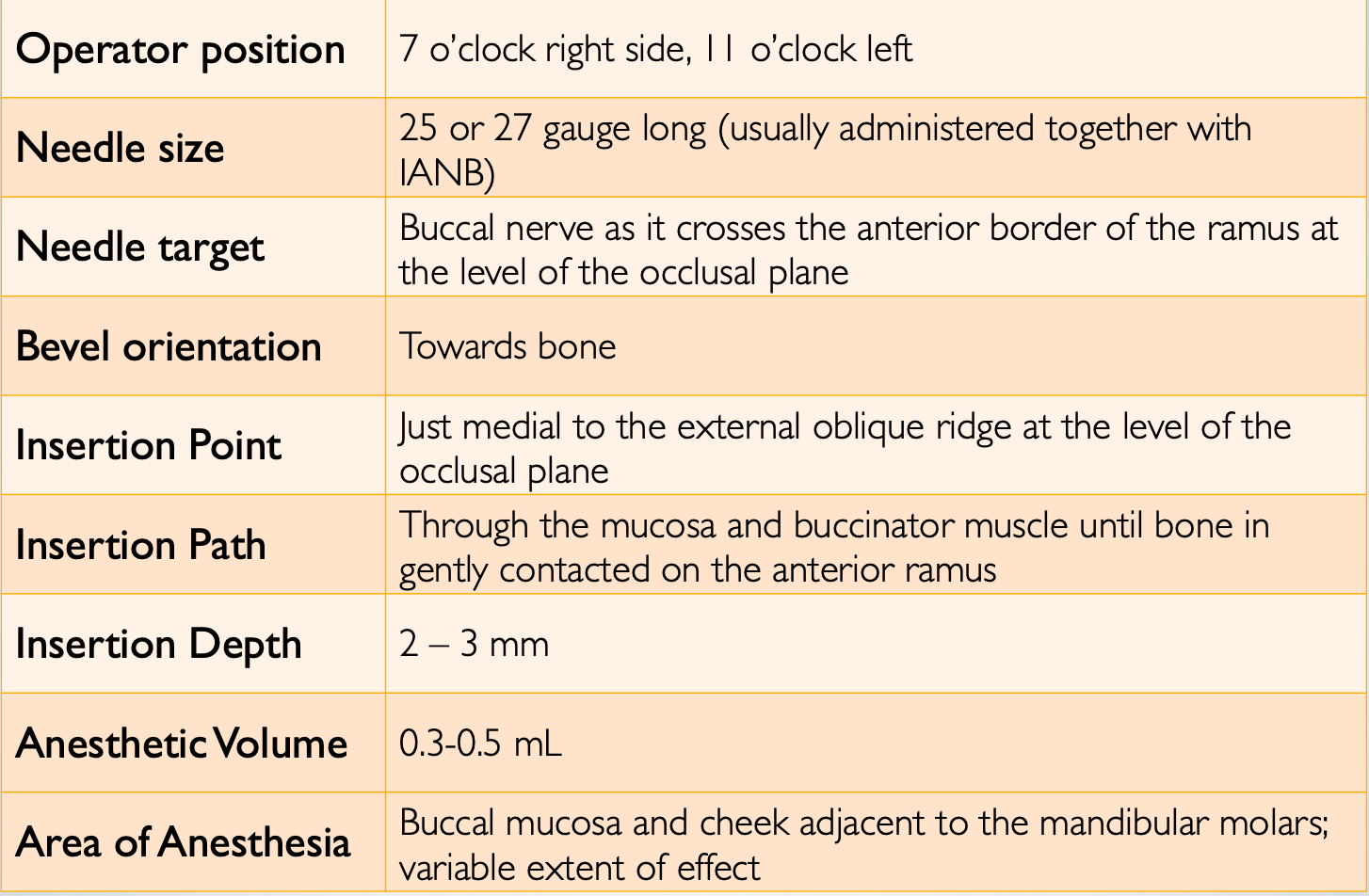
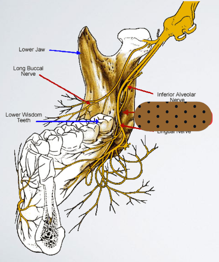
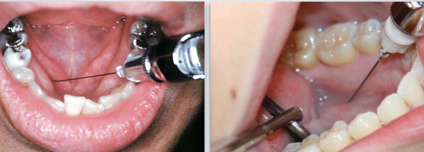
(long) buccal nerve block
Area Anesthetized: soft tissue and periosteum buccal to the mandibular molar teeth
(long) buccal nerve block
(long) buccal nerve block
(long) buccal nerve block
(long) buccal nerve block
Accessory innervation
Can provide portion of pulpal innervation to mandibular teeth (most commonly in the mesial portion of the mandibular 1st molar or premolars)
(BLANK) block can be a useful supplement to inferior alveolar block when it appears to be inadequate
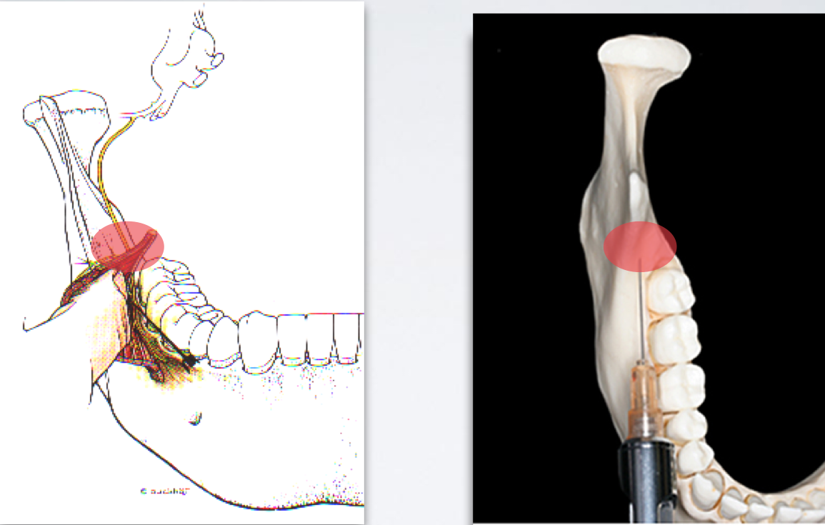
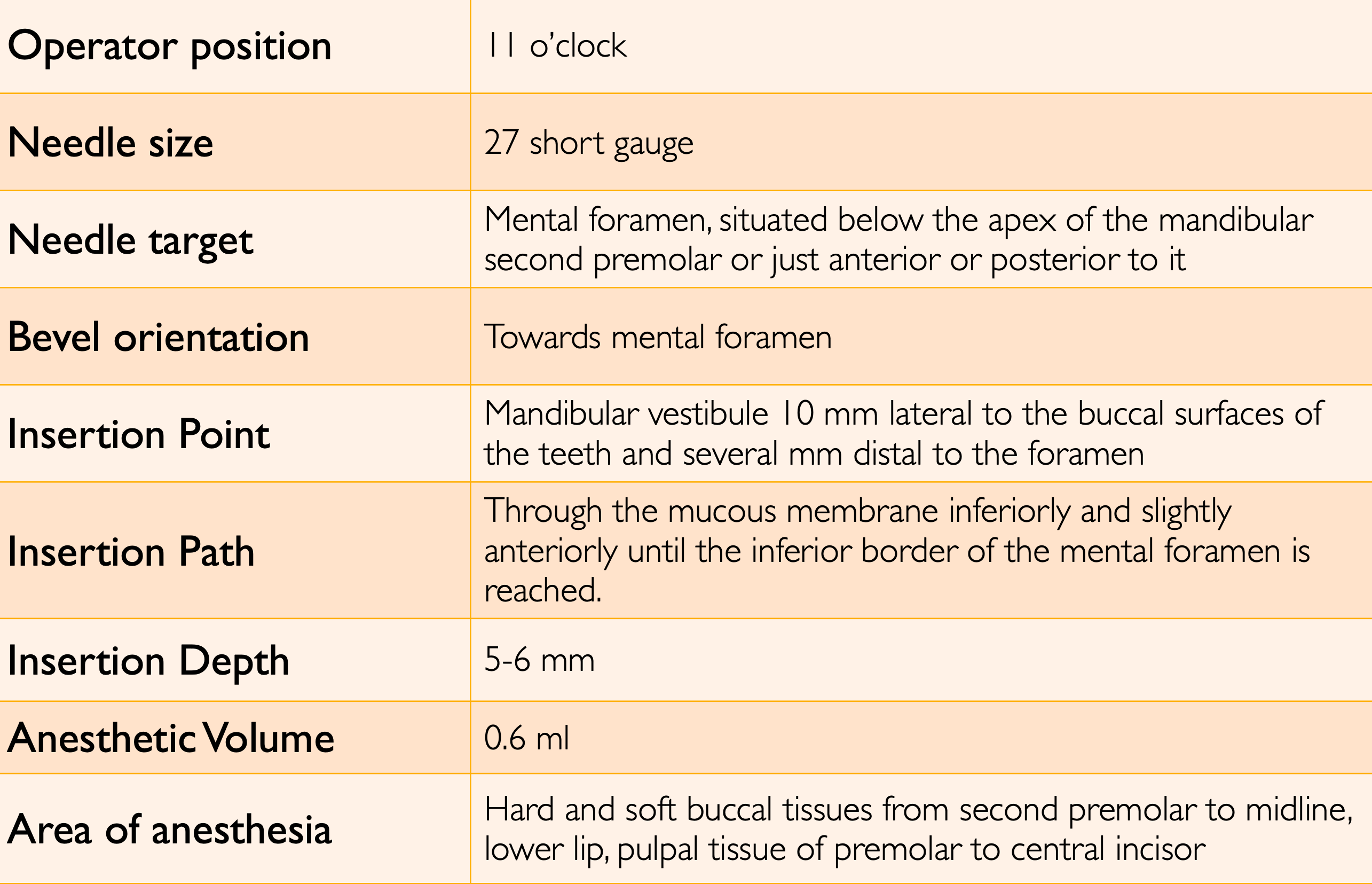
mylohyoid nerve block
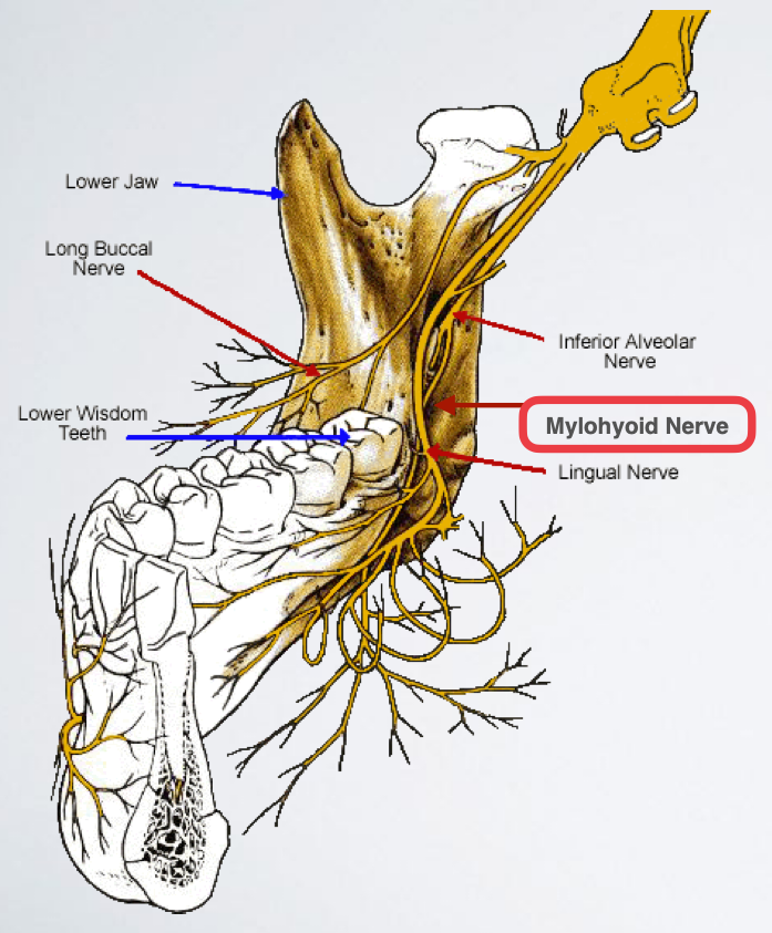
Use 25 gauge long needle
Retract the tongue
Direct the syringe from the opposite side
Direct needle tip to the apical region of the tooth immediately posterior to the tooth in question, until bone is contacted
Aspirate and deposit ~0.6 ml of solution
mylohyoid nerve block
A “true” mandibular nerve block
Nerves Anesthetized:
Inferior alveolar nerve
Mental/incisive nerve
Lingual nerve
Mylohyoid nerve
Auriculotemporal nerve
Buccal nerve (in most cases)
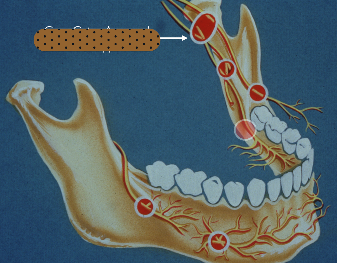
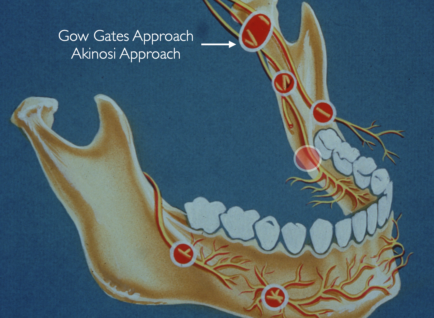
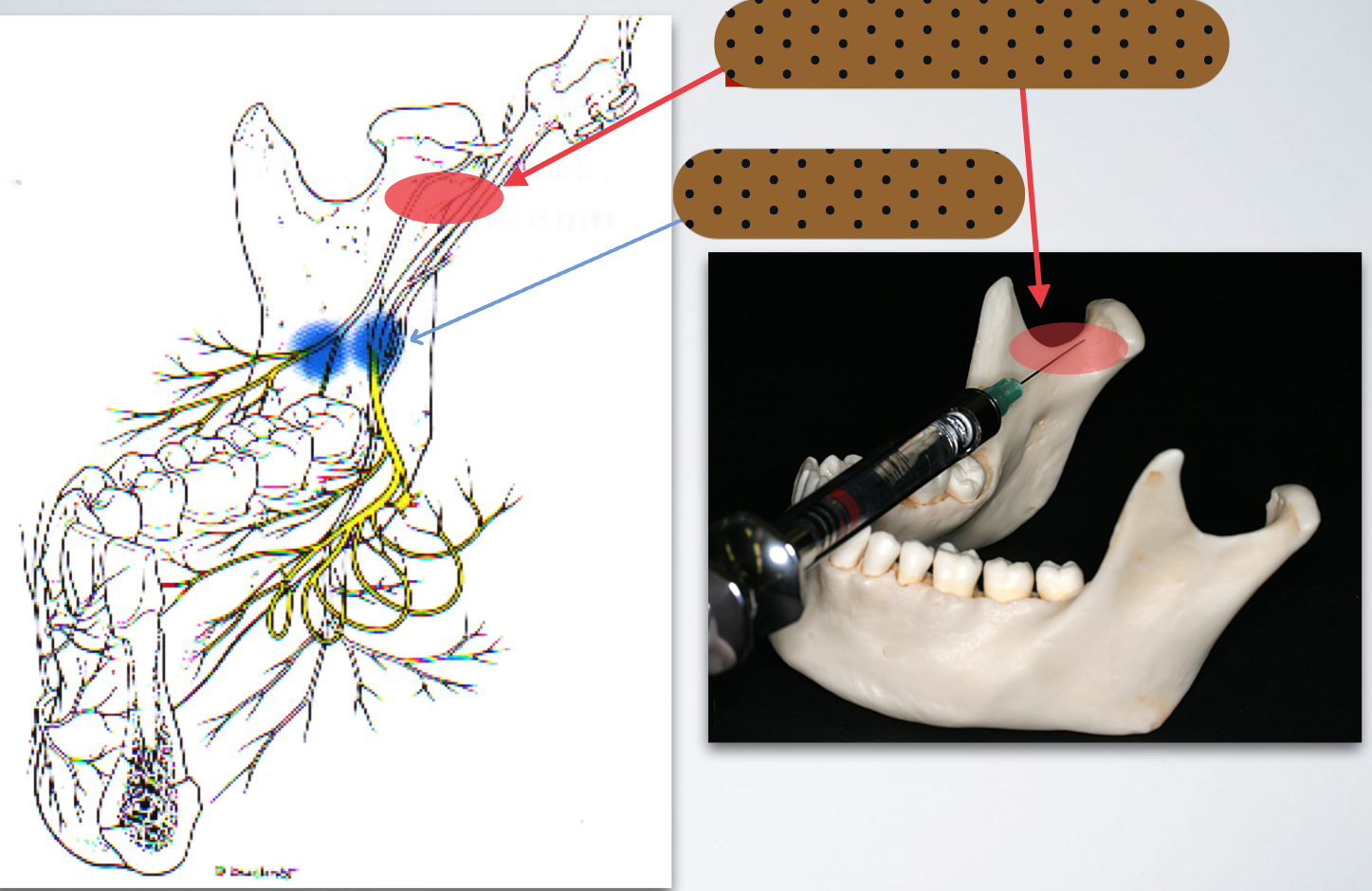
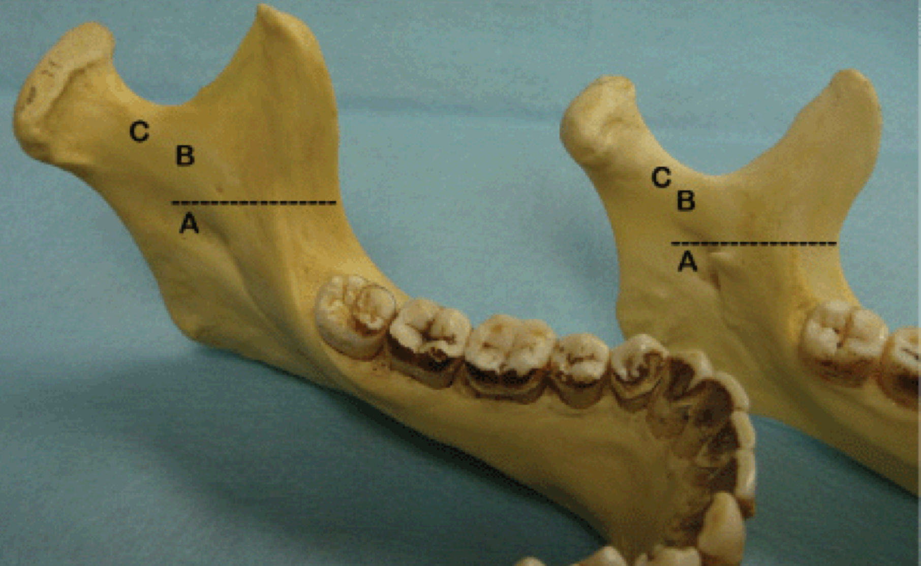
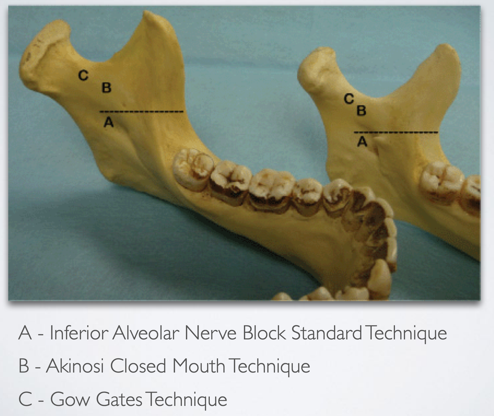
Gow-gates mandibular technique
Area of Anesthesia:
All mandibular teeth on the side of injection
Surrounding periodontium and alveolar
Buccal and lingual soft tissue
Anterior two thirds of the tongue and floor of oral cavity
Floor of the mouth
Body of the mandible, inferior portion of the ramus
Skin over the zygoma, posterior portion of cheek, and the temporal regions
Gow-gates mandibular technique
Indication
Multiple procedures on mandibular teeth
When buccal and lingual soft tissue anesthesia is required
When a classic inferior alveolar nerve block is unsuccessful
Contraindication
Infection/acute inflammation in the area of injection (rare)
Patient who might bite/lacerate lip or tongue (ex: pediatric or special needs patients)
Patient who are unable to open their mouth wide (ex: trismus)
Gow-gates mandibular technique
Advantage
Requires only one injection for total mandibular anesthesia
Higher success rate (>95%) than standard technique
Less positive aspiration (2%) than standard technique (10-15%)
Fewer post injection complication (trismus)
Absence of problems with accessory innervation Bony contact
Disadvantage
Lingual and lower lip anesthesia is uncomfortable for many patients
The time to onset of anesthesia is longer (5 minutes) than standard technique
Learning curve to achieve high success
Gow-gates mandibular technique
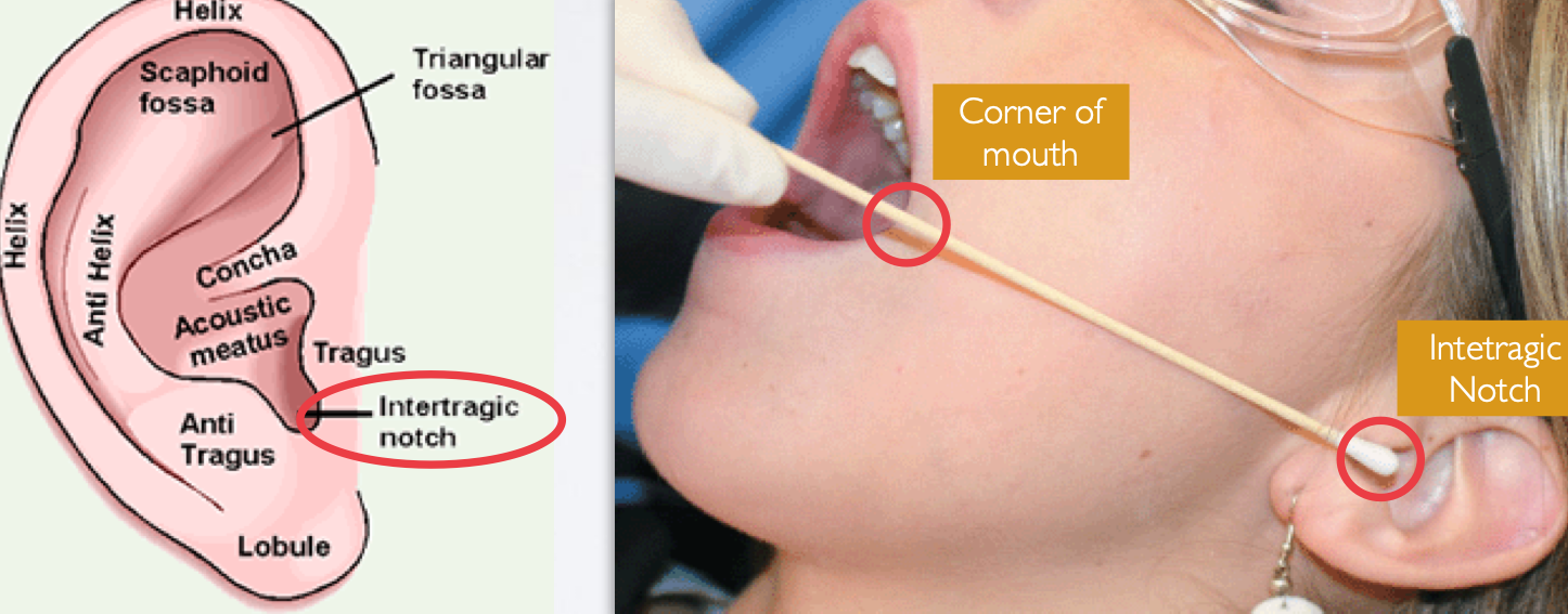
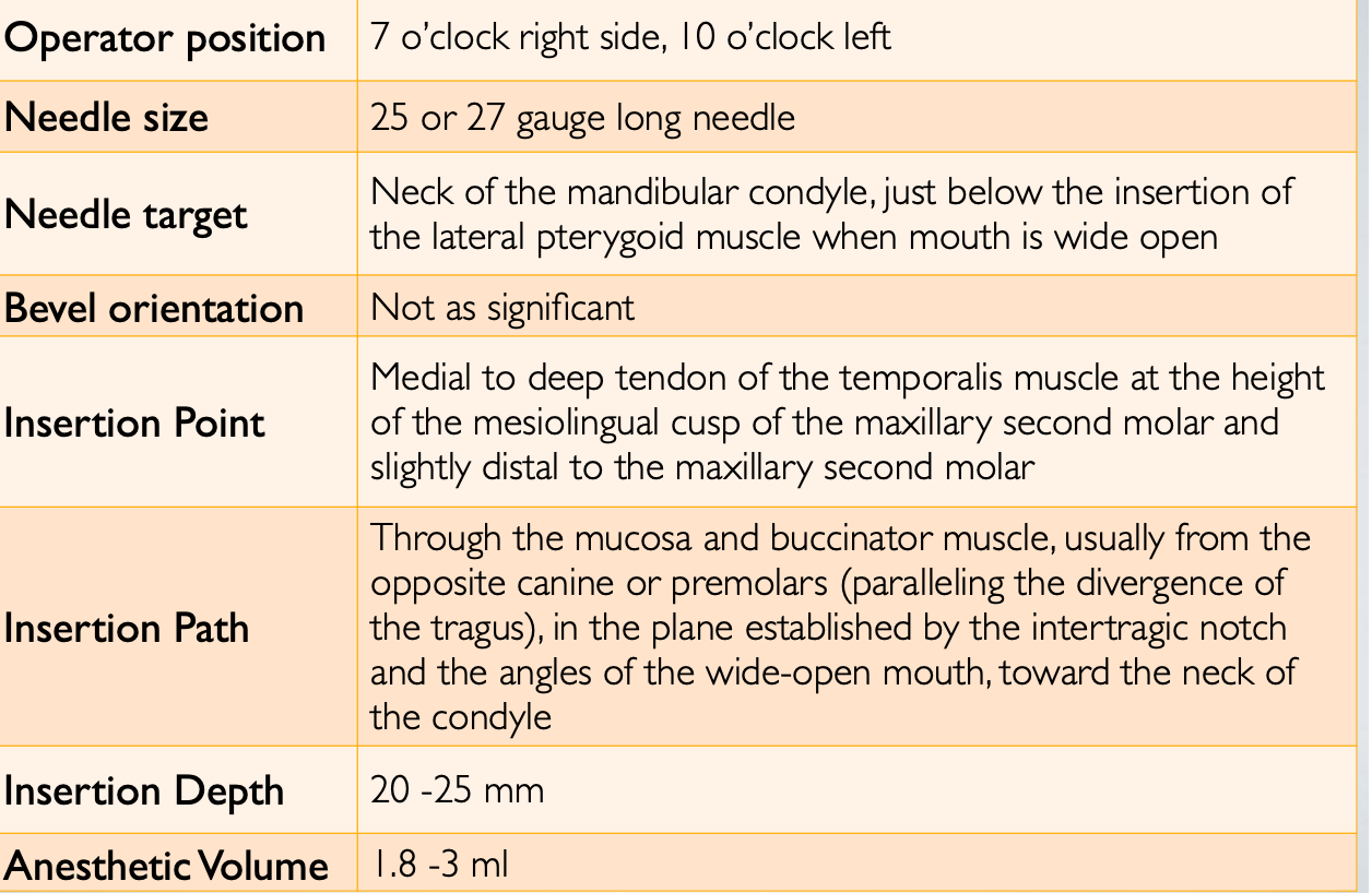
Insertion Point:
At the height of the ML cusp of maxillary 2nd molar, penetrate just distal to the maxillary 2nd molar
Target Point:
Lateral aspect of condylar neck
Use both intraoral and extraoral landmarks to
establish the path of insertion
Gow-gates mandibular technique
Gow-gates mandibular technique
Gow-gates mandibular technique
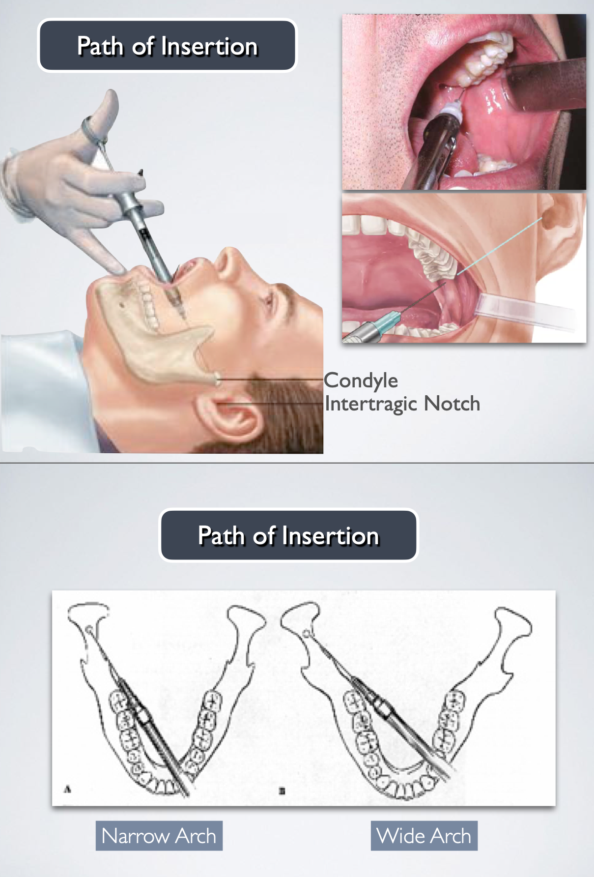
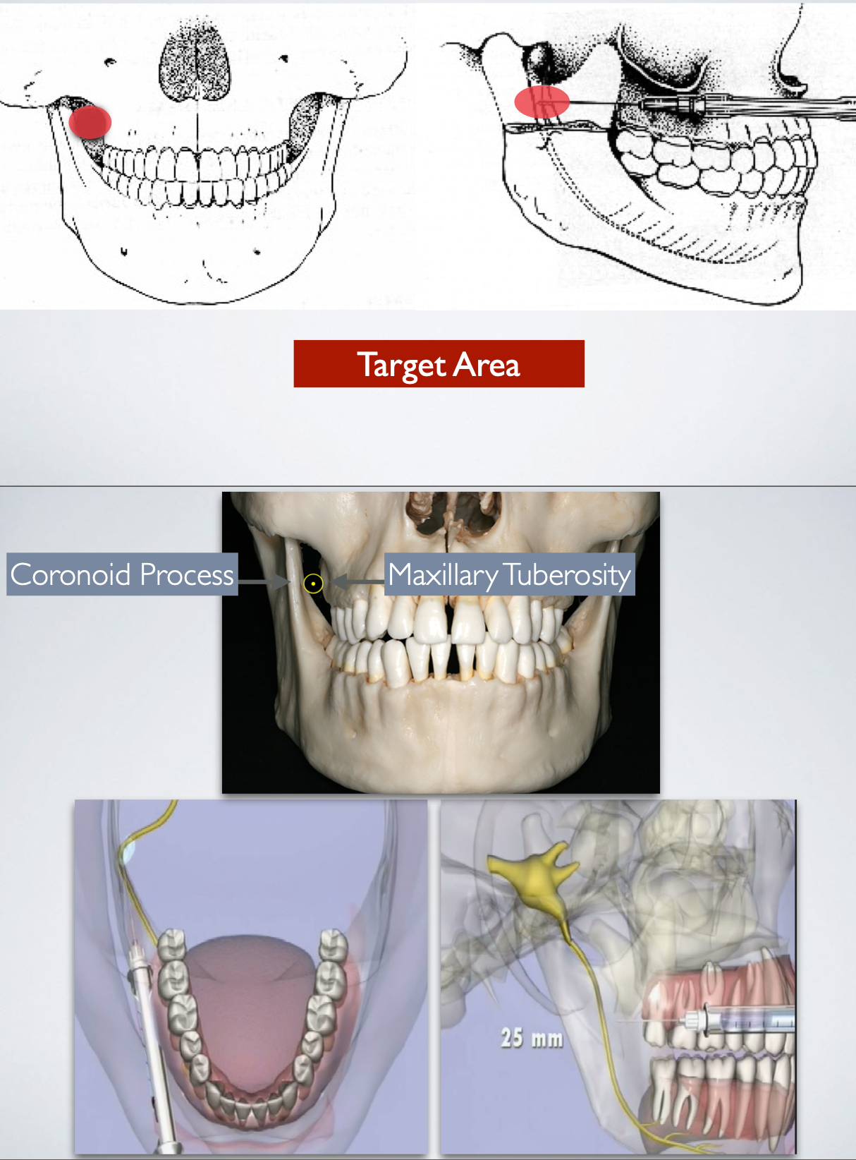
Thumb palpating the anterior border of the ramus Index finger over the intertragic notch of the ear
This line forms the path of insertion
Advance the needle slowly until bone is contacted
Target: The bone contacted is the neck of the condyle
Average depth of needle insertion is about 25 mm
Gow-gates mandibular technique
Gow-gates mandibular technique
Nerves anesthetized:
Inferior alveolar nerve
Mental/Incisive nerve
Lingual nerve
Mylohyoid nerve
akinosi closed mouth mandibular technique
Areas Anesthetized:
Mandibular teeth to the midline
Body of the mandible
Buccal soft tissue served by mental nerve
Anterior two thirds of tongue
Floor of oral cavity
Lingual soft tissue
akinosi closed mouth mandibular technique
Advantage
Relatively atraumatic
Patient does not need to open the mouth
Less post injection complications
Lower aspiration rate (<10%)
Disadvantage
Difficult to visualize path of insertion/depth of insertion
No bony contact
akinosi closed mouth mandibular technique
akinosi closed mouth mandibular technique
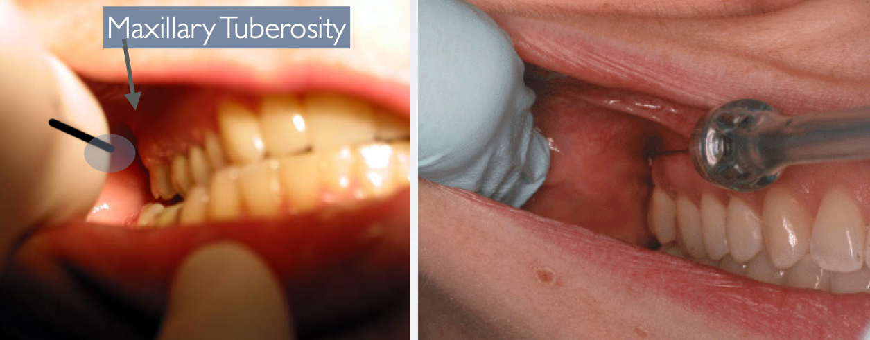
Height of Insertion: Mucogingival Junction of Maxillary 2nd or 3rd Molar Insertion depth: ~25 mm (measured from the maxillary tuberosity)
akinosi closed mouth mandibular technique
akinosi closed mouth mandibular technique
Buccal soft tissue anterior to the mental foramen (around the 2nd premolar to the midline)
Skin of lower lip
Pulps of premolars, canines, and incisors
mental/incisive nerve block
Advantage
If treatment on mandibular anterior teeth, this block negates the need for bilateral inferior alveolar blocks
Does not block lingual nerve = no tongue numbness
High success rate
Disadvantage
Midline difficulty, may require a supplemental injections
mental/incisive nerve block
Target Site: Mental nerve as it exits mental foramen (between apices of the 1st and 2nd premolar)
1 injection 1 technique mental and incisive soft tissue and anteriors
mental/incisive nerve block
mental foramen
mental/incisive nerve block
mental/incisive nerve block
mental/incisive nerve block
mental/incisive nerve block