Identification of bacteria
1/21
There's no tags or description
Looks like no tags are added yet.
Name | Mastery | Learn | Test | Matching | Spaced |
|---|
No study sessions yet.
22 Terms
Identification of bacteria
Colonies from culture samples can be transferred to a microscope slide and stained for microscopic analysis
Staining colours the cells improving visibility under the microscope
GRAM’s Stain
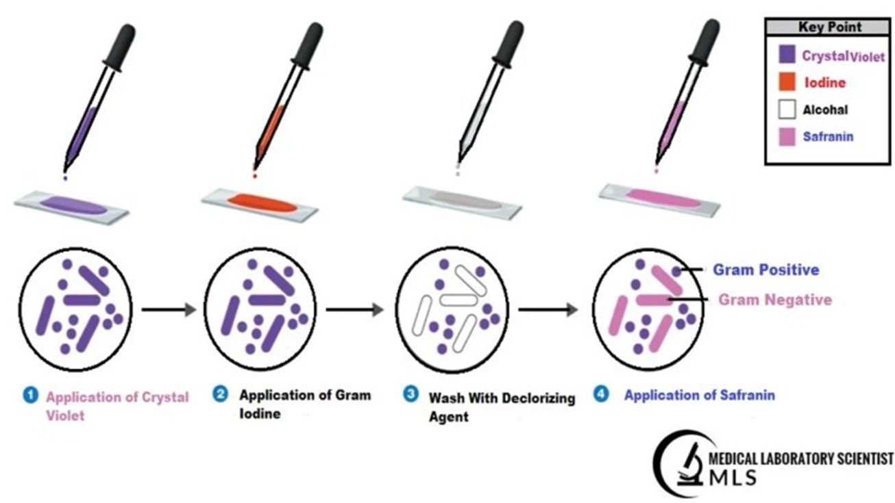
1 - Application of Crystal Violet
2 - Application of Gram Iodine
3 - Wash with Decolorising Agent
4 - Application of Safranin
Staining with a suitable stain, for example ..
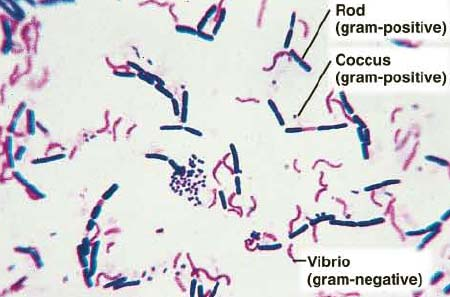
Gram stain allows cell morphology to be observed
Bacteria can be classified by the thickness or composition of the cell wall structure
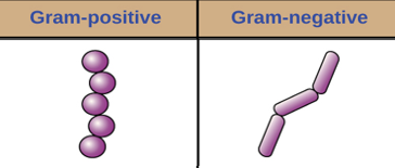
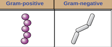
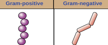
Gram’s positive
Gram’s negative
Acid fast

Gram’s stain
This is the most commonly used stain in microbiology that provides the first step in identifying bacteria
Bacteria are categorised by the colour that they stain

Gram’s positive
Blue/Purple

Gram’s negative
Red/Pink
Gram stain process - Step 1
Crystal Violet - Primary stain added to specimen smear
Cell effects - Stains cell purple or blue


Gram stain process - Step 2
Iodine - Mordant makes dye less soluble so it adhere to cell walls
Cell effects - Cells remain purple or blue

Gram stain process - Step 3
Alcohol - Decolouriser washes away stain from gram-negative cell walls
Cell effects - Gram-positive cells remain purple or blue. Gram-negative cells are colourless

Gram stain process - Step 4
Safranin - Counterstain allows dye adherence to gram-negative cells
Cell effects - Gram-positive cells remain purple or blue. Gram-negative cells appear pink or red.
Alternative bacterial stains - Simple/structural stains (methylene blue)
Colours cells allowing shape, size and arrangement to be observed
Alternative bacterial stains - Differential stains (Gram’s stain, Ziehl-Neelsen)
This is a combination of fixative and 2 dyes, a primary and counter stain, that allows differentiation of cells
Alternative bacterial stains - Structural stains
This stains certain parts of the cell, e.g. Flagella, microspore and capsule also aiding in identification of the bacterium
Gram positive bacteria have a thick, porous peptidoglycan layer (outer layer of the cell) which absorbs both stains (will also absorb toxic substances so much easier to destroy), but the purple crystal violet stain is most visible. For example ..
Several staphylococcal and streptococcal spp.
Gram’s negative bacteria have a think peptidoglycan layer which is protected by a further impenetrable outer membrane. Have small holes ‘porins’ are contained within the outer membrane. The think peptidoglycan layer does not retain the crystal violet stain but absorbs the pink safranin stain. For example ..
Pseudomonus aeruginosa
Gram’s negative bacteria - Slime layer
Have an outer membrane and ‘slime layer’ which repel the stain and disinfectant, therefore are much more difficult to destroy
Slime layer also provide camouflage to the antigen, which is what is detected by the immune system to identify it as a foreign body
Gram-positive organisms stain violet / purple, for example ..
Clostridia Spp
Staphylococcus Spp
Listeria
Gram-negative organism stain pink / red, for example ..
Some species of Escherichia coli
Salmonella
Pseudomonas
Acid fast bacteria
Rarely diagnosed in vet practice
Have a waxy (mycolic acid) outer layer
Highly resistant to staining and treatment
Gram positive bacteria - do not stain well with this method
Ziehl-Neelsen stain required (red on blue background)
All other organisms stain blue
Can be zoonotic and cause very significant illness

Acid fast bacteria examples
Mycoplasma
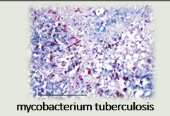
Mycobacterium tuberculosis
M leprae
Nocardia
Procedure for Ziehl-Neelsen Staining
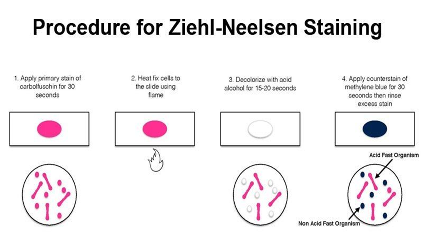
1 - Apply primary stain of carbolfuchsin for 30 seconds
2 - Heat fix cells to the slide using flame
3 - Decolourise with acid alcohol for 15-20 seconds
4 - Apply counterstain of methylene blue tor 30 seconds then rinse excess stain