HSC 214 exit quiz 12
1/67
There's no tags or description
Looks like no tags are added yet.
Name | Mastery | Learn | Test | Matching | Spaced | Call with Kai |
|---|
No analytics yet
Send a link to your students to track their progress
68 Terms
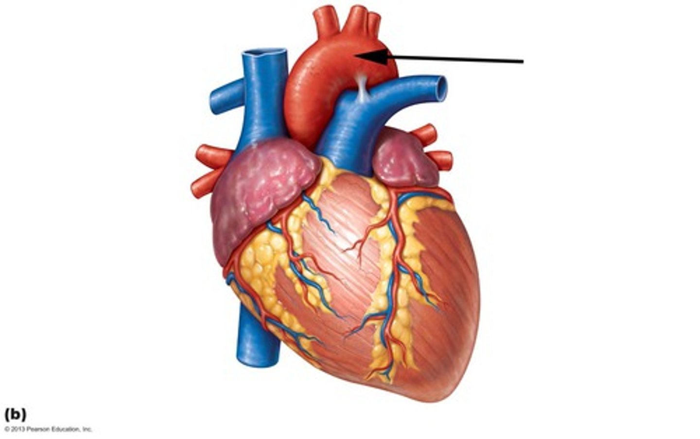
Ascending aorta
Area supplied: Body
Terminal branch: Aortic arch
Relationship: Immediately extending superiorly from the heart
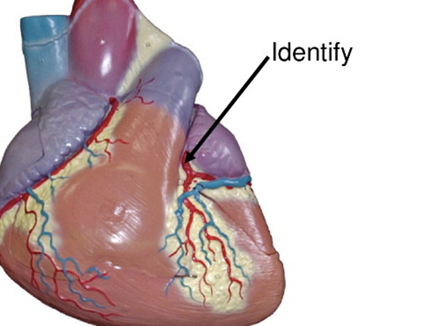
Right coronary artery
Area supplied: Right and left ventricles, right atrium, and interventricular septum
Terminal branch: Posterior interventricular and right marginal arteries
Relationship: Passes between the pulmonary trunk and right auricle
Left coronary artery
Area supplied: Right and left ventricles, left atrium, and interventricular septum
Terminal branch: Anterior interventricular and circumflex arteries
Relationship: Passes between the pulmonary trunk and left auricle
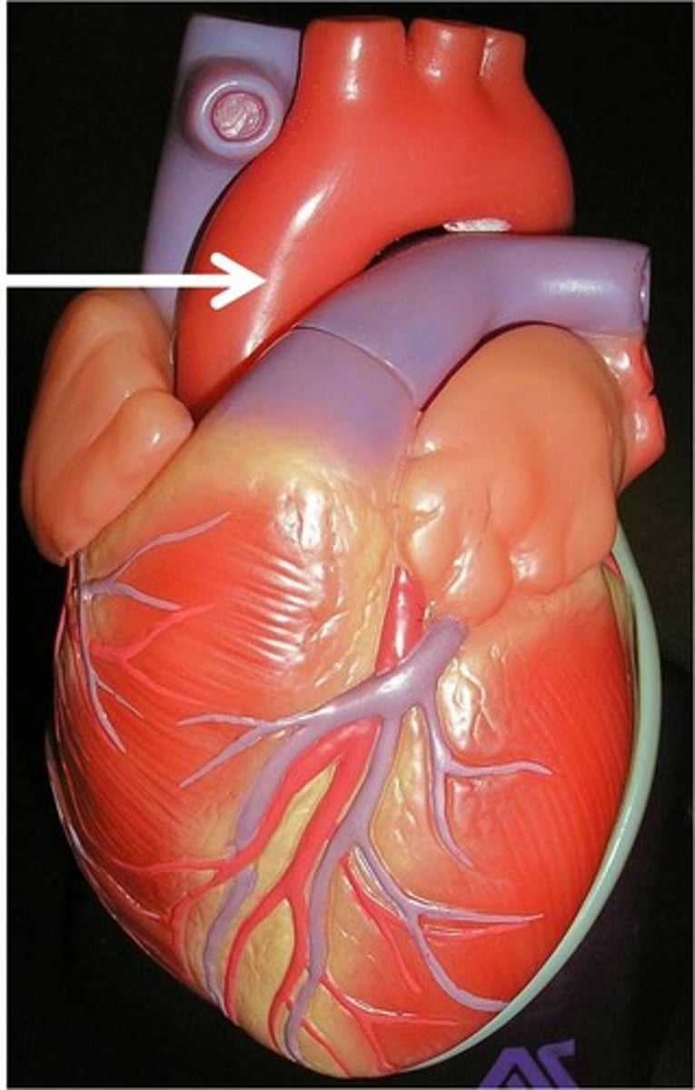
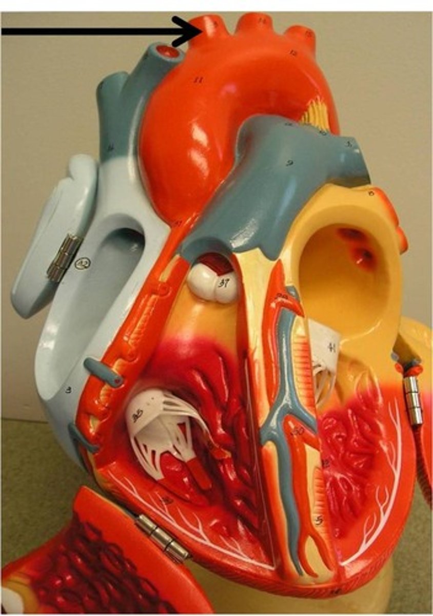
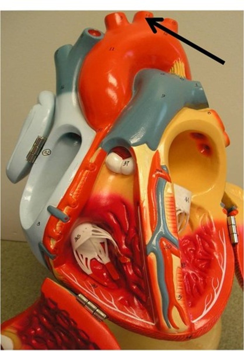
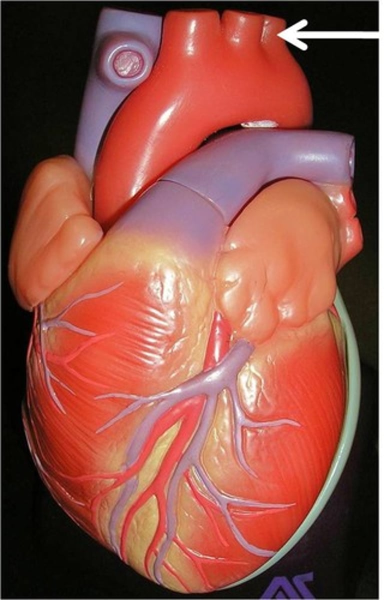
Aortic arch
Area supplied: Systemic circulation
Terminal branch: Thoracic (descending) aorta
Relationship: Gives rise to the brachiocephalic trunk, left common carotid, and left subclavian arteries
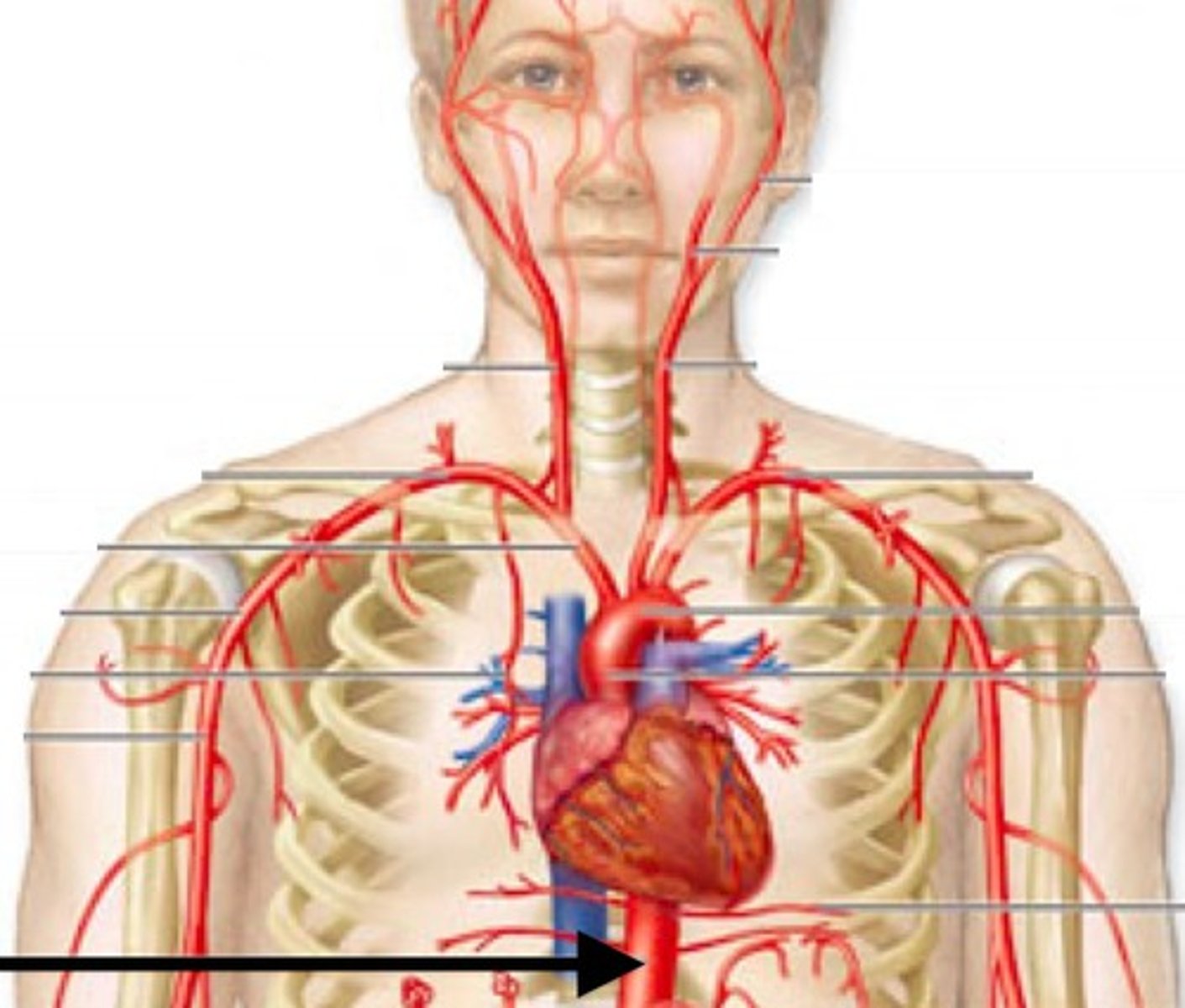
Thoracic aorta
Area supplied: Lungs, thoracic wall, and body above the diaphragm
Terminal branch: Abdominal aorta
Relationship: Located on the left side of the posterior mediastinum in the thoracic cavity
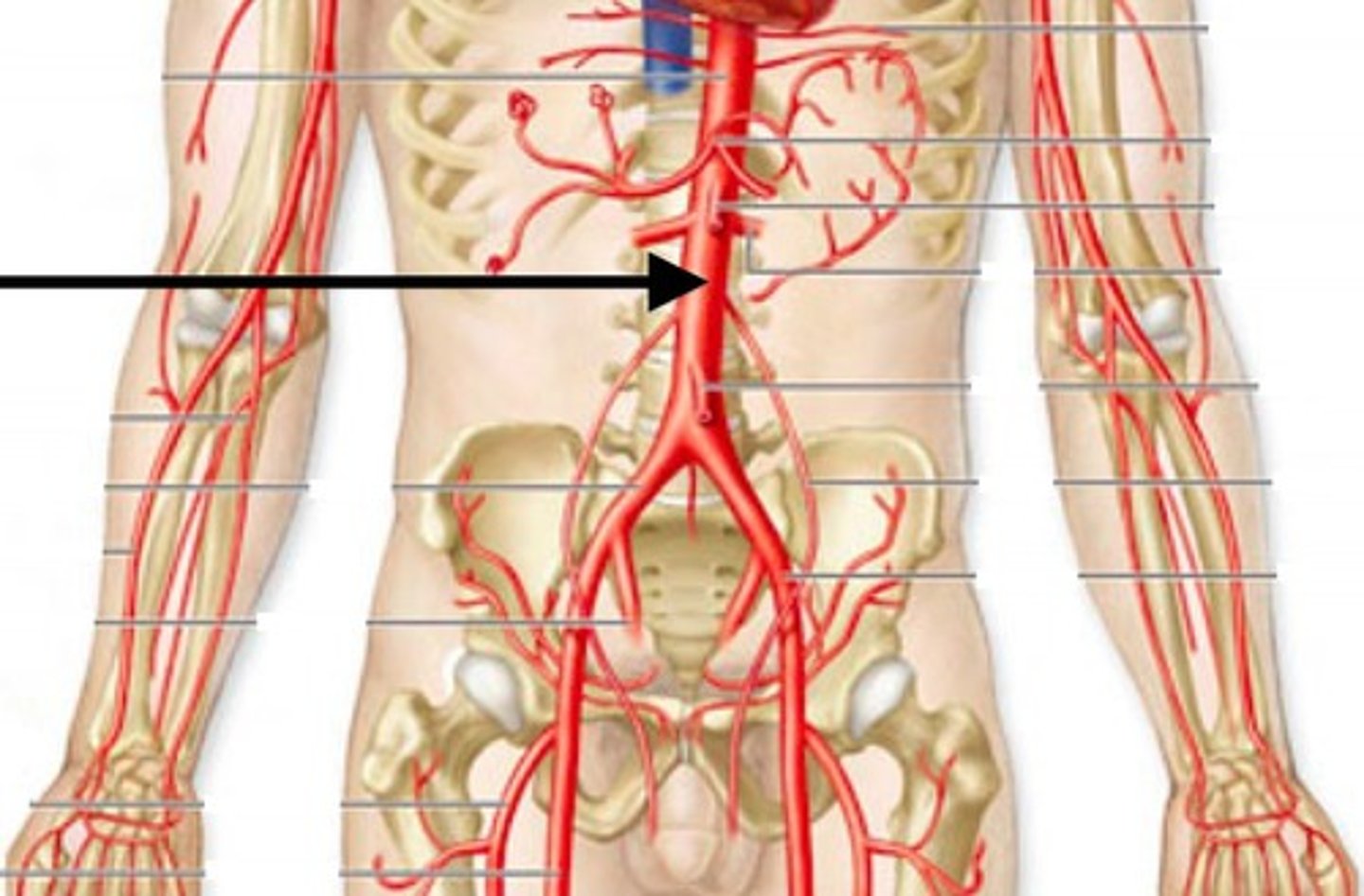
Abdominal aorta
Area supplied: Everything below the diaphragm
Terminal branch: Right and left common iliac arteries
Relationship: Passes along the anterior aspect of the vertebral bodies
Brachiocephalic trunk artery
Area supplied: Right side of the head, neck, and upper arm
Terminal branch: Right common carotid and right subclavian arteries
Relationship: First branch of the aortic arch
Right common carotid artery
Area supplied: Right side of the head and neck
Terminal branch: Right internal and external carotid arteries
Relationship: Bifurcates at the level of the thyroid gland
Right subclavian artery
Area supplied: Right side of the neck, chest, upper arm
Terminal branch: Right axillary artery
Relationship: Passes under (sub-) the clavicle and between the scalene muscles

Left common carotid artery
Area supplied: Left side of the head and neck
Terminal branch: Left internal and external carotid arteries
Relationship: Bifurcates at the level of the thyroid gland
Left subclavian artery
Area supplied: Left side of the head, neck, brain, chest, upper arm
Terminal branch: Left axillary artery
Relationship: Passes under (sub-) the clavicle and between the scalene muscles
External carotid artery
Area supplied: Face, meninges, and neck structures
Terminal branch: Superficial and temporal arteries
Relationship: Anterior branch of the common carotid artery
Internal carotid artery
Area supplied: Eye, cerebrum, cerebral arterial circle
Terminal branch: Anterior and middle cerebral arteries
Relationship: Posterior branch of the common carotid artery. Enters the cranial cavity through the carotid canal
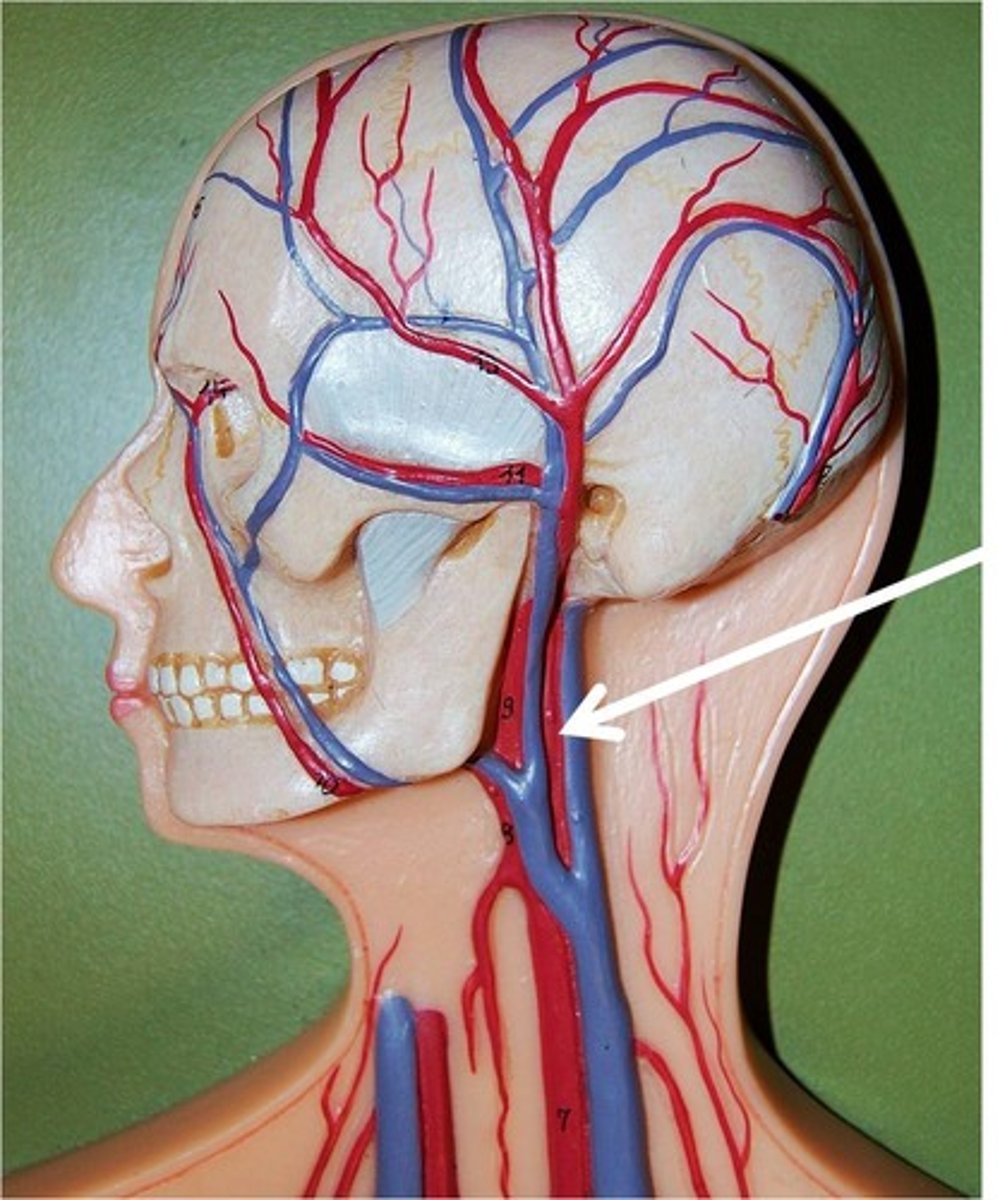
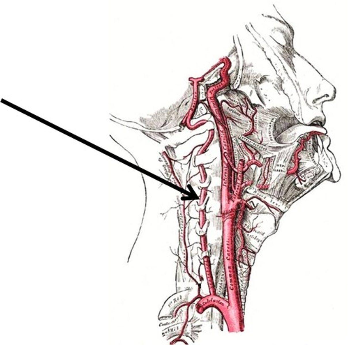
Vertebral artery
Area supplied: Brain, spinal cord, and vertebral column
Terminal branch: Basilar artery
Relationship: Originates from each subclavian artery. Passes through the transverse foramen of cervical vertebrae
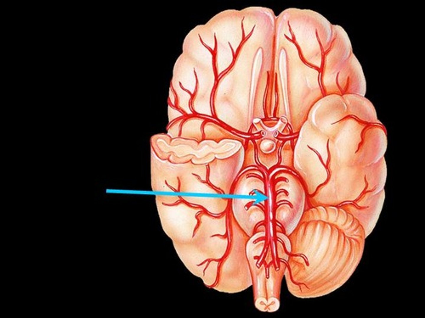
Basilar artery
Area supplied: Pons and cerebellum
Terminal branch: Cerebral arterial circle
Relationship: Midline vessel that ascends on the anterior surface of the pons
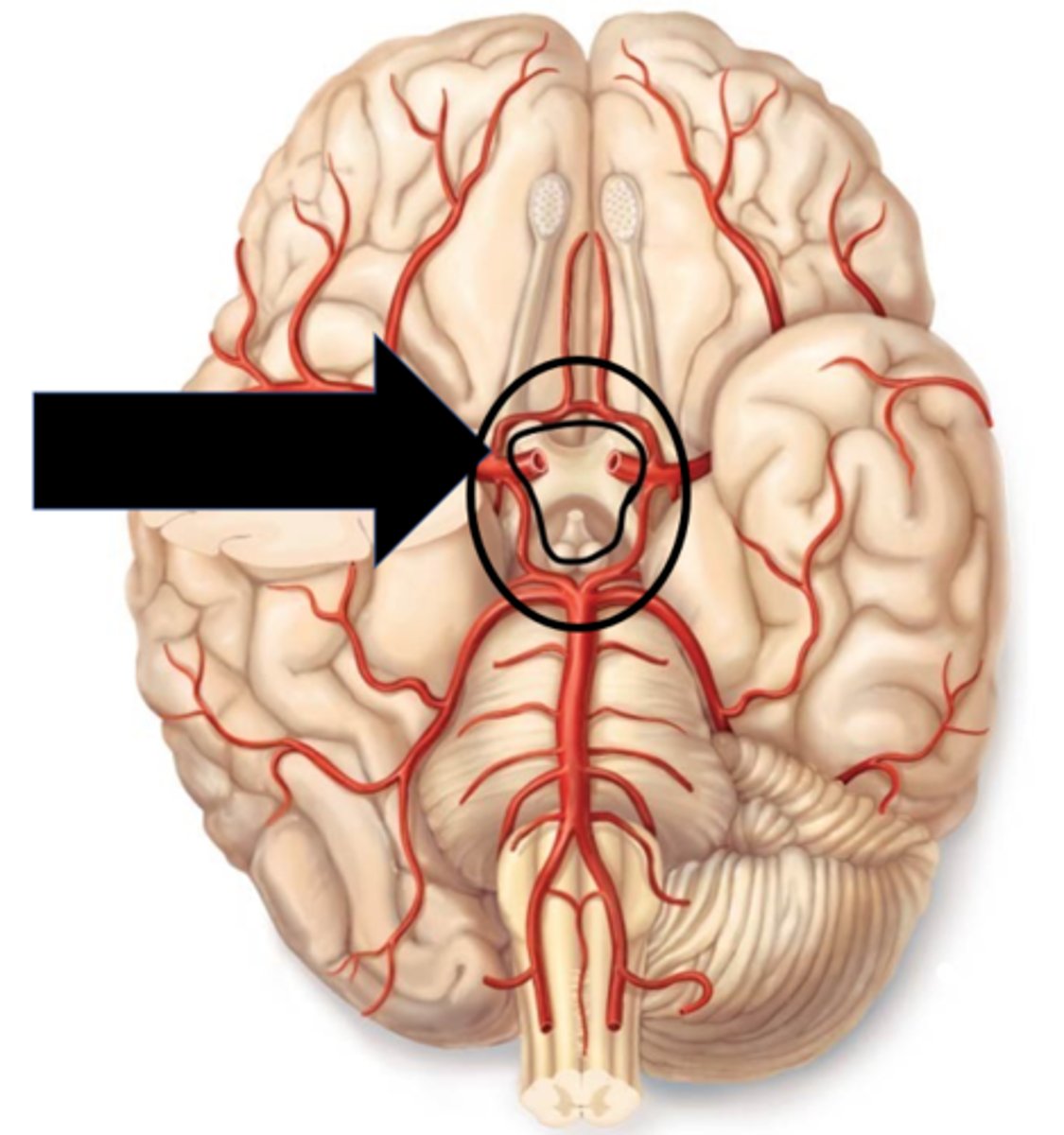
Cerebral arterial circle (circle of willis)
Area supplied: Brain by many branches off circle
Terminal branch: N/A
Relationship: Circular anastomosis on the ventral surface of the brain is supplied by left and right internal carotid and basilar arteries
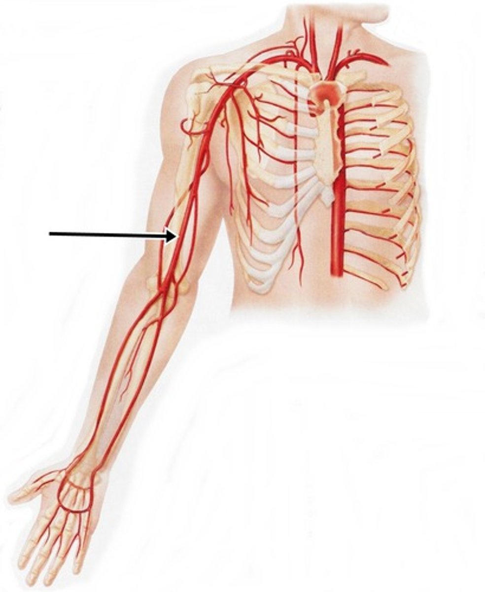
Axillary artery
Area supplied: Pectoral and scapular regions
Terminal branch: Brachial artery
Relationship: Begins at the 1st rib and ends at the teres minor muscle
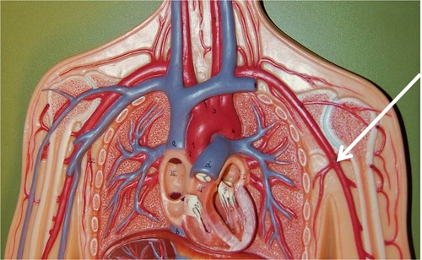
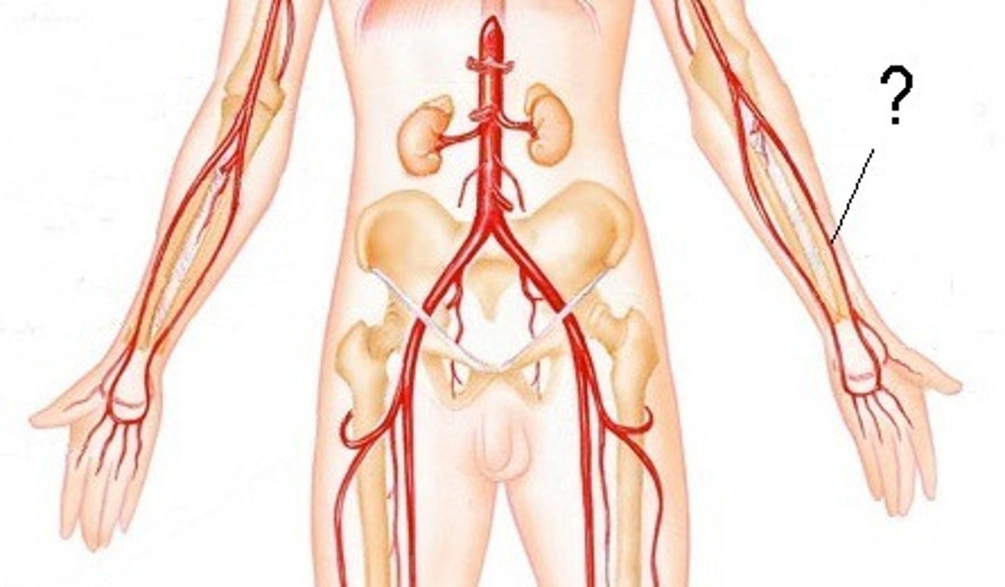
Brachial artery
Area supplied: Arm muscles and elbow joint
Terminal branch: Radial and ulnar arteries
Relationship: Passes inferiorly along the medial aspect of the arm deep to the biceps brachii muscle
Radial artery
Area supplied: Superficial posterior forearm muscles, deep hand muscles, and elbow joint
Terminal branch: Continues as the deep palmar arterial arch
Relationship: Passes distally along the lateral aspect of the forearm
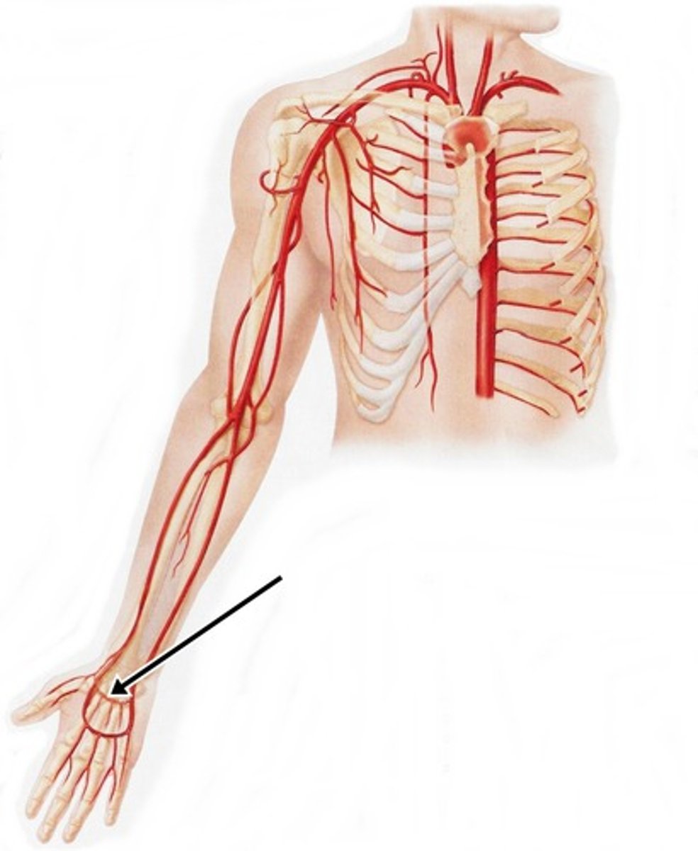
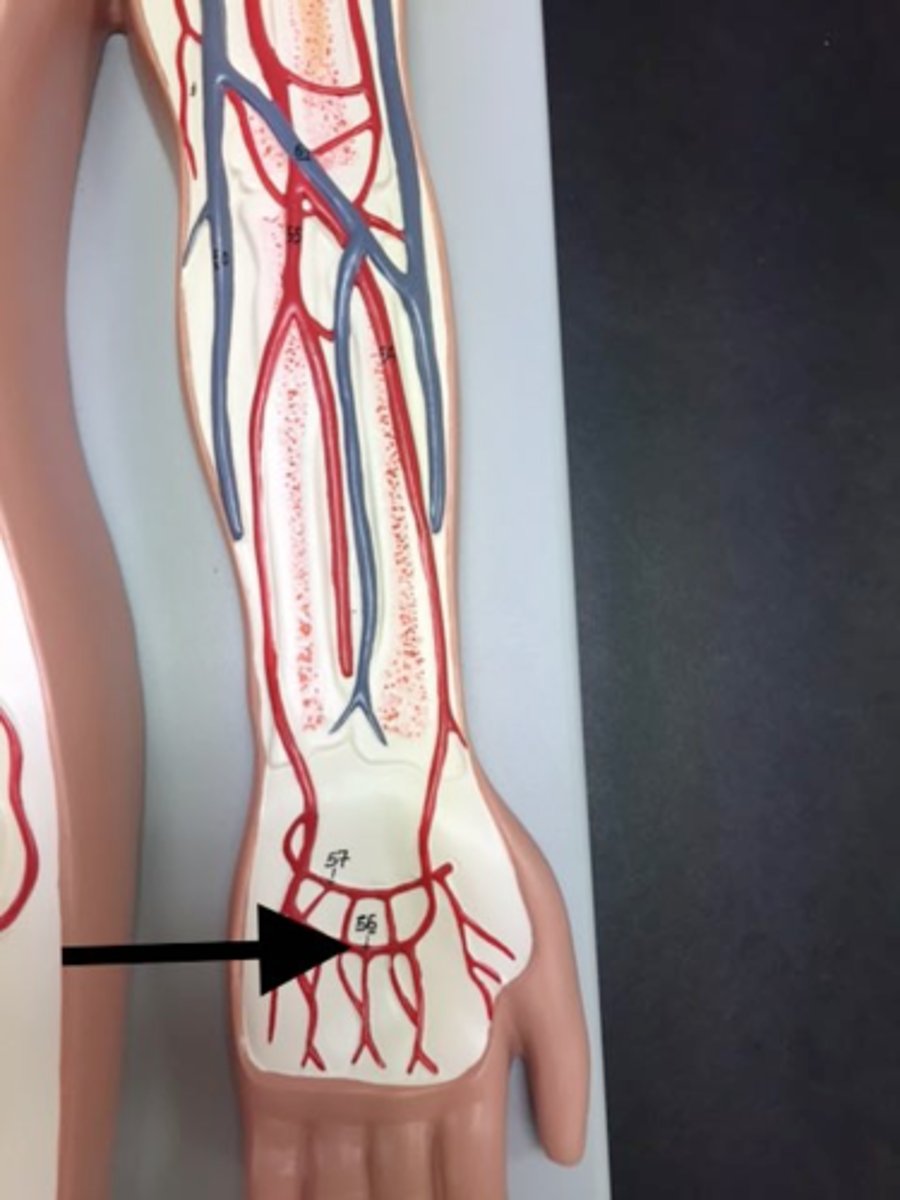
Deep palmar arch
Area supplied: Hand
Terminal branch: N/A
Relationship: Arches in the palm over the bases of the metacarpal bones to unite with a branch of the ulnar artery
Ulnar artery
Area supplied: Medial forearm muscles, hand muscles, and elbow joint
Terminal branch: Continues as the superficial palmar arterial arch
Relationship: Passes distally along the medial aspect of the forearm
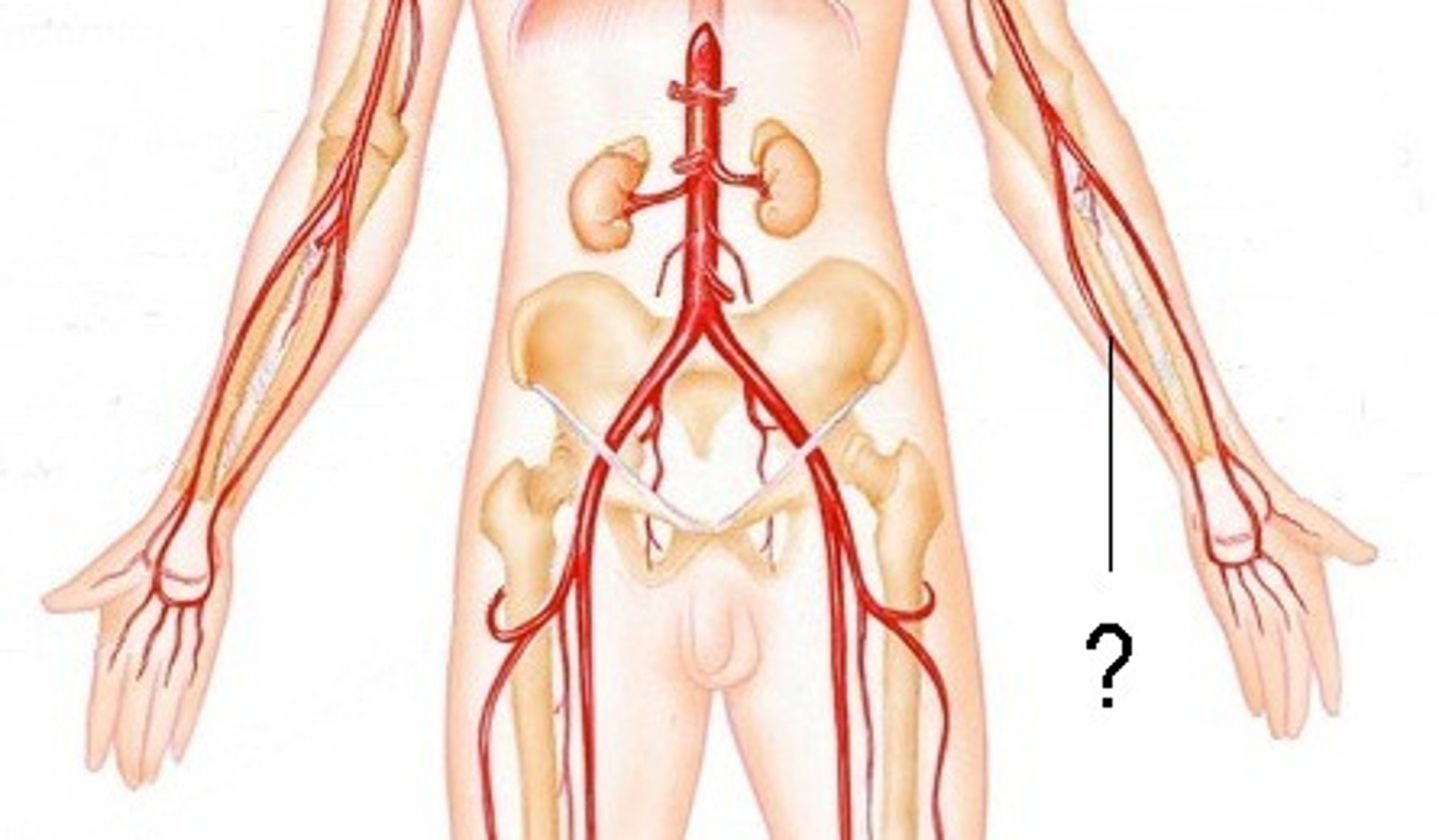
Superfical palmar arch
Area supplied: Hand
Terminal branch: N/A
Relationship: Arches in the palm over the middle of the metacarpal bones to unite with a branch of the radial artery
Thoracic (descending) aorta
Area supplied: Thoracic cavity
Terminal branch: Abdominal aorta
Relationship: Located on the left side of the posterior mediastinum in the thoracic cavity
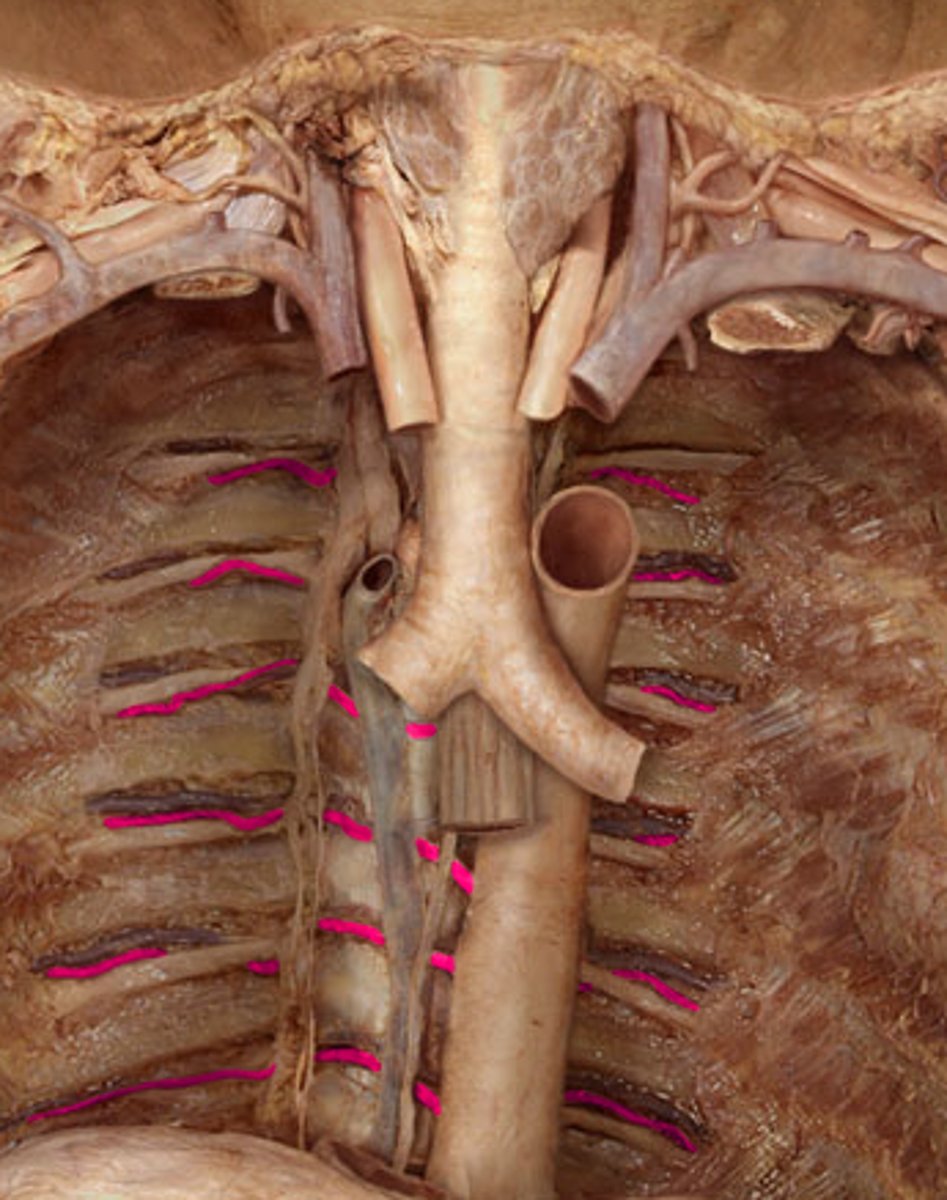
Posterior intercostal artery
Area supplied: Thoracic and abdominal walls, breast, spinal cord, and meninges
Terminal branch: N/A
Relationship: Sits in the costal groove of the ribs
Abdominal aorta
Area supplied: Abdominopelvic cavity
Terminal branch: Common iliac arteries
Relationship: Passes along the anterior aspect of the vertebral bodies

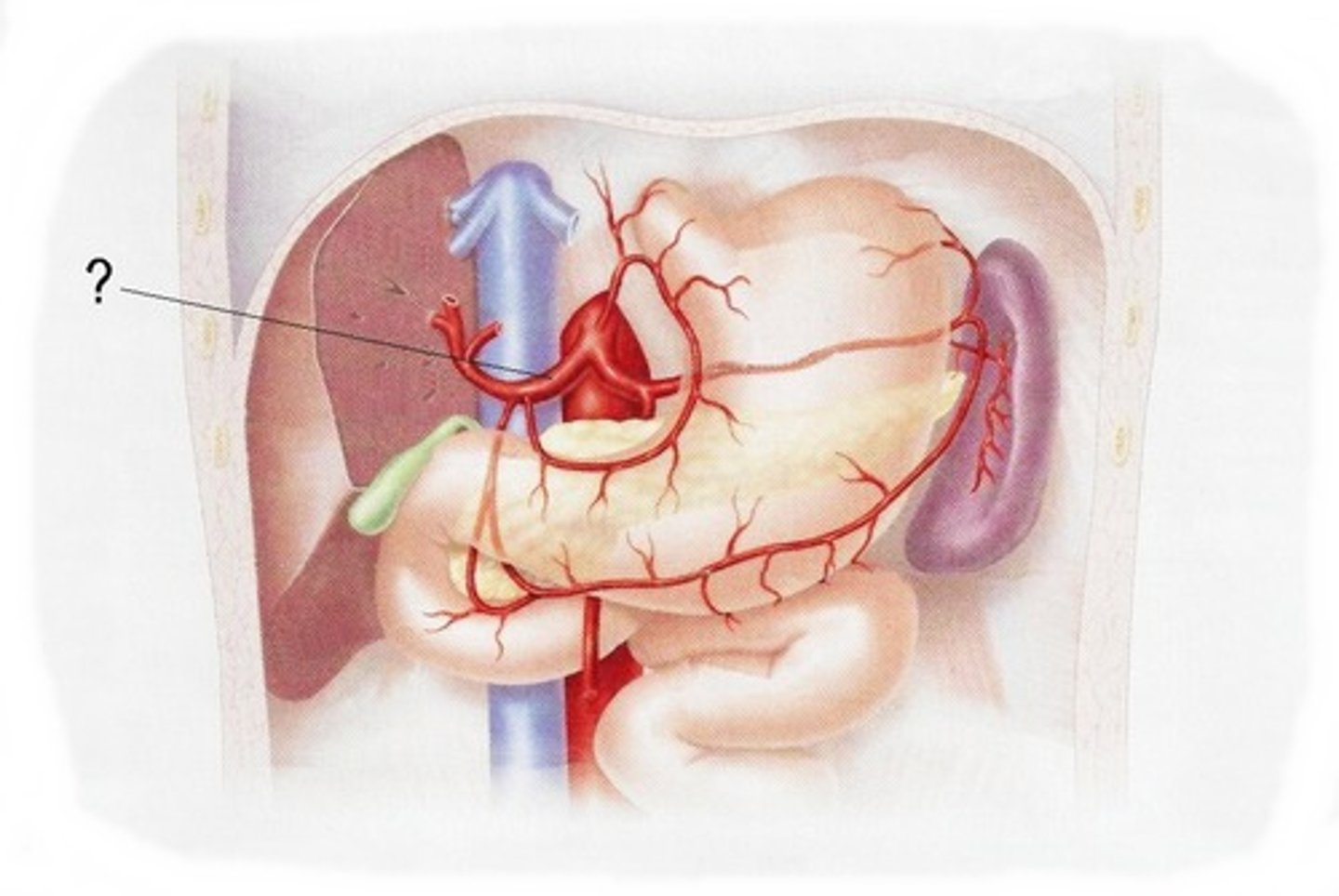
Celiac trunk
Area supplied: Esophagus, stomach, duodenum, liver, gallbladder, pancreas, and spleen
Terminal branch: Left gastric, common hepatic and splenic arteries
Relationship: Superior-most anterior branch of the abdominal aorta
Left gastric artery
Area supplied: Stomach and esophagus
Terminal branch: N/A
Relationship: Short branch passing along the lesser curvature of the stomach

Common hepatic artery
Area supplied: Liver, pancreas, stomach, gallbladder, and duodenum
Terminal branch: N/A
Relationship: Branch passing anterior and to the right to enter the lesser omentum
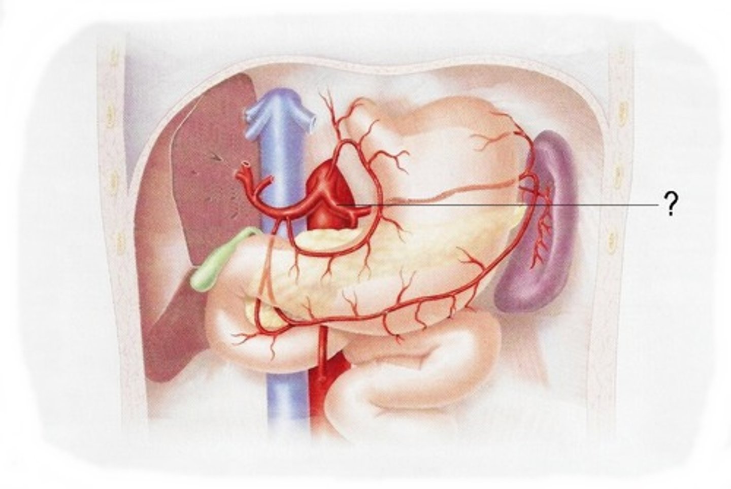
Splenic artery
Area supplied: Spleen, pancreas, and stomach
Terminal branch: N/A
Relationship: "Snake-like" branch passing left along the superior border of the pancreas
Superior mesentreric artery
Area supplied: Small intestines, pancreas, cecum, appendix, ascending and transverse colon
Terminal branch: N/A
Relationship: Middle anterior branch of the abdominal aorta immediately inferior to the celiac trunk
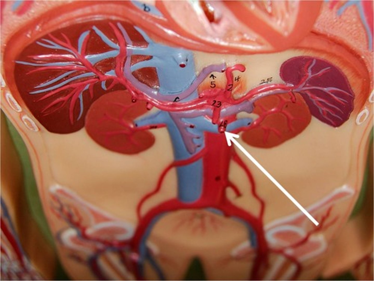
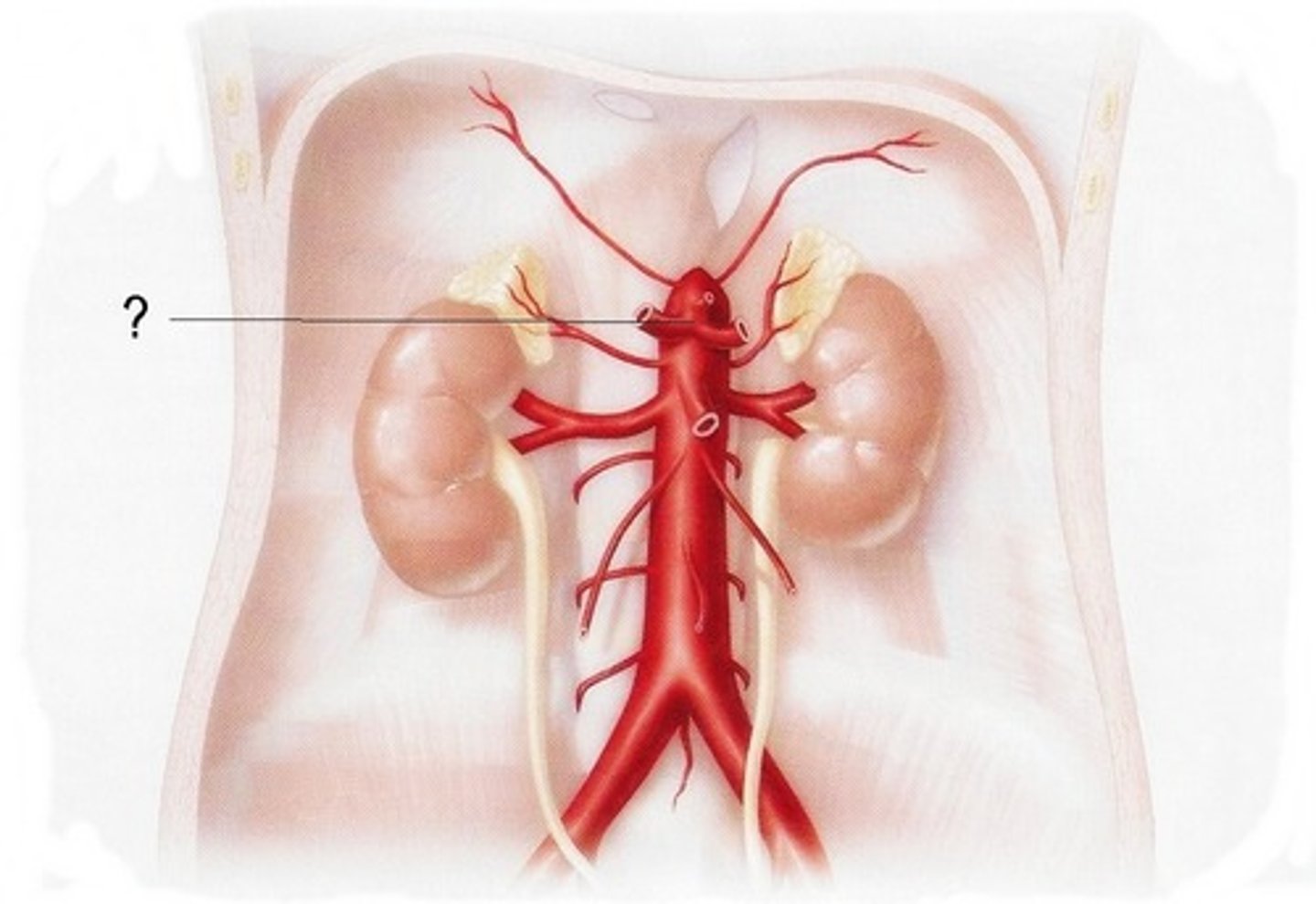
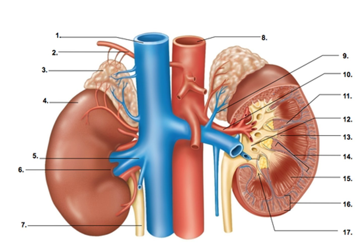
Renal artery
Area supplied: Kidneys, ureter, and adrenal glands
Terminal branch: N/A
Relationship: Lateral branches of the abdominal aorta
Inferior mesenteric artery
Area supplied: Transverse, descending, and sigmoid colon, rectum, and anal canal
Terminal branch: N/A
Relationship: Inferior-most anterior branch of the abdominal aorta
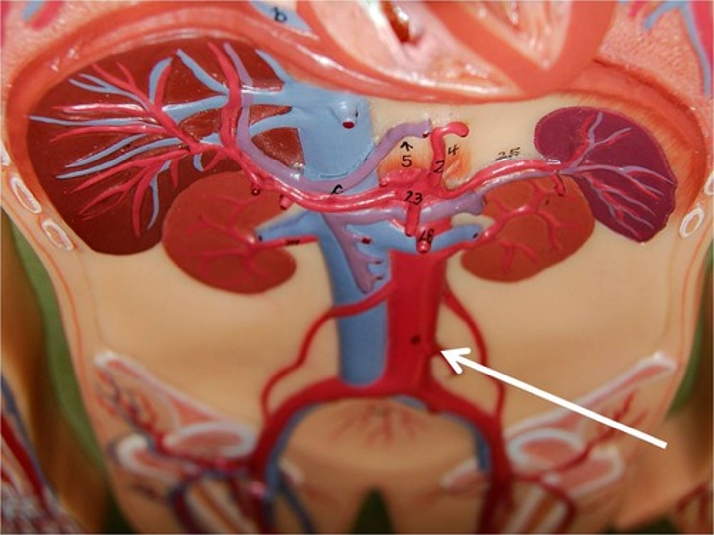
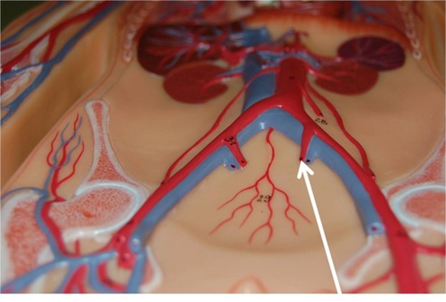
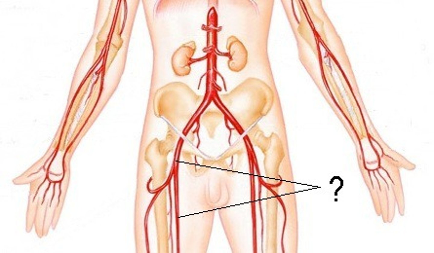
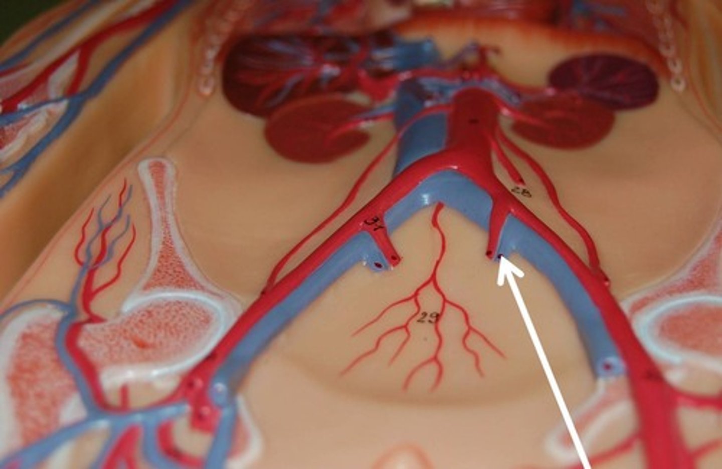
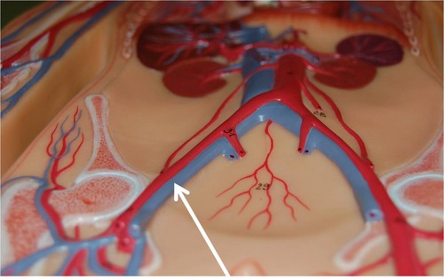
Common iliac artery
Area supplied: Pelvis, posterior abdominal wall, perineum, and lower limb
Terminal branch: External and internal iliac arteries
Relationship: Terminal branches of the abdominal aorta
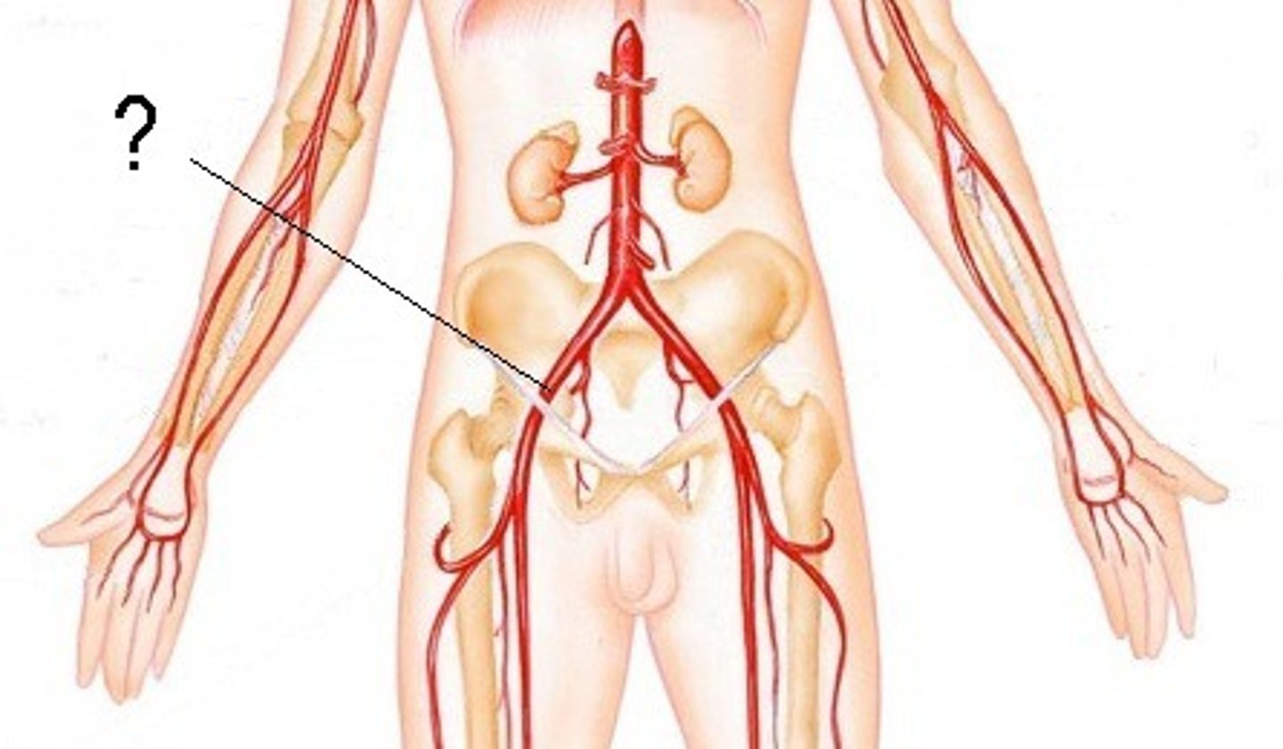
External iliac artery
Area supplied: Lower limb and abdominal wall
Terminal branch: Femoral artery
Relationship: Descends laterally along the medial border of the psoas major muscle
Internal iliac artery
Area supplied: Pelvis, posterior abdominal wall, perineum, and gluteal region
Terminal branch: N/A
Relationship: Descends deep and posterior into the pelvis
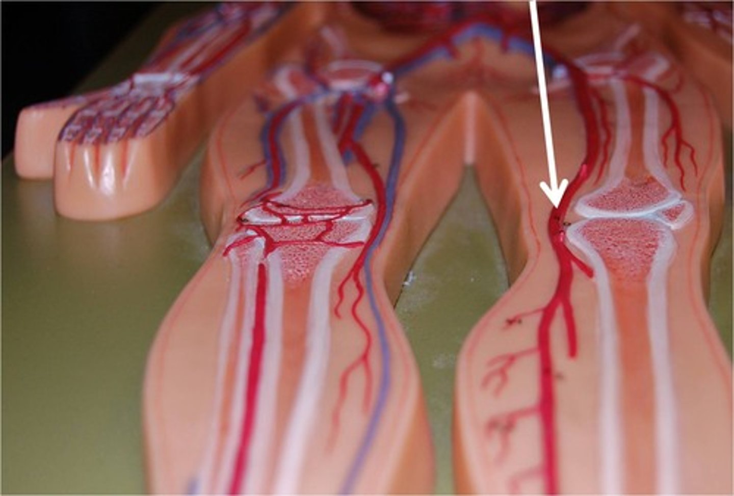
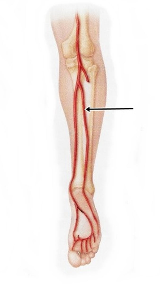
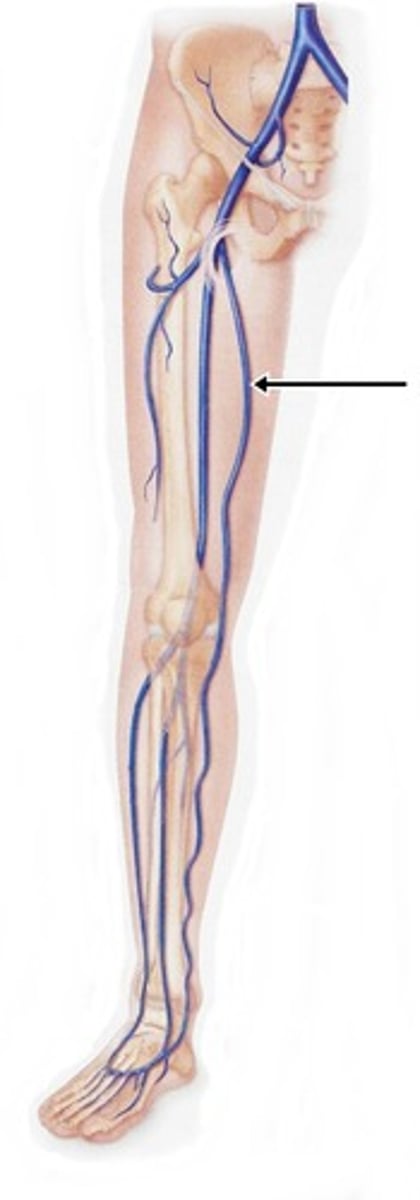
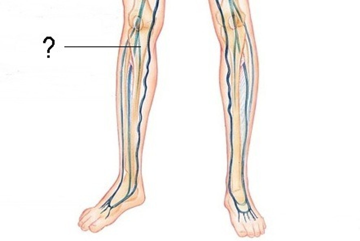
Femoral artery
Area supplied: Abdominal wall, thigh muscles, external genitalia, and hip and knee joints
Terminal branch: Popliteal artery
Relationship: Descends along the anterior and medial thigh in the inguinal triangle
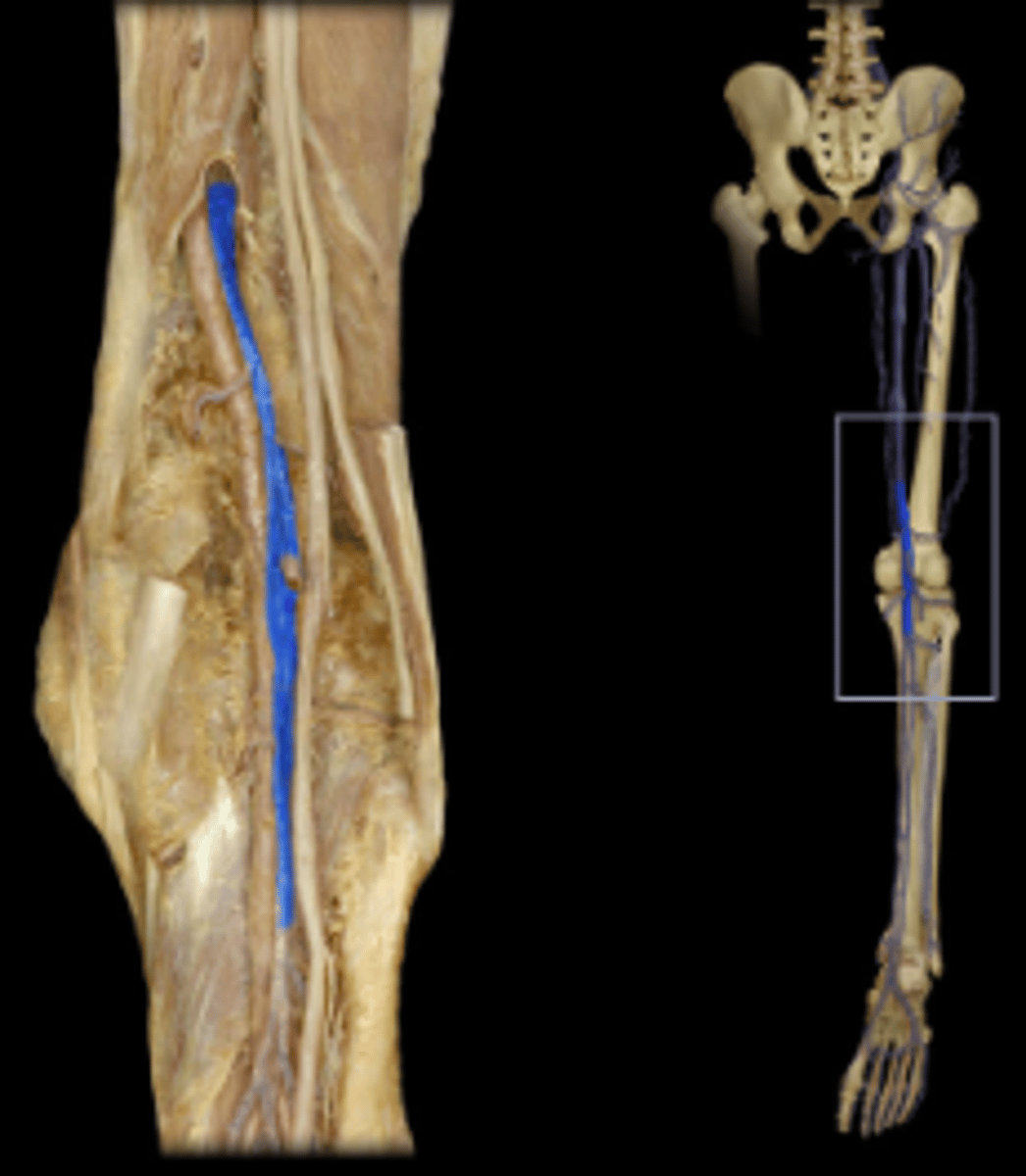
Popliteal artery
Area supplied: Knee joint
Terminal branch: Anterior and posterior tibial arteries
Relationship: Deepest structure in the popliteal fossa
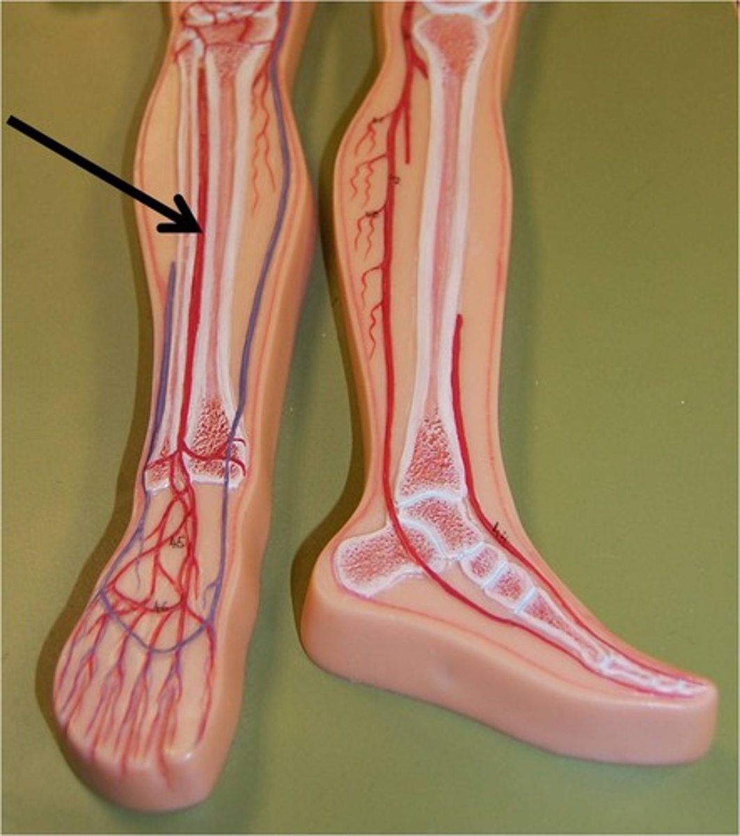
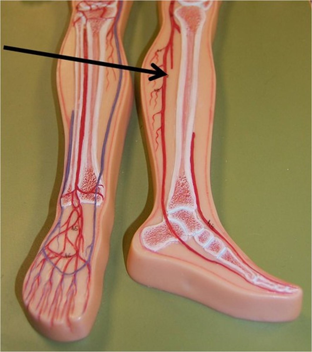
Anterior tibial artery
Area supplied: Anterior compartment leg muscles
Terminal branch: Dorsalis pedis artery
Relationship: First branch of the popliteal artery descending deep to the tibialis anterior muscle
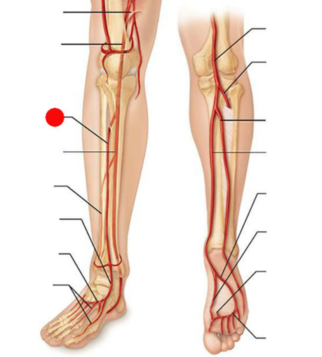
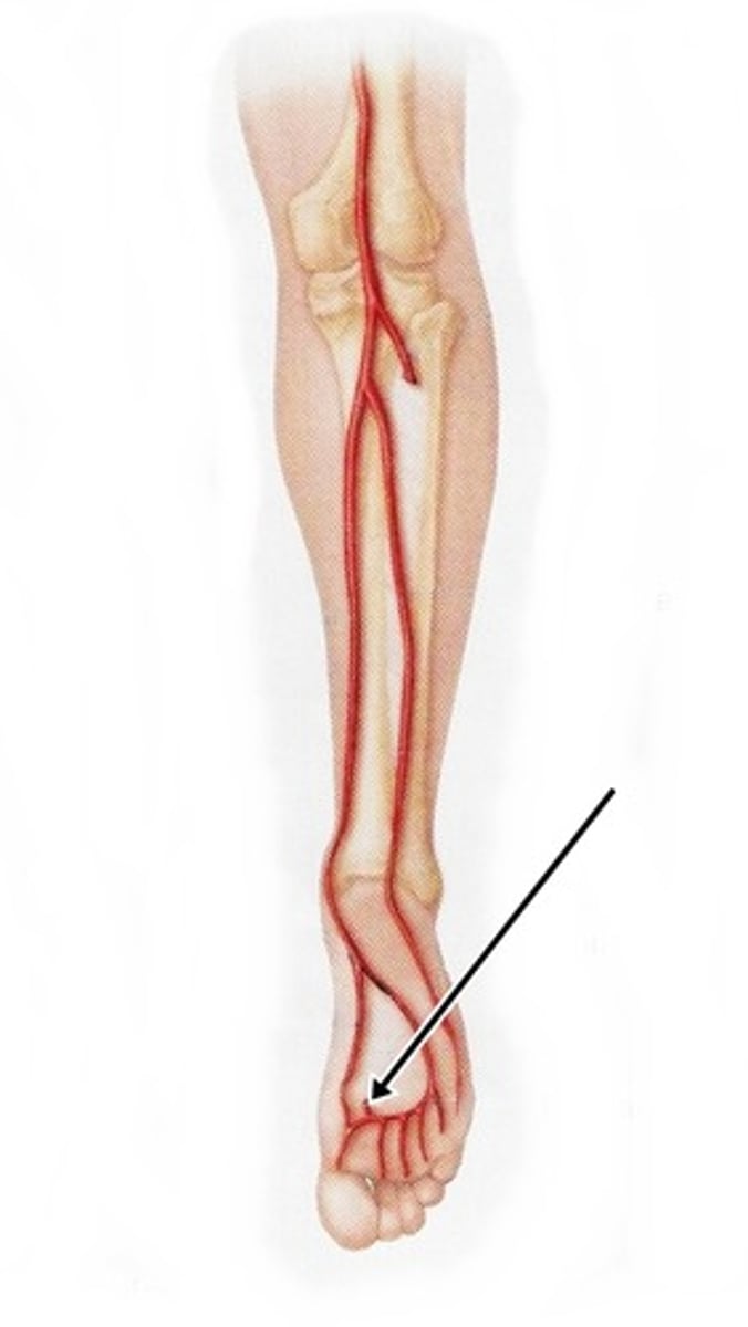
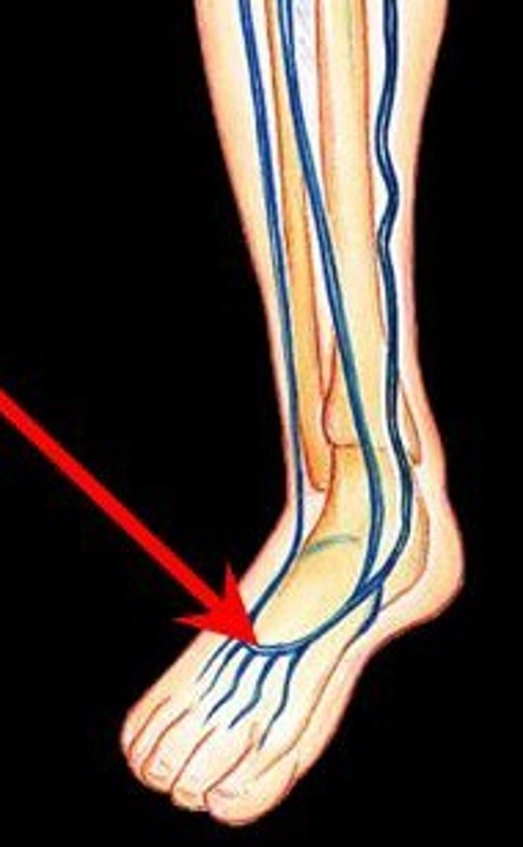
Dorsalis pedis artery
Area supplied: Dorsum and plantar foot muscles
Terminal branch: N/A
Relationship: Location of the foot pulse where the artery crosses the ankle
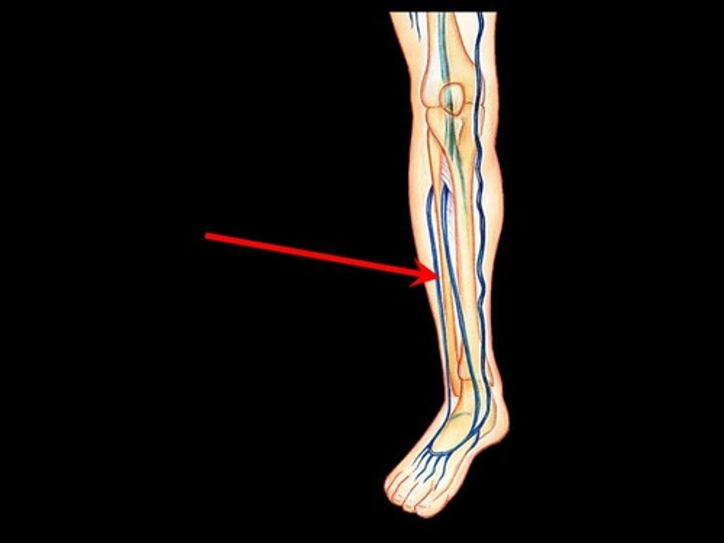
Posterior tibial artery
Area supplied: Posterior compartment leg muscles
Terminal branch: Medial and lateral plantar arteries
Relationship: Continuation of the popliteal artery descending through the posterior lef to the ankle
Fibular artery
Area supplied: Lateral compartment leg muscles and the flexor hallucis longus muscle
Terminal branch: N/A
Relationship: Descends through the deep compartment of the posterior leg
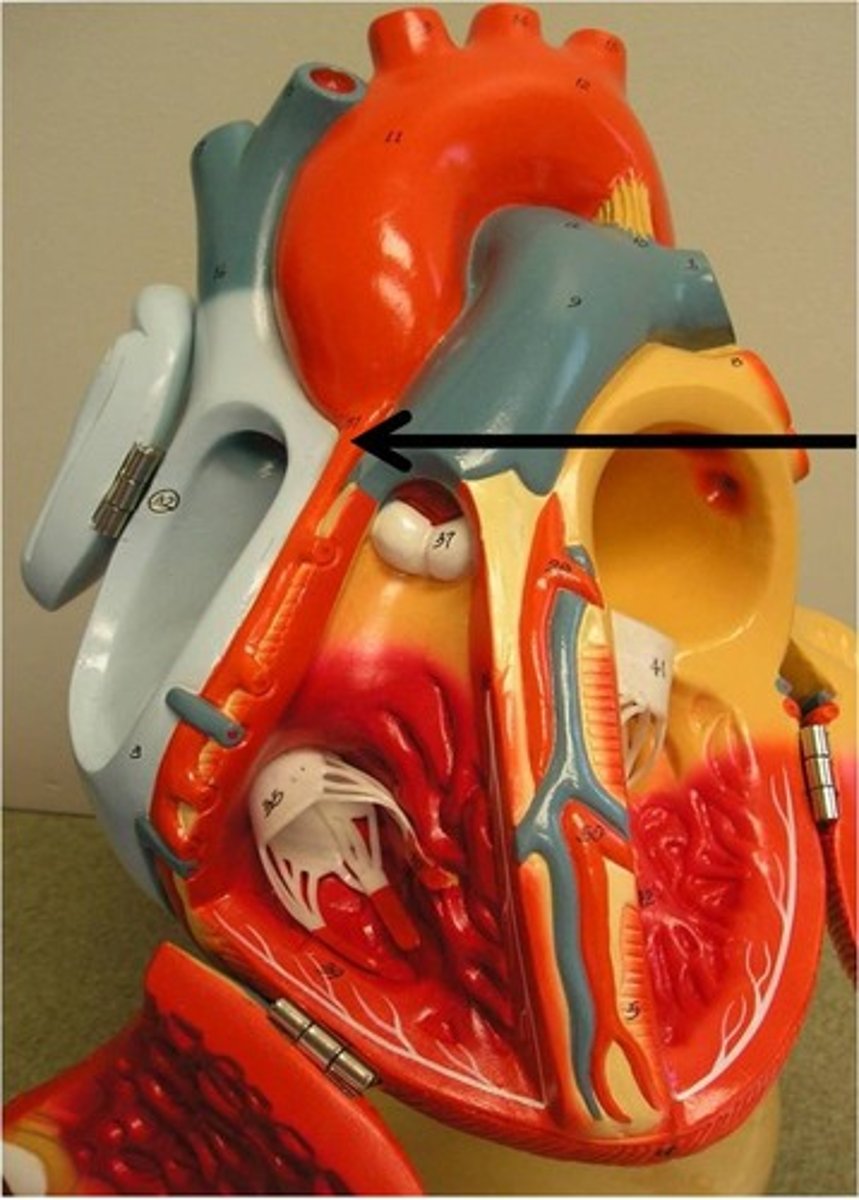
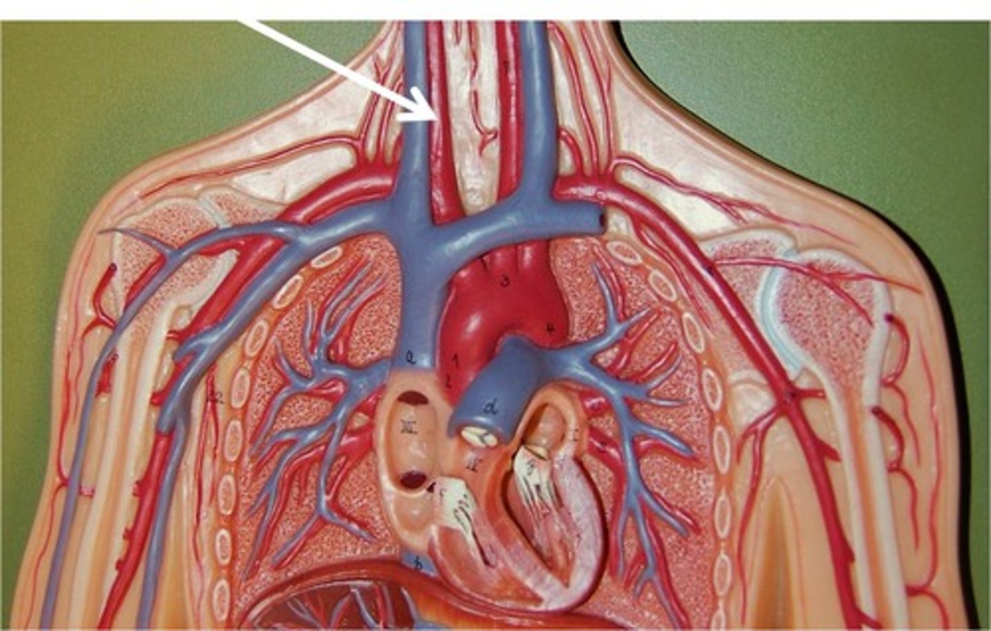
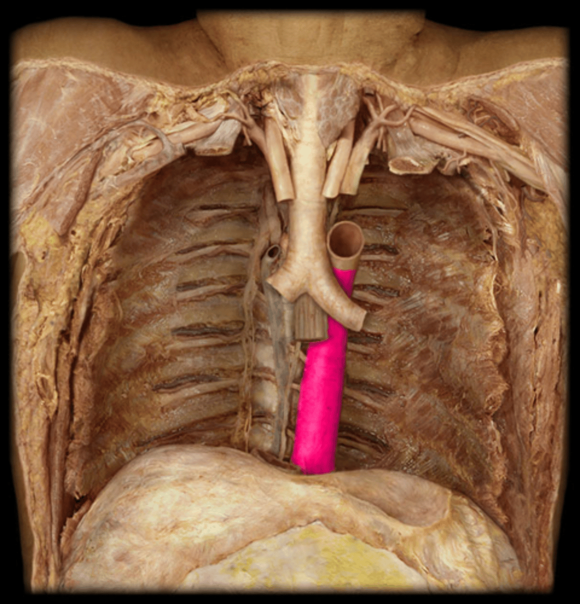
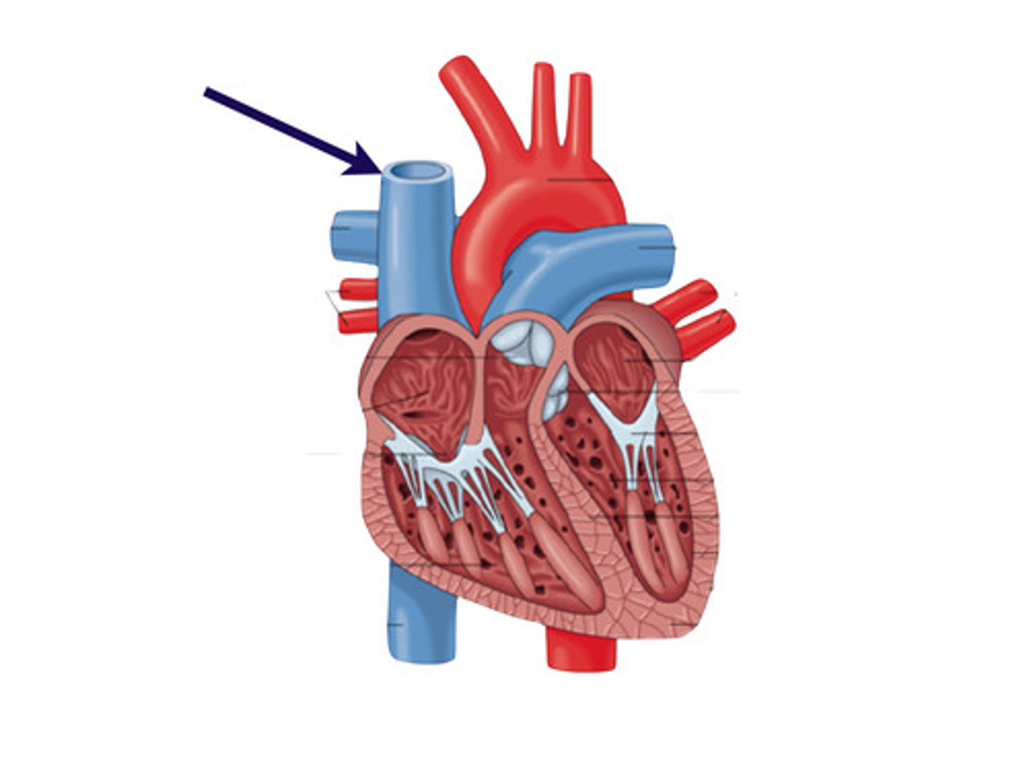
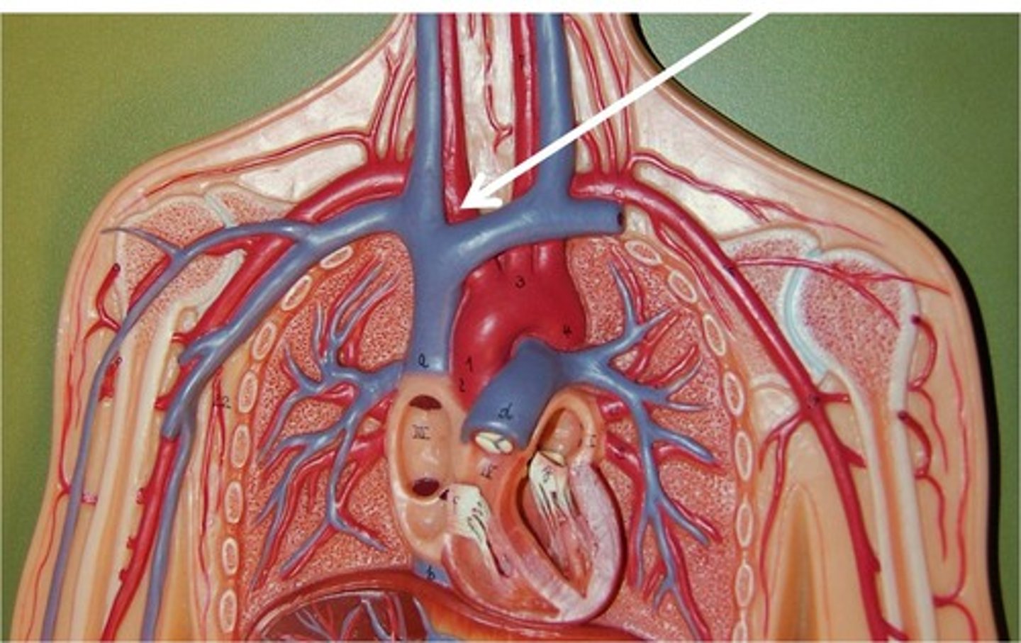
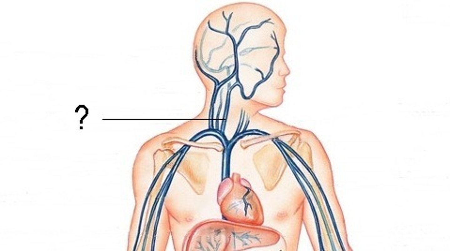
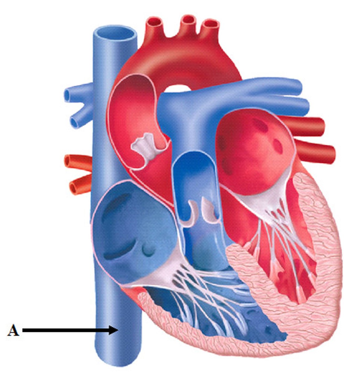
Superior vena cava
Area drained: Upper limb, head, and neck
Drains into: Right atrium of the heart
Relationship: Formed by the union of the left and right brachiocephalic veins
Brachiocephalic vein
Area drained: Upper limb, head, and neck
Drains into: Superior vena cava
Relationship: Left brachiocephalic vein is longer than the right
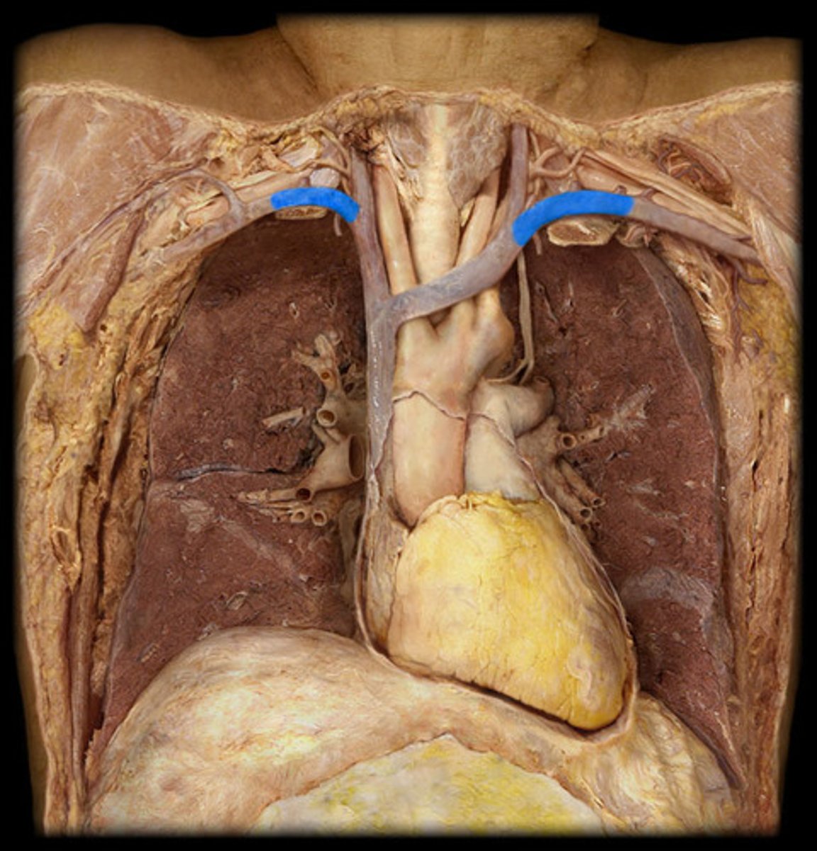
Subclavian vein
Area drained: Upper limb and neck
Drains into: Brachiocephalic vein
Relationship: Unites with the internal jugular vein to form the brachiocephalic vein
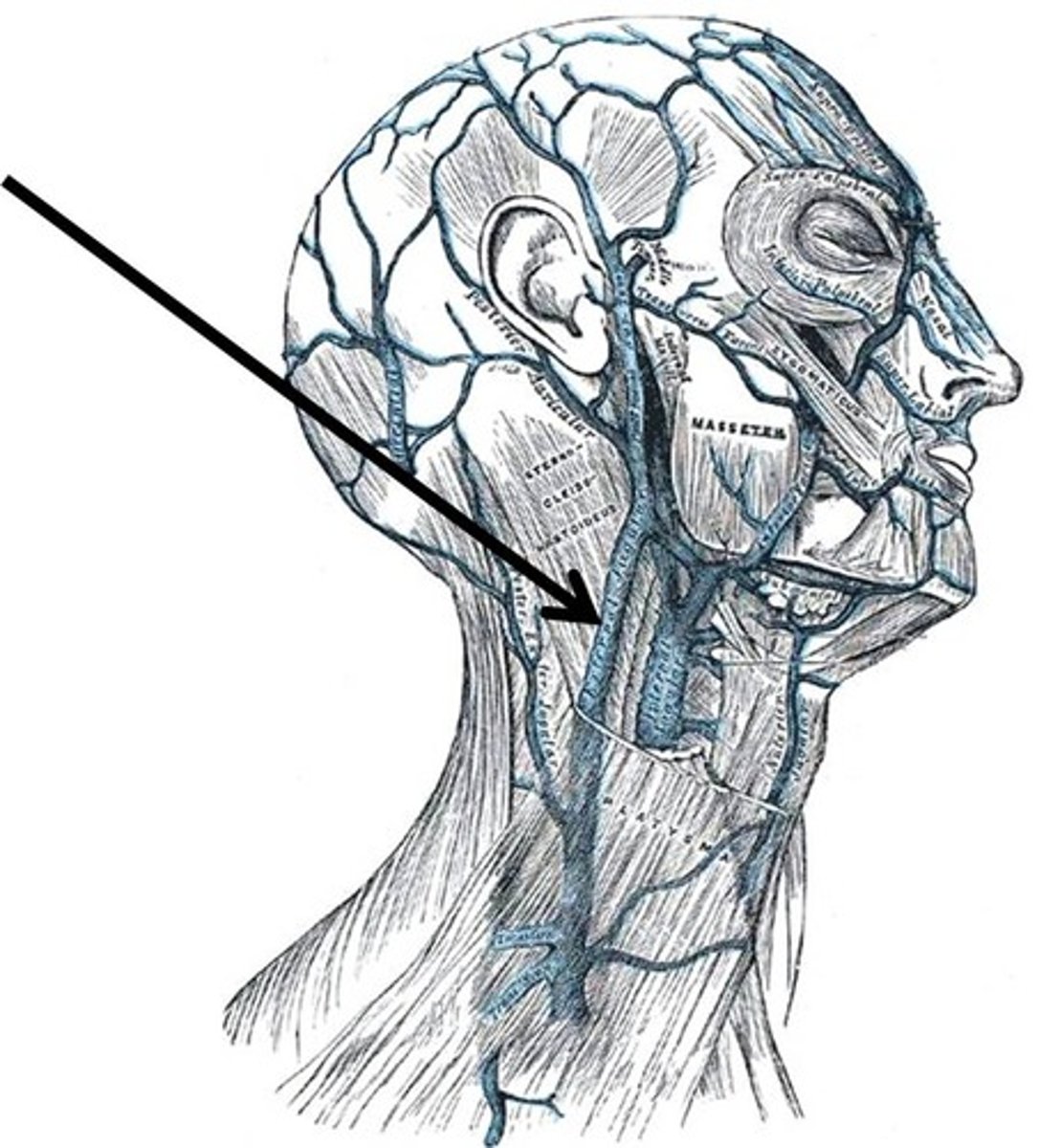
External jugular vein
Area drained: Scalp and face
Drains into: Subclavian vein
Relationship: Superficial to the sternocleidomastoid muscle
Vertebral vein
Area drained: Neck
Drains into: Subclavian vein
Relationship: Passes through the transverse foramen of the cervical vertebrae
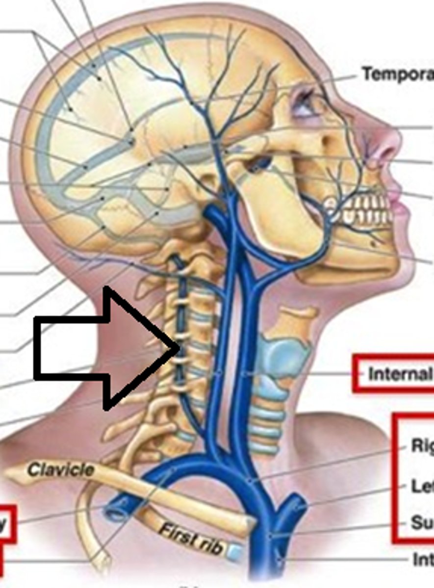
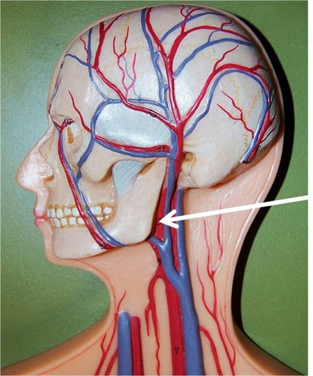
Internal jugular vein
Area drained: Brain, face, and neck
Drains into: Brachiocephalic vein
Relationship: Unites with the subclavian vein to form the brachiocephalic vein. Deep to the sternocleidomastoid muscle
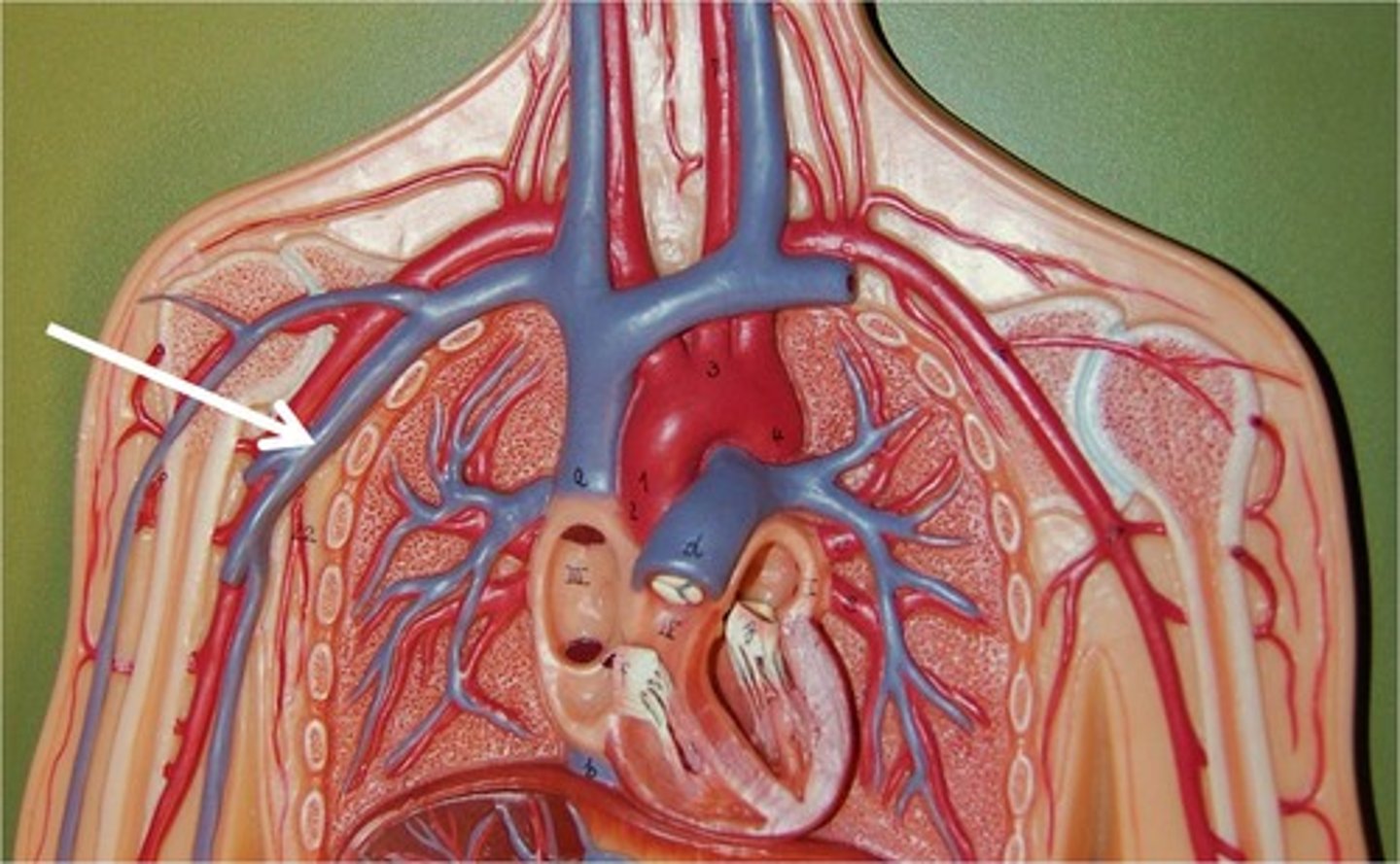
Axillary vein
Area drained: Pectoral, shoulder, and scapular regions
Drains into: Subclavian vein
Relationship: Continues as the subclavian vein at the lateral border of the first rib
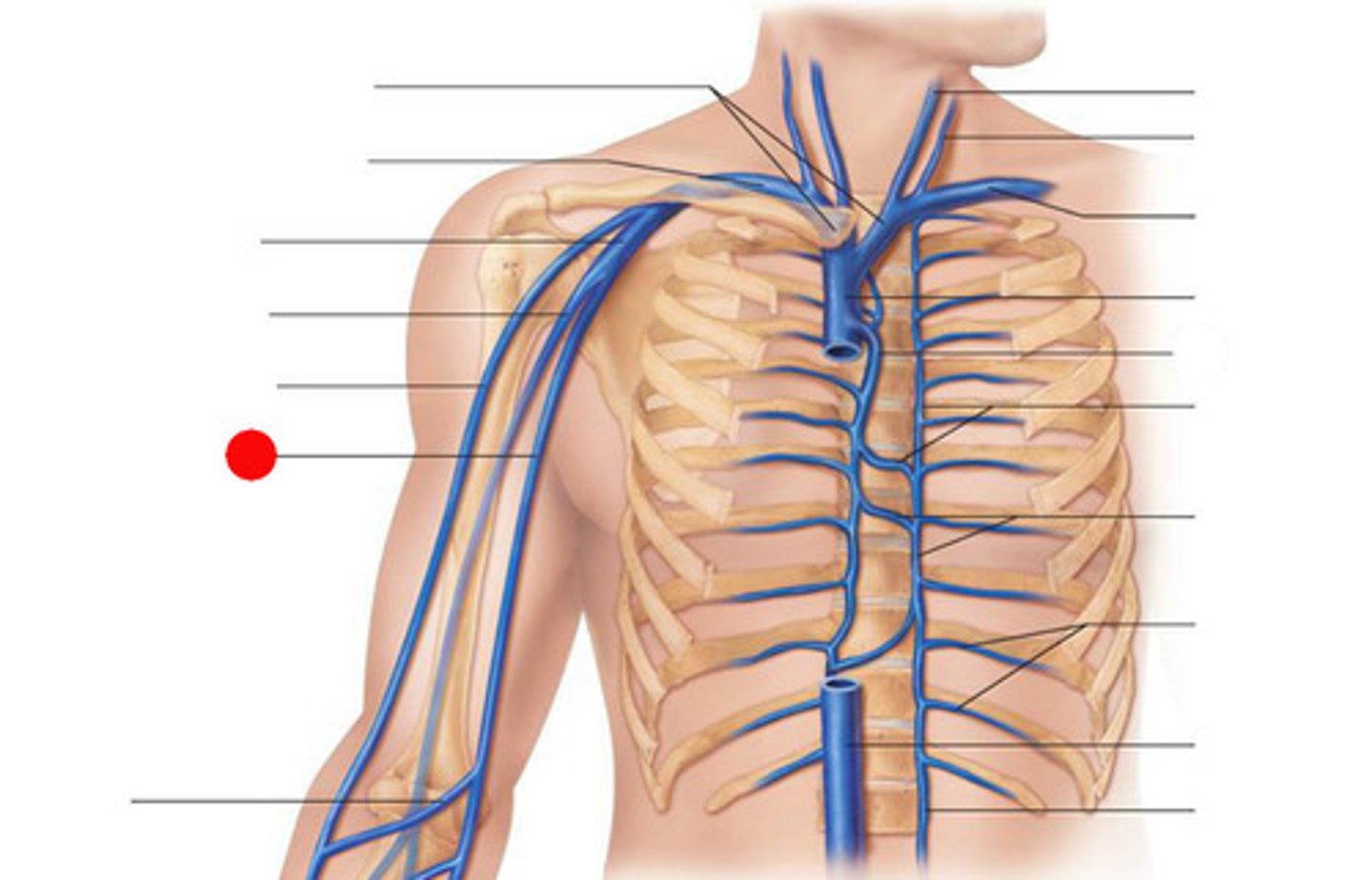
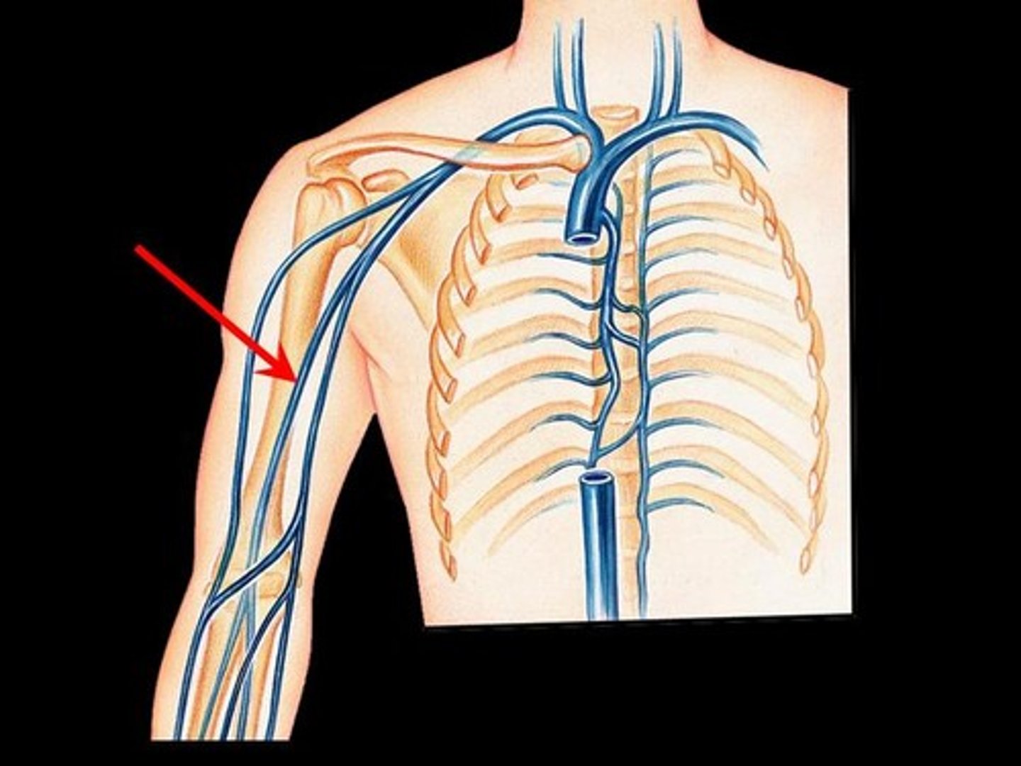
Basilic vein
Area drained: Superficial structures of the medial and dorsal hand, forearm, and arm
Drains into: Axillary vein
Relationship: Ascends on the medial aspect of the arm superficial to the muscles
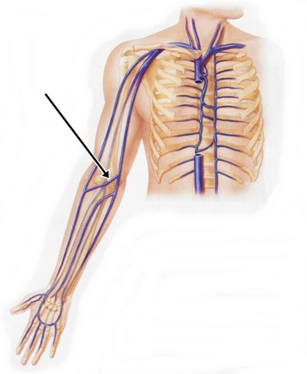
Median cubital vein
Area drained: Superficial forearm
Drains into: Basilic vein
Relationship: Connects the basilic and cephalic veins together across the cubital fossa. Use frequently for drawing blood
Cephalic vein
Area drained: Superficial structures of the lateral and dorsal hand, forearm, and arm
Drains into: Axillary vein
Relationship: Ascends on the lateral aspect of the arm superficial to the muscles. Passes between the pectoralis major and deltoid muscles
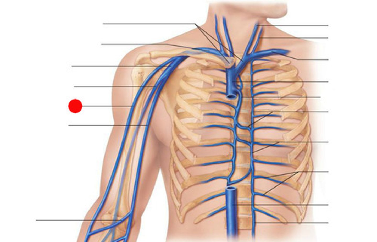
Brachial vein
Area drained: Arm muscles and elbow joint
Drains into: Axillary vein
Relationship: Located deep to the muscles of the arm
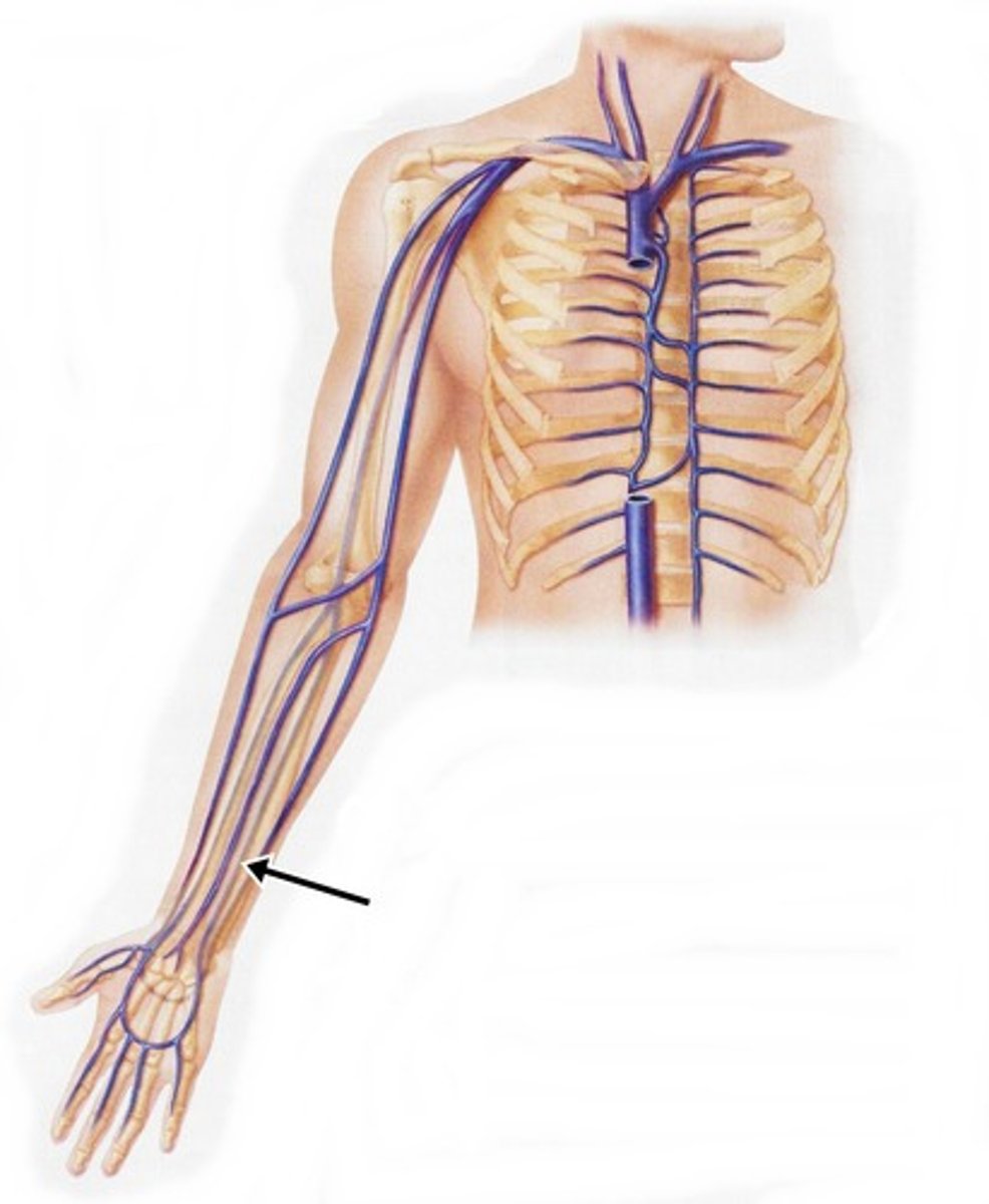
Radial vein
Area drained: Lateral aspect of the forearm
Drains into: Brachial vein
Relationship: Located deep to the muscles on the lateral aspect of the forearm
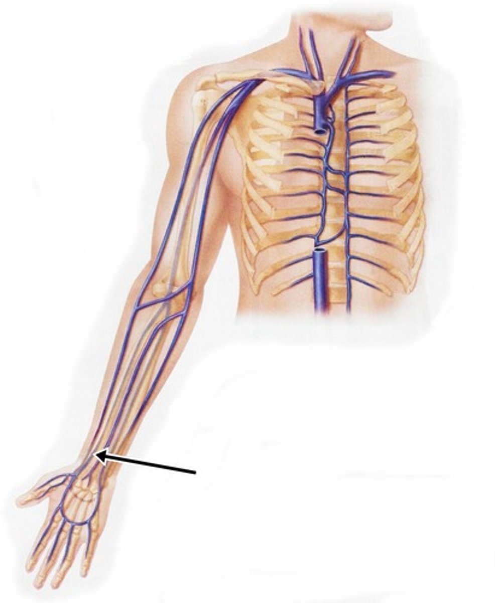
Ulnar vein
Area drained: Medial aspect of the forearm
Drains into: Brachial vein
Relationship: Located deep to the muscles on the medial aspect of the forearm
Inferior vena cava
Area drained: Everything inferior to the diaphragm, except the posterior abdominal wall
Drains into: Right atrium
Relationship: Large vein of the body
Hepatic vein
Area drained: Liver
Drains into: Inferior vena cava
Relationship: Transports deoxygenated blood back to the inferior vena cava after being detoxified by the liver
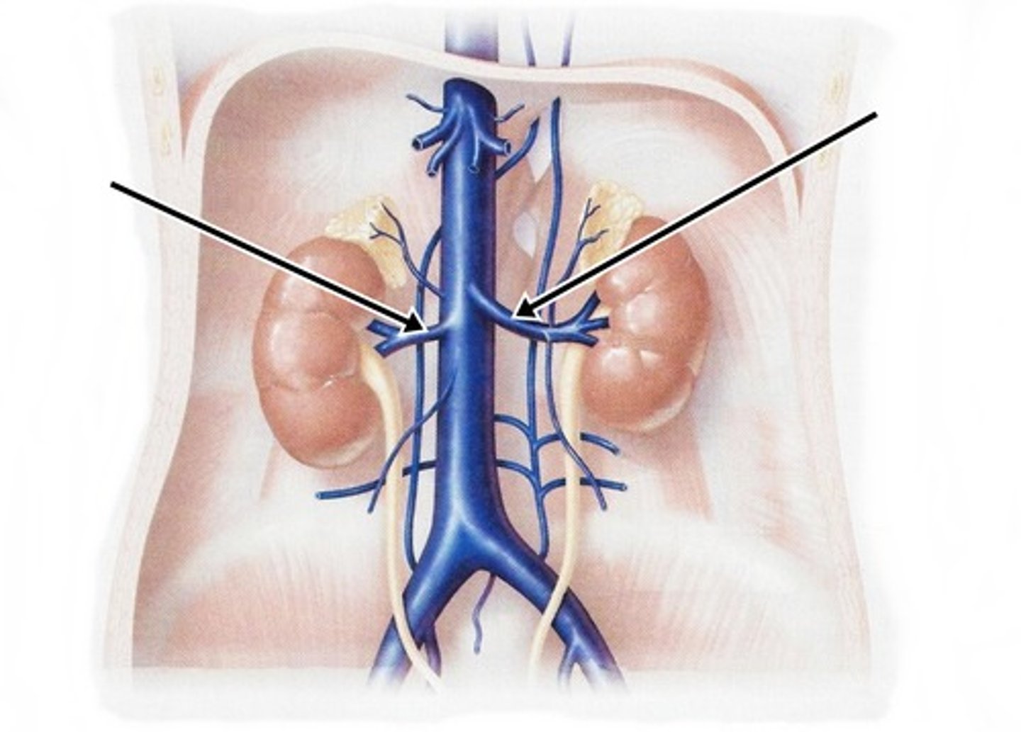
Renal vein
Area drained: Kidneys, ureters, adrenal glands, and testis/ovaries
Drains into: Inferior vena cava
Relationship: Passes medially from the kidney to enter the inferior vena cava. Left renal vein is longer than the right
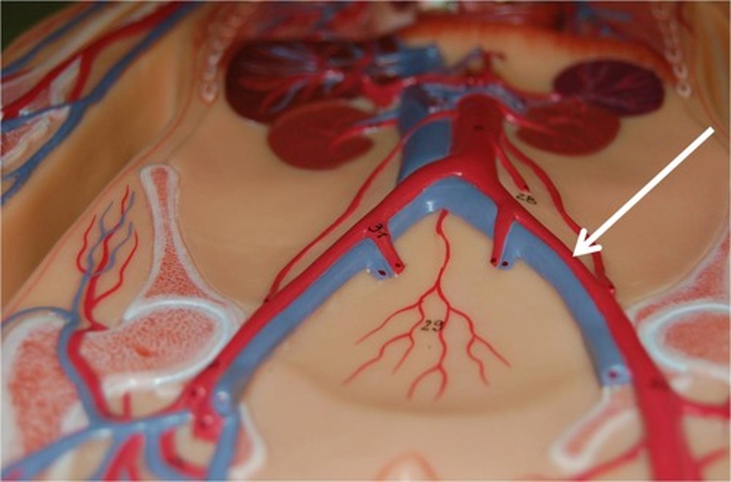
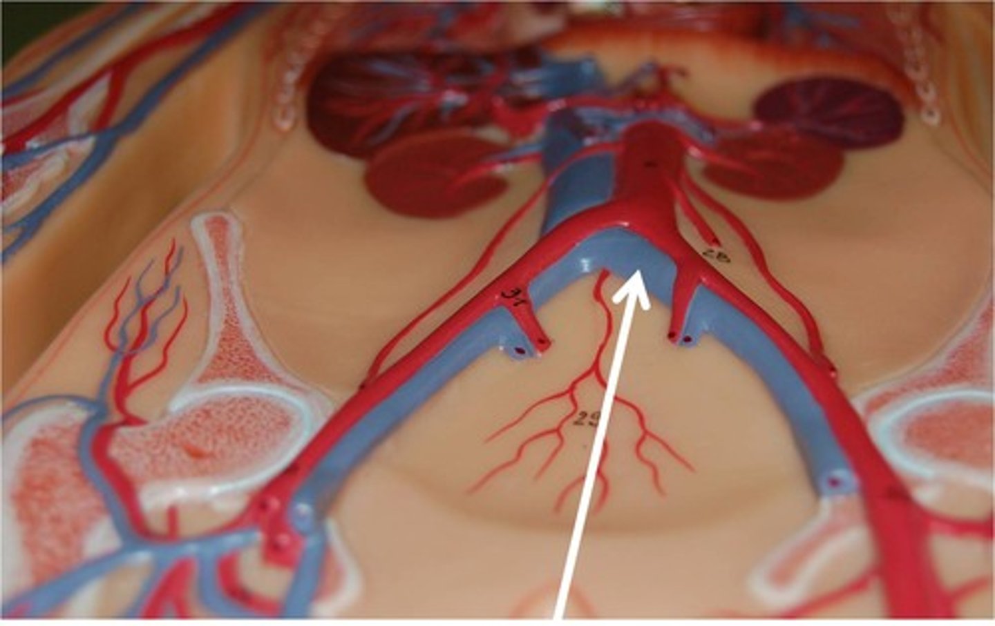
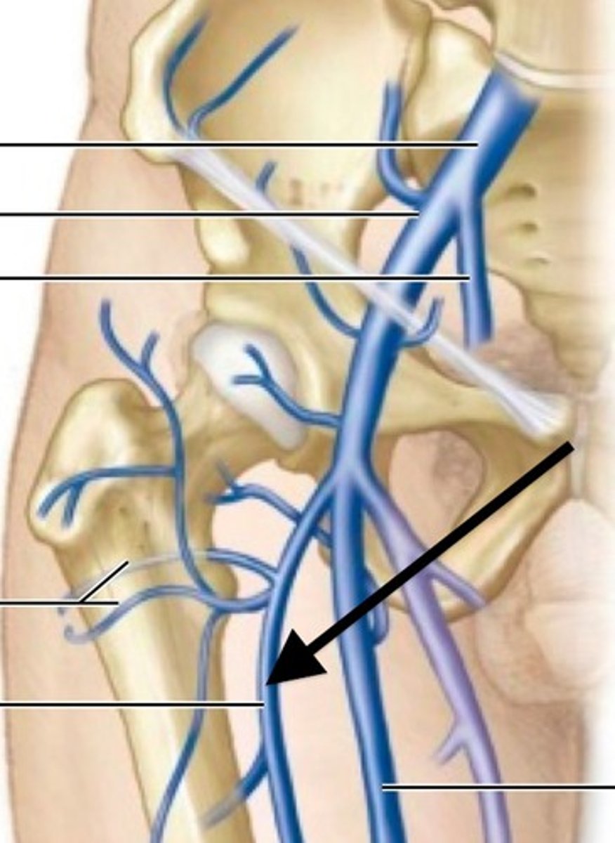
Common iliac vein
Area drained: Internal and external iliac veins
Drains into: Inferior vena cava
Relationship: At the bifurcation of the inferior vena cava
Internal iliac vein
Area drained: Pelvis, perineum, gluteal region, and posterior abdominal wall
Drains into: Common iliac vein
Relationship: Ascends along the lateral pelvis
External iliac vein
Area drained: Lower limb and anterior abdominal wall
Drains into: Common iliac vein
Relationship: Begins above the inguinal ligament
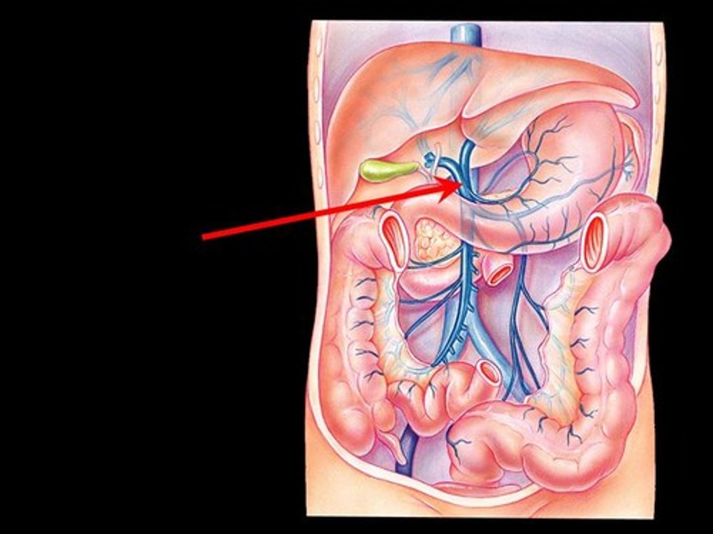
Hepatic portal vein
Area drained: Digestive tract, esophagus, spleen, pancreas, and gallbladder
Drains into: Liver
Relationship: Formed by the superior mesenteric, inferior mesenteric, and splenic veins. Passes behind the pancreas to enter the inferior side of the liver
Femoral vein
Area drained: Thigh muscles, external genitalia, and hip joint
Drains into: External iliac vein
Relationship: Most medial structure in the inguinal region passing through the inguinal ligament
Great saphenous vein
Area drained: Dorsum of foot and medial leg and thigh
Drains into: Femoral vein
Relationship: Longest vein in the body. Ascends on the superficial and medial aspect of the leg and thigh
Dorsal venous arch vein
Area drained: Toes and dorsum of foot
Drains into: Great saphenous vein
Relationship: Also known as the dorsalis pedis vein
Politeal vein
Area drained: Knee joint
Drains into: Femoral vein
Relationship: Lies in the popliteal fossa. Formed by the union of posterior and anterior tibial veins and passes through adductor magnus muscle to become femoral vein
Posterior tibial vein
Area drained: Posterior leg muscles
Drains into: Popliteal vein
Relationship: Passes down the entire length of the posterior leg
Fibular vein
Area drained: Lateral compartment of the leg and the flexor hallucis longus muscle
Drains into: Posterior tibial vein
Relationship: Stays on the posterior aspect of the leg
Anterior tibial vein
Area drained: Anterior compartment of leg
Drains into: Popliteal vein
Relationship: Passes between the tibia and fibula to the anterior side of the leg