Circulatory system
1/26
Earn XP
Description and Tags
Too much stuff
Name | Mastery | Learn | Test | Matching | Spaced | Call with Kai |
|---|
No analytics yet
Send a link to your students to track their progress
27 Terms
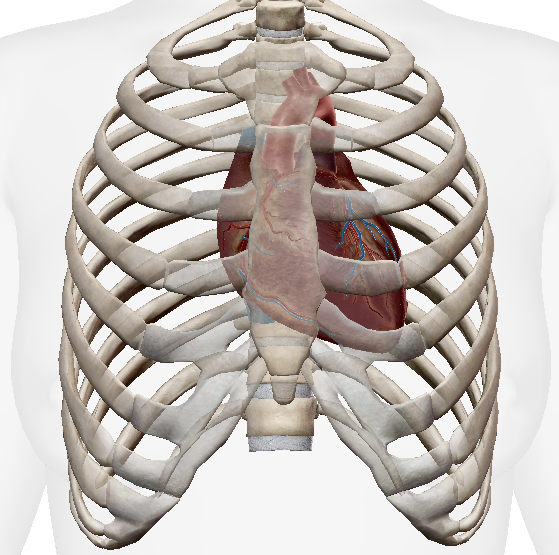
Where is the location of the heart?
Between R02 and R05, posterior to sternum.
Inside the thoraxic cage and cavity, surrounded by a double-membrane pericardium (1 fibrous, 2 serous layers)
Pathway of coronary arteries?
Aorta → Various places of the heart → Right Atrium
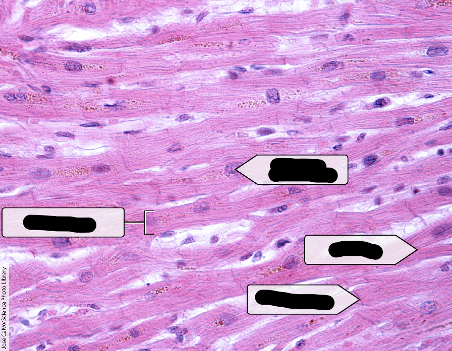
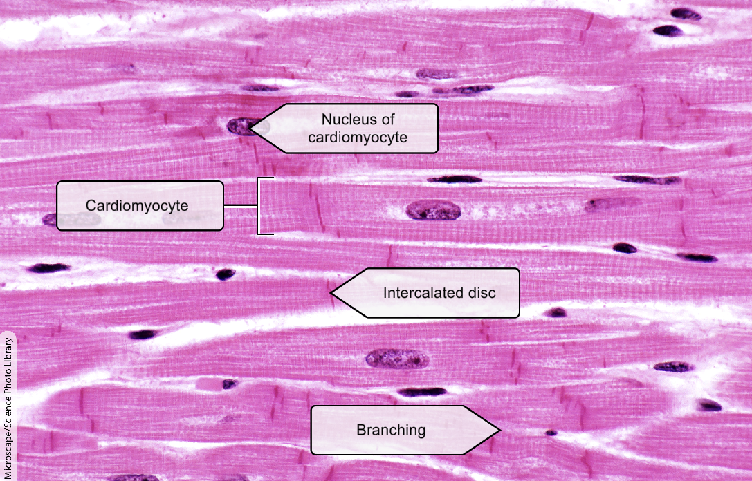
What is this tissue?
Cardiomyocyte
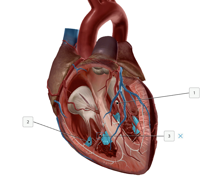
Name these structures
Chordae Tendineae
Papillary muscles
Moderator Band
What are these?
SA node
Internodal Bundle
AV node
Bundle of His/AV bundle
Bundle branch + Purkinje fiber
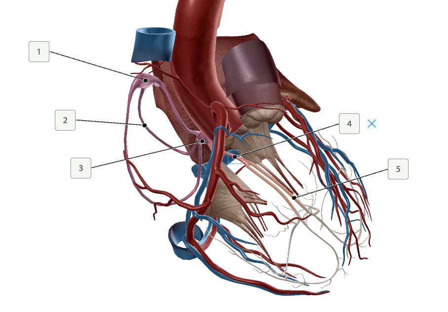
Name these structures
Right coronary artery
Left coronary artery
Anterior interventricular branch
Circumflex branch
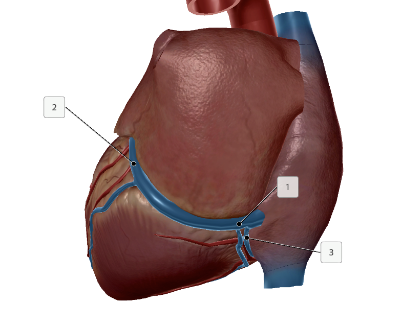
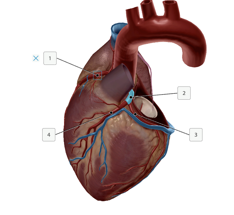
Name these structures
Coronary vein
Coronary sinus
Great cardiac vein
Small cardiac vein
Define Ventricular Septal Defect (VSD)
Definition: a gap in the septum, caused by errors in development preventing the Muscular Ridge (bottom) and Membraneous Region (top) from meeting.
What are some common diseases VSD is associated with?
Other cardiac disease
Down syndrome
Fetal alcohol syndrome
Symptoms of VSD?
Uneven pressure of blood (LV is higher) causes mixing of blood, into the right ventricle (instead of circulating body).
Higher oxygen saturation in RV, Pul. arteries.
Heard as a heart murmur (Holosystolic Murmur)
Smaller VSD may be asymptomatic
Pulmonary Hypertension
In servere cases, Blood flow of VSD reveses and supplies aorta with deox blood.
Cyanosis - blue lips and fingers
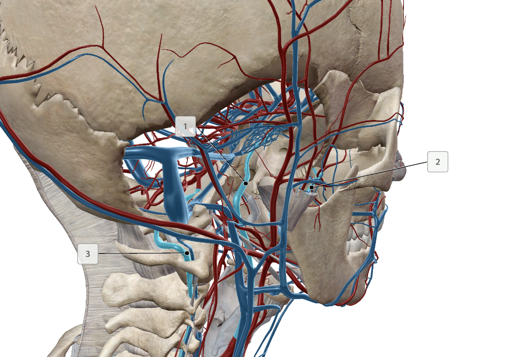
What are these
Deep Arteries of the Head/Neck
Internal Carotid Artery
Maxillary Artery
Vetebral Artery
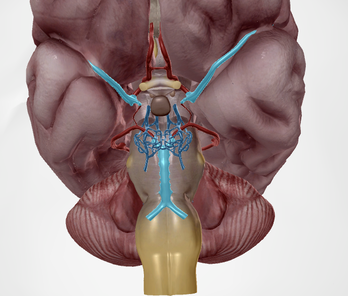
What are these (blood vessels and highlighted part)?
Vessel: Circle of Willis
Top: Middle Cereberal Artery
Bottom: Basilar Arteries
Note: the Circle of Willis of most people is incomplete, and this knowledge is important for medical imaging.
What are these arteries?
Subclavian (Left is arched, R is not)
Auxillary Artery
Brachial Artery
Deep Brachial Artery
What are these?
Vessels that supply the forearm
Ulnar
Radial
Deep Palmar Arch
Superficial Palmar Arch
What are these?
Pelvic arteries
Common Iliac
Internal Iliac
External Iliac
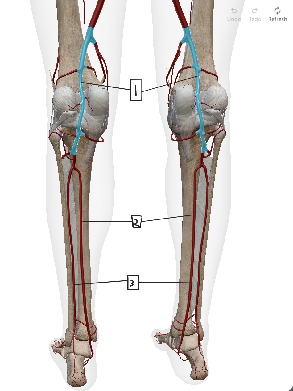
What are these?
Leg arteries
Popliteal
Posterior Tibial
Fibular
Note: Anterior tibial also exists. Not shown here
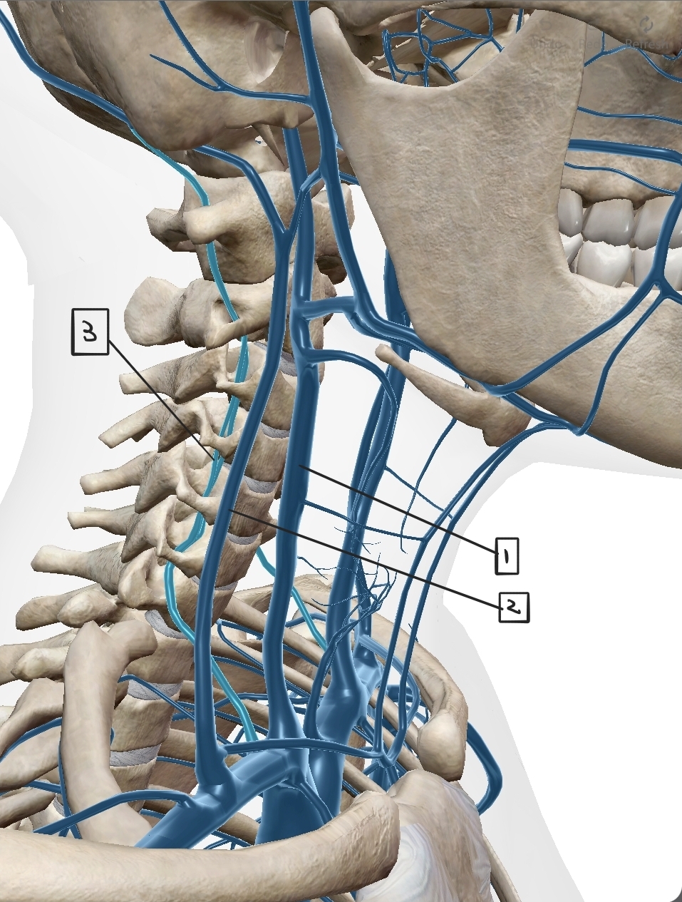
What are these?
Head-draining veins
Internal Juglar
External Juglar
Vertebral.
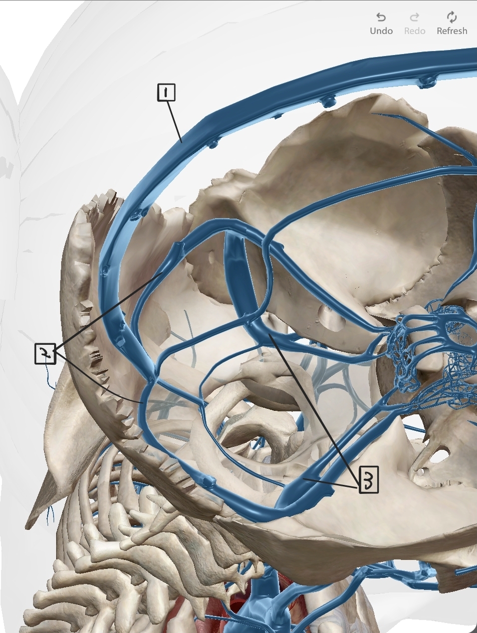
What are these?
Sinus of the head
Superficial sinus
Transverse Sinus
Sigmoid Sinus
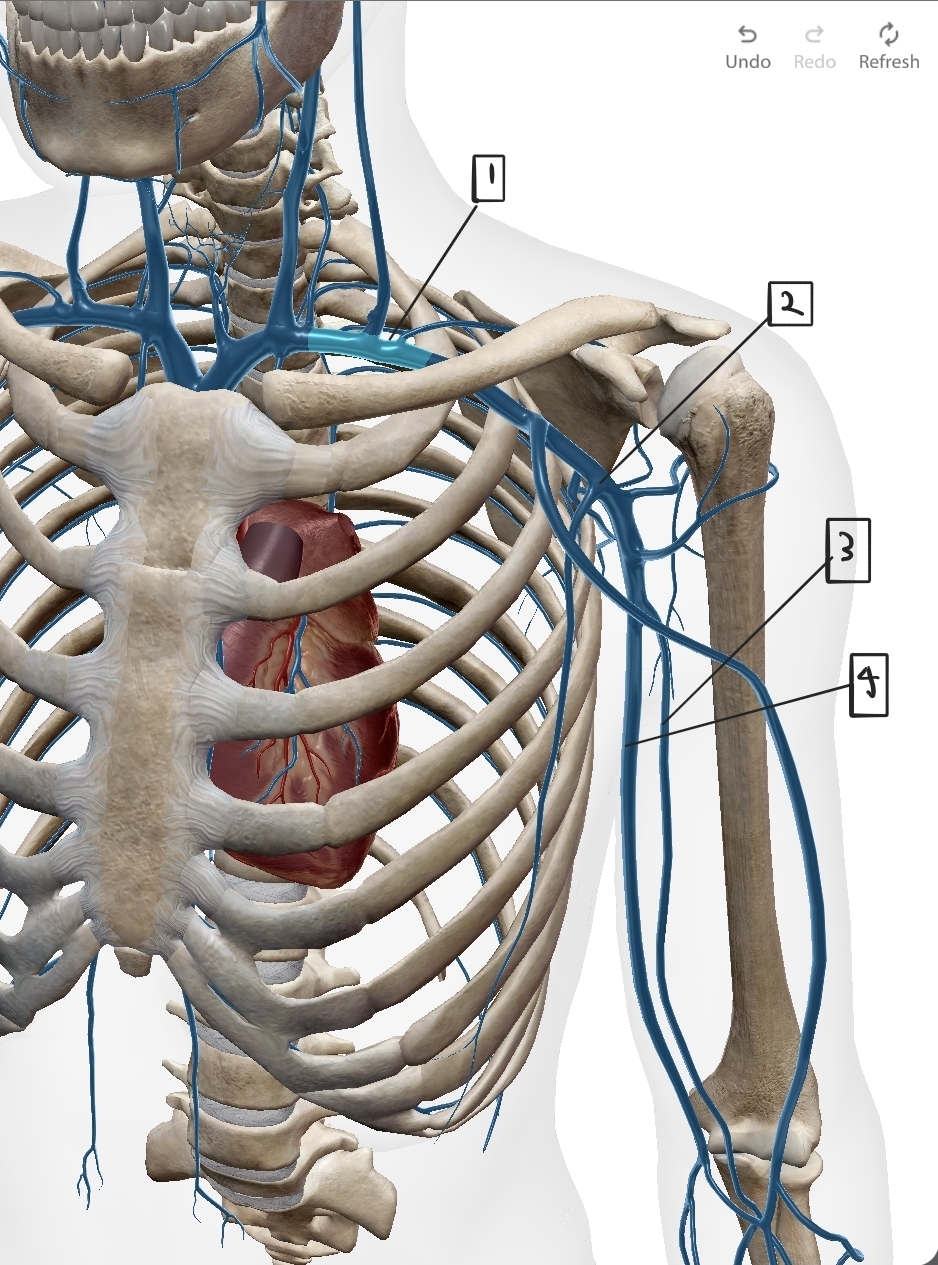
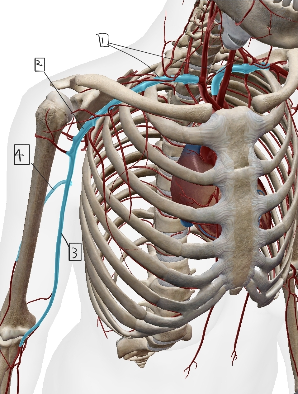
What are these?
Upper arm Veins
Subclavian
Auxillary
Brachial
Basilic
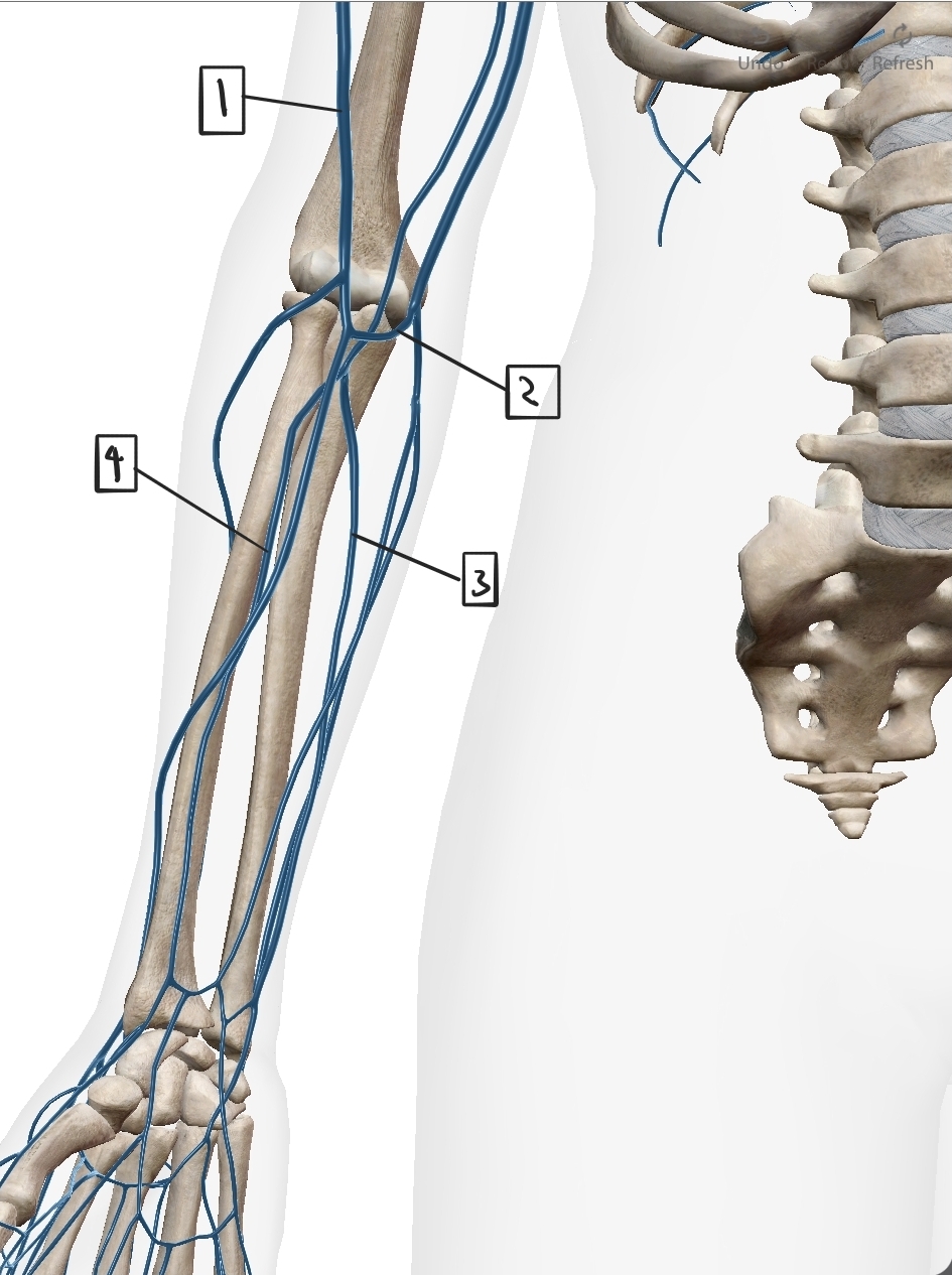
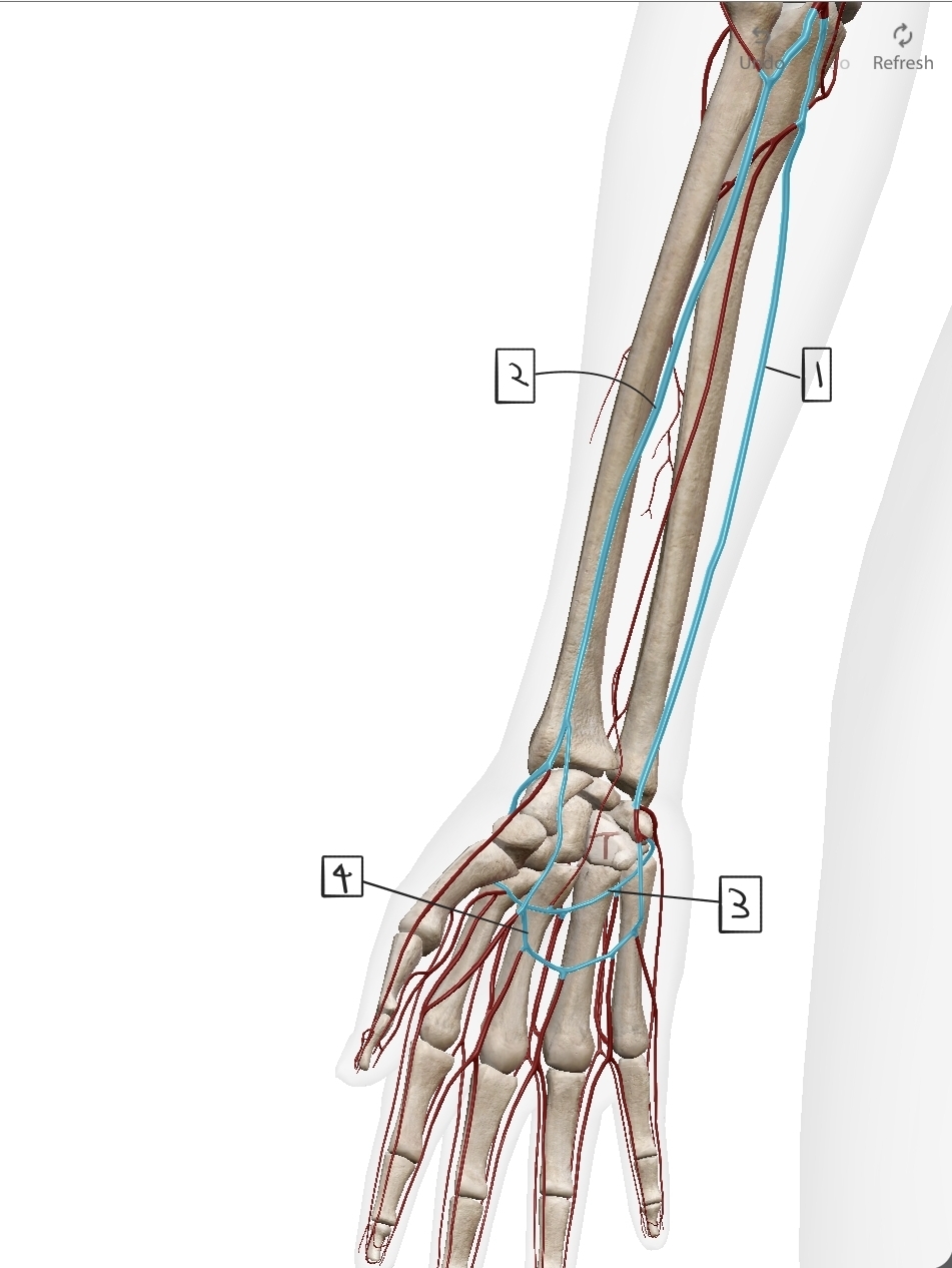
What are these?
Veins of the Forearm
Cephalic
Median Cubital
Ulnar
Radial
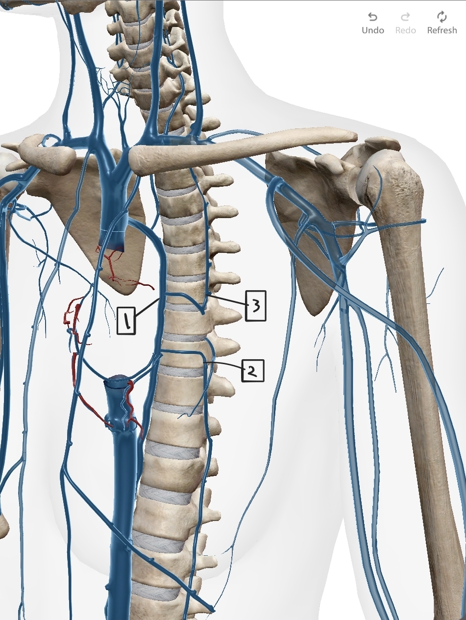
What are these?
The Azygos System (drain thorax wall)
Azygos (Into superior Vena Cava)
Hemiazygos
Accessory Azygos
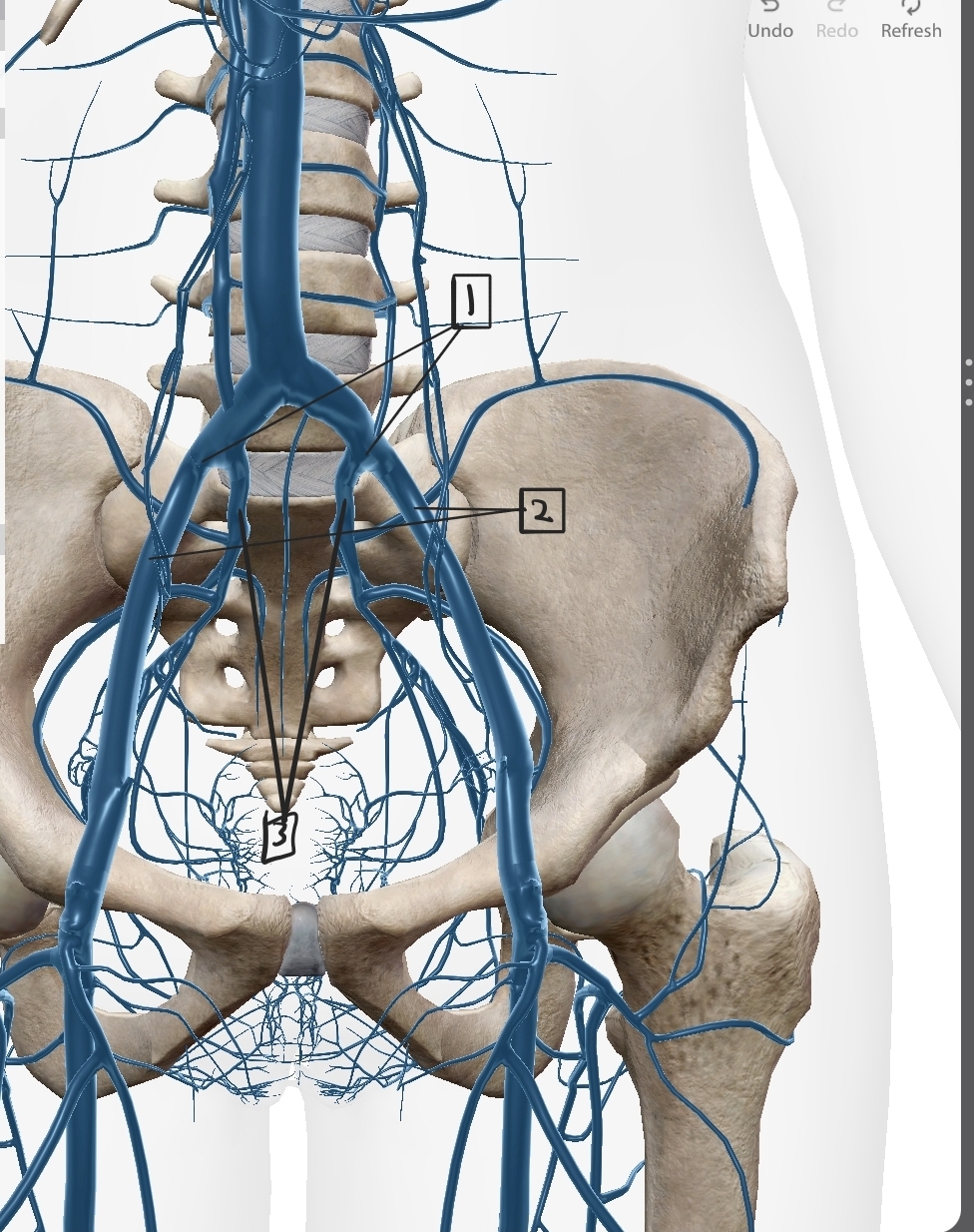
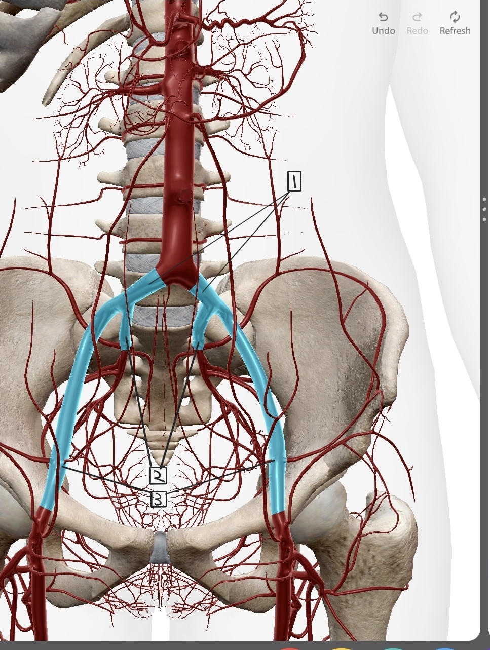
Iliac veins
Common
External
Internal
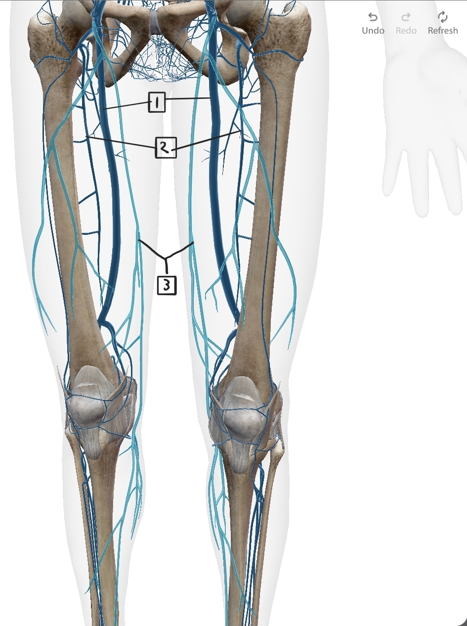
Upper leg veins
Commen Femoral
Deep Femoral
Great Saphenous
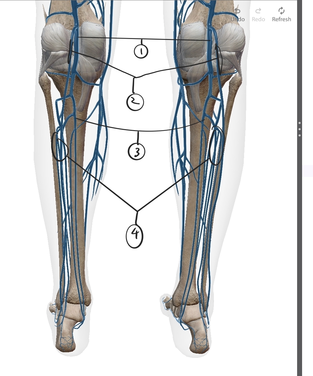
Lower Leg Veins
Popliteal
Small Saphenous
Posterior Tibial
Fibular
Note: (not listed) Anterior Tibial
Define athrosclerosis
Def: Buildup of plaque under arterial endothelium due to LDL buildup.
Buildup causes obstruction in artery.
Buildup is capped with collagen, and narrows lumen
Symptoms of athrosclerosis
Fatty streats in arteries, by invasion of LDL causing inflammation
Invade →Immune cell enter →form core of plaque
Thrombosis - when a fatty streak bursts and mixes with blood, completely blocking blood flow.
Heal-flow cycle can eventually cause it to break off, and clot elsewhere. Can cause strokes, etc if caught in an important vessel.
Strokes
Ischemic: a piece of athrosclerosis breaks off, blocking bloodflow to part of brain.
Hemorrhagic: Blood vessel burts, causing blood to flow into brain and damage it.