Lower Extremity Venous Anatomy and Evaluation
1/27
There's no tags or description
Looks like no tags are added yet.
Name | Mastery | Learn | Test | Matching | Spaced | Call with Kai |
|---|
No analytics yet
Send a link to your students to track their progress
28 Terms
Which veins in the lower extremity contain ZERO valves?
Common iliac veins
Internal iliac veins
IVC
What are metatarsal veins?
Vessels that drain blood from feet before terminating at deep venous arches
What are the anterior tibial veins (ATV)
Paired vessels that originate from plantar arches and drain blood from anterior calf
What are the posterior tibial veins (PTVs)?
Paired vessels that originate at plantar arches and drain blood from posterior calf
What are the peroneal veins?
Paired vessels that drain blood from lateral calf
What is the popliteal vein?
Vessel that originates from anterior tibial veins and tibioperoneal trunk that courses posterior to popliteal artery
What is the femoral vein?
Continuation of popliteal vein that courses posterior to femoral artery
What is the deep or profunda femoral vein?
Vessel that joins femoral vein and drains blood from thigh muscles
What is the common femoral vein (CFV)?
Vessel that originates from femoral vein and deep or profunda femoral vein
What is considered to be the longest vein in the body?
GSV
What is the greater saphenous vein (GSV)?
Superficial vessel that originates at dorsal venous arch and terminates at saphenofemoral junction in groin
What is the anterior accessory saphenous vein (AASV)?
Superficial vessel that joins GSV at groin
What is the small saphenous vein (SSV)?
Superficial vessel that originates at dorsal venous arch and joins popliteal vein in distal thigh
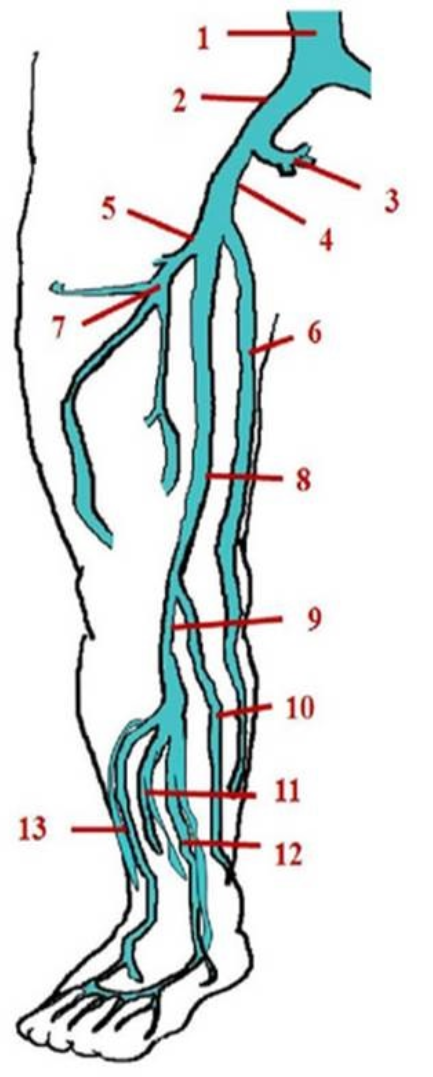
Identify this image.
IVC
Common iliac vein
Internal iliac vein
External iliac vein
Common femoral vein
GSV
Deep femoral vein
Femoral vein
Popliteal vein
SSV
Peroneal veins
Posterior tibial veins
Anterior tibial veins
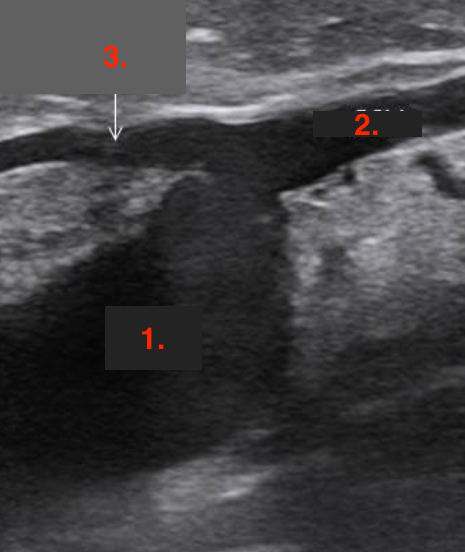
Identify this image.
Saphenofemoral junction (SFJ)
CFV
GSV
Superficial epigastric vein
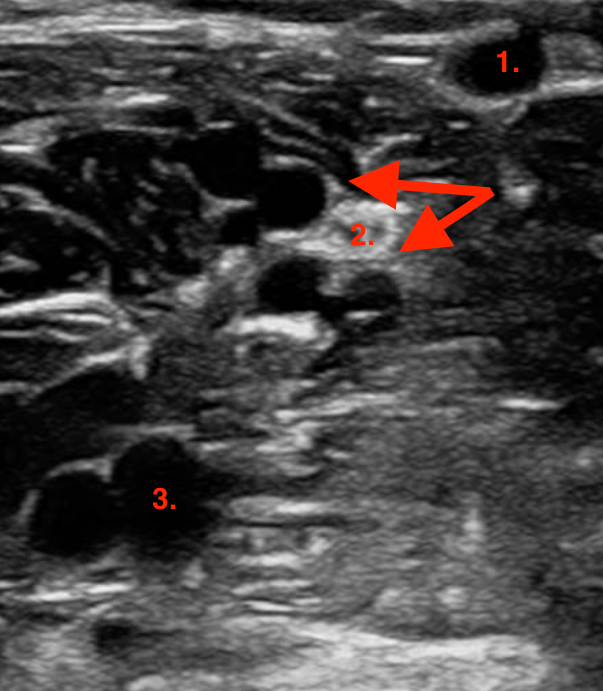
Identify this image.
Calf veins
SSV
Gastrocnemius veins
Popliteal vein
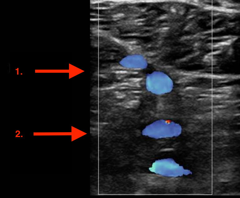
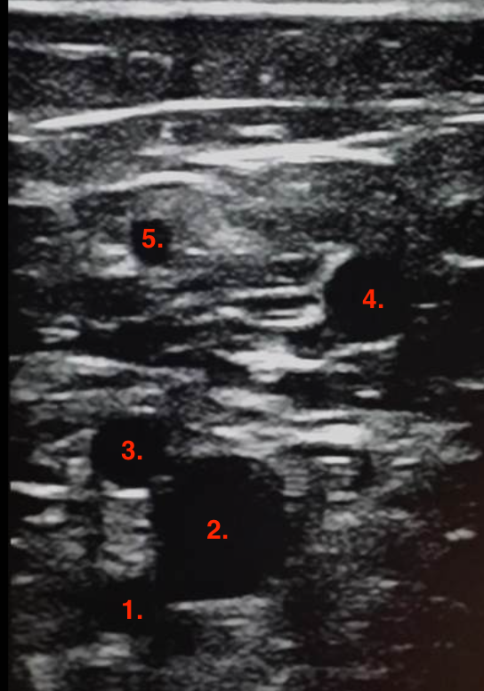
Identify this image.
Calf veins
Posterior tibial veins
Peroneal veins
Identify this image.
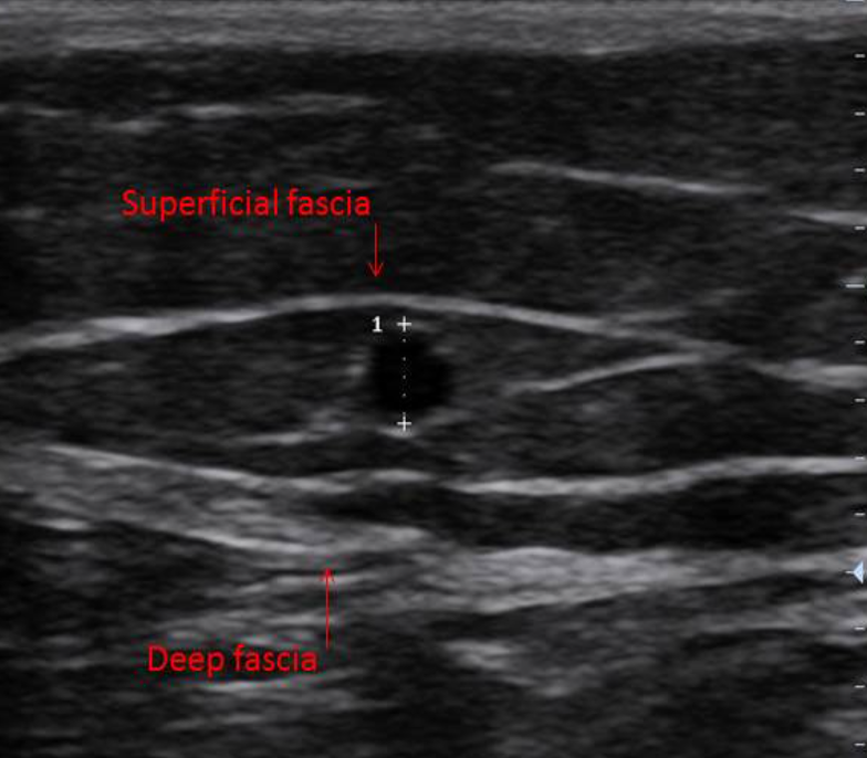
Eye sign used to distinguish saphenous veins from tributaries
Identify this image.
Deep femoral artery
CFV
SFA
GSV
Anterior accessory vein
What are perforator veins?
Vessels that connect superficial and deep venous systems
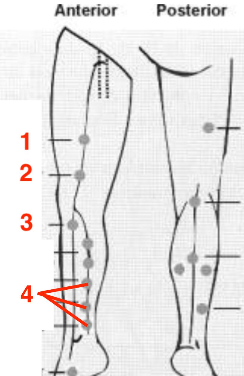
Identify this image.
Dodd perforators that connect GSV to FV
Hunterian perforators that connect GSV to FV
Boyd or paratibial perforators that connect GSV to posterior tibial veins
Cockett’s or posterior tibial perforators that form around ankle
What are communicating veins?
Vessels that connect great and small saphenous veins
What is the posterior arch vein?
Vessel that connects GSV to posterior tibial or Cockett’s perforators
What is the vein of Giacomini?
Vessel that originates at saphenopopliteal junction and becomes posterior circumflex vein
What are sural veins?
Vessels that serve as blood reservoirs and terminate at popliteal vein
What is the soleal plexus or sinus?
Vessels that serve as blood reservoirs that terminate at posterior tibial or peroneal veins
How is a lower extremity venous exam performed?
Evaluate all vessels in reverse Trendelenburg or semi-erect position
Patient should externally rotate leg with knee slightly bent
What is the normal sonographic appearance of an lower extremity venous exam?
Deep veins of thigh demonstrate spontaneous and phasic flow
Calf veins DO NOT demonstrate spontaneous flow
NO CARDIAC PULSATILITY