BSCI 1511 - Neurons, synapses and signaling
1/47
There's no tags or description
Looks like no tags are added yet.
Name | Mastery | Learn | Test | Matching | Spaced | Call with Kai |
|---|
No analytics yet
Send a link to your students to track their progress
48 Terms
Sensory input
Receives information – sensing the external environment (e.g., light) or internal conditions (e.g., blood pressure)
PNS
detection of stimuli by sensory neurons.
Integration
Processes information – processing sensory input in context
CNS
processing of information by interneurons in the CNS.
Motor input
Transmits information directing a physiological or behavioral response – e.g, activation of muscle, gland, etc
response via motor neurons to muscles or glands.
Learning & Memory
Provides a mechanism for using experience to modify the response
CNS
How do neurons transmit information?
Receives information, transmits it along an axon, and sends it to other cells via synapses.
Uses electrical pulses for long‑distance transmission.
Uses chemical signals (neurotransmitters) for short‑distance communication at synapses.
Interpretation occurs in the brain or ganglia.
Name the key structures of a neuron and their functions.
Dendrites – receive signals.
Cell body (soma) – integrates incoming signals.
Axon – conducts action potentials away from the cell body.
Axon terminals (synaptic terminals) – release neurotransmitters to communicate with other cells.
Describe the three functional types of neurons.
Sensory neurons: Transmit information regarding internal and external stimuli.
◦ Interneurons: Integrate information and form circuits within the brain or ganglia.
◦ Motor neurons: Trigger muscle or gland activity
What is the difference between the CNS and the PNS?
Central Nervous System (CNS) - The brain and the spinal cord, neurons carry out integration
Peripheral Nervous System (PNS) - All nerves and ganglia located outside the brain and spinal cord, Neurons that carry information into and out of the CNS
CNS (central nervous system) = brain and spinal cord (integration).
PNS (peripheral nervous system) = nerves carrying information into and out of the CNS.
What are the three functional types of neurons?
Sensory neurons – transmit information about external/internal stimuli.
Interneurons – integrate information (most neurons in the brain).
Motor neurons – trigger responses (muscle or gland activity).
Describe the step-by-step process of how a neuron receives, conducts, and transmits information.
Neuron receives info from the external environment or other neurons. The input is collected by the cell body and dendrites
The neuron processes this incoming sensory input in context. This integration determines whether the signal is strong enough to be passed along. All information travels toward the axon hillock, which acts as the trigger zone for the next step
If the stimulus is strong enough to reach a specific threshold (typically -55mV in mammals), the neuron generates an action potential
The action potential travels like a pulse of electrical current down the neuron's single, long axon
When the signal reaches the synaptic terminals, it must cross a junction called a synapse to reach the next cell.
• Chemical Signal: The electrical impulse triggers the release of chemical messengers called neurotransmitters.
• Response: These chemicals cross the gap and bind to receptors on the postsynaptic cell (which could be another neuron, a muscle, or a gland), directing a specific physiological or behavioral response
Nerves
Axons of neurons bundled together
What are glial cells? and what are there functions?
The neuron’s supporting cells
Nourish neurons
Insulate axons
Immune protection
Regulate the extracellular fluid surrounding neurons
List the major types of glial cells and their specific roles
Ependymal cells: Line brain ventricles.
◦ Astrocytes: Nourish neurons, regulate extracellular fluid, and facilitate information transfer.
◦ Oligodendrocytes: Myelinate axons in the CNS.
◦ Schwann cells: Myelinate axons in the PNS.
◦ Microglia: Serve as immune cells in the CNS
What is membrane potential, and what is the specific value for a resting neuron?
Membrane potential is the charge difference (voltage) across the plasma membrane caused by the attraction of opposite charges. For a resting neuron, this is between -60 and -80 mV
What is the resting potential of a neuron?
The membrane potential of a neuron not sending a signal.
Typically between ‑60 mV and ‑80 mV.
Inside of the cell is negative relative to the outside.
Maintained by ion concentration gradients and selective permeability.
How are ions distributed across a resting neuron membrane?
K⁺ – higher inside, lower outside.
Na⁺ – higher outside, lower inside.
Cl⁻ – higher outside, lower inside.
Large anions (proteins) trapped inside.
What does resting potential result from?
ATP-dependent sodium-potassium ion pumps create concentration gradients across the plasma membrane: more K+ inside, more Na+ outside 2. The plasma membrane, because it has more leak channels for K+ than for Na+ , is more permeable to K+ than Na+A
Why does K⁺ leave the cell despite the negative charge inside?
K⁺ flows out down its chemical concentration gradient (higher inside). This stops when the electrical gradient pulls it back in at equilibrium.
What maintains the Na⁺ and K⁺ concentration gradients?
The Na⁺/K⁺ ATPase pump pumps 3 Na⁺ out for every 2 K⁺ in, using ATP.
What two factors establish the resting potential?
Na⁺/K⁺ ATPase pump creates concentration gradients (high K⁺ inside, high Na⁺ outside).
Membrane is more permeable to K⁺ than Na⁺ (more K⁺ leak channels).
Why is the resting potential (‑60 to ‑80 mV) not exactly equal to Eₖ (‑90 mV)?
Because a small number of Na⁺ ions leak into the cell (via Na⁺ leak channels), making the membrane potential slightly less negative than Eₖ.
Equilibrium Potential (E)
The magnitude of a cell’s membrane voltage at equilibrium and is calculated using the Nernst equation
Nernst Equation
Contrast hyperpolarization and depolarization in terms of ion movement.
Hyperpolarization increases membrane potential magnitude (more negative) by opening gated K+ channels. Depolarization decreases membrane potential magnitude (more positive) by opening gated Na+ channels
What happens when gated K⁺ channels open? What happens when gated Na⁺ channels open?
Gated K⁺ channels open → hyperpolarization (more negative).
Gated Na⁺ channels open → depolarization (less negative / more positive).
Action potential
A consequence of the sequential opening and closing of voltage-gated ion channels for sodium and potassium
What cells produce myelin in the CNS and PNS?
CNS – oligodendrocytes.
PNS – Schwann cells.
Why are action potentials propagated in only one direction along an axon?
Because of the brief refractory period caused by the inactivation gate of voltage‑gated Na⁺ channels.
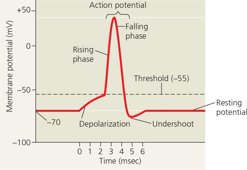
List the four phases of an action potential.
Resting state: Before an action potential begins, the neuron is at its resting membrane potential of approximately –70 millivolts, with voltage‑gated sodium channels closed and voltage‑gated potassium channels closed.
Depolarization: When a stimulus depolarizes the membrane to threshold (about –55 millivolts), voltage‑gated sodium channels open rapidly, allowing sodium ions to rush into the cell, which makes the inside of the membrane positive (up to about +40 millivolts).
Repolarization: As the membrane potential becomes positive, voltage‑gated sodium channels inactivate and close, stopping sodium influx, while voltage‑gated potassium channels open slowly, allowing potassium ions to flow out of the cell, which brings the membrane potential back down toward resting level.
Hyperpolarization (undershoot): The voltage‑gated potassium channels close slowly, so excess potassium ions leave the cell, causing the membrane potential to become temporarily more negative than resting (about –80 to –90 millivolts).
Return to resting state: The voltage‑gated potassium channels finally close, and the sodium‑potassium pump gradually restores the original ion gradients, returning the membrane potential to approximately –70 millivolts.
What are graded potentials?
These are small changes in membrane potential that either depolarize or hyperpolarize the cell. By themselves, they only travel a few millimeters before dying out
What causes an action potential to be generated?
An action potential is generated as Na+ flows inward across the membrane; this influx depolarizes the neighboring region, reaching the threshold and reinitiating the action potential there
How do graded potentials differ from action potentials?
Graded potentials – small changes in membrane potential (depolarization or hyperpolarization); travel a few mm and die out.
Action potentials – rapid, large depolarization followed by repolarization; propagate long distances without losing strength; all‑or‑none.
Continuous conduction
Invertebrate animals: insects etc
• Axons are not insulated
• Voltage-gated channels throughout
• Transfer speed: 5 cm to 30 m per sec
• To increase speed of conduction, axons are made thicker
Saltatory conduction
Vertebrate animals
Schwann cells in the PNS and oligodendrocytes in the CNS insulate the axons using myelin
• Voltage-gated ion channels are only in the gaps between the myelin sheaths, called nodes of Ranvier
• Transfer speed: 100 m/sec or faster
• Faster conduction – fewer ion channels need to be activated and deactivated
Extra note: increasing the diameter of the axon increases speed
Chemical synapses
A junction between two neurons where Chemical neurotransmitters released by the presynaptic neuron are received by the postsynaptic cell
• Most common
Electrical synapses
Special junctions between neurons, where electric currents flow from one neuron to another
Use for rapid behaviors such as heart contractions
Not common, probably wont be on exam
What triggers neurotransmitter release at a chemical synapse?
An action potential depolarizes the presynaptic terminal → opens voltage‑gated Ca²⁺ channels → Ca²⁺ influx → synaptic vesicles fuse → release neurotransmitter.
What are ionotropic receptors?
Ligand‑gated ion channels that open when a neurotransmitter binds, causing a postsynaptic potential.
Excitatory (EPSP) – permeable to Na⁺ and K⁺ → depolarization.
Inhibitory (IPSP) – permeable to K⁺ or Cl⁻ → hyperpolarization.
Ionotropic receptors: Excitatory and Inhibitory
It is a receptor that is a ligand gated ion channel
Binding results in a graded potential called a postsynaptic potential
Excitatory: When the channel is permeable to both K + and Na + , it depolarizes • Excitatory postsynaptic potential (EPSP)
Inhibitory: When the channel is permeable to only K + or only Cl - , it hyperpolarizes • Inhibitory postsynaptic potential (IPSP), inhibits transfer of info
What is an ionotropic receptor?
A ligand‑gated ion channel that opens directly when a neurotransmitter binds, causing rapid (milliseconds) changes in ion flow across the membrane. No G protein or second messenger is involved.
What is a metabotropic receptor?
A G‑protein coupled receptor (GPCR) that, when activated, triggers a G protein and second messenger cascade, leading to slower (seconds) but longer‑lasting effects.
How do metabotropic receptors differ from ionotropic receptors?
Metabotropic receptors are G‑protein coupled receptors (GPCRs) that activate second messenger systems, leading to slower, longer‑lasting effects.
What is summation?
The addition of postsynaptic potentials (EPSPs and IPSPs). Two types:
Temporal summation – rapid, repeated firing from one synapse.
Spatial summation – simultaneous firing from multiple synapses.
Temporal summation, spatial summation, Spatial summation of IPSP and EPSP
Temporal summation: Two rapid An (Excitatory Postsynaptic Potential )EPSPs at the same synapse add up to trigger an action potential
Spatial summation: Two simultaneous EPSPs at different synapses add up to trigger an action potential
Spatial summation of EPSP and IPSP: IPSPs cancel out the reception of an EPSP, can even go below the threshold
How to stop the signaling of neurotransmitters?
Enzymatic hydrolysis of the neurotransmitter
Recapture by the presynaptic neuron for reuse
Simple diffusion: diffuses out of the synapse
Acetylcholine
Muscle stimulation, memory formation, and learning
• At the neuromuscular junction, it binds an ionotropic receptor and induces skeletal muscle contraction; it is degraded by acetylcholinesterase
• In cardiac muscle, it binds a metabotropic receptor and reduces the heart rate
Glutamate
In CNS
Formation of long-term memory
Dopamine and Serotonin
Affect sleep, mood, attention, and learning
Nitric oxide
Relaxes smooth muscle vasodilation