KIN 1Y03 Lecture 31: Skeletal Muscle Physiology pt 1 (sliding muscle contraction)
1/14
There's no tags or description
Looks like no tags are added yet.
Name | Mastery | Learn | Test | Matching | Spaced | Call with Kai |
|---|
No analytics yet
Send a link to your students to track their progress
15 Terms
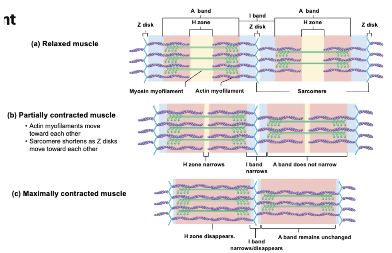
sliding filament mechanism
explains how actin/myosin are moving relative to each other
relaxed muscles in sliding filament mechanism
Relaxed muscles
Myofibrils run entire length of muscle
Only amount of overlap btw myofilament (actin and myosin) changes ⇒ not its length

partially contracted muscle in sliding filament mechanism
Partially contracted muscle
Myosin myofilaments stay in center of sarcomere
Myosin head attach to the actin adn pull towards the M-line ⇒ actin myofilaments move towards each other)
Sarcomere shortens as Z-disks move towards each other
H-zones get small (more overlap)
I-band decreases bc myosin heads are moving towards Z-disks

maximally contracted muscles in sliding filament mechanism
Maximally contracted muscles
Actin myofilaments are pulled so close that theyre overlapping @ center of sarcomere
H-zone = GONE (bc no more, only myosin myofilaments)
I-band = GONE / narrow bc myosin heads acc reach Z-disk
A-bands = unchanged (where after time ⇒ length of sarcomere = A-band)
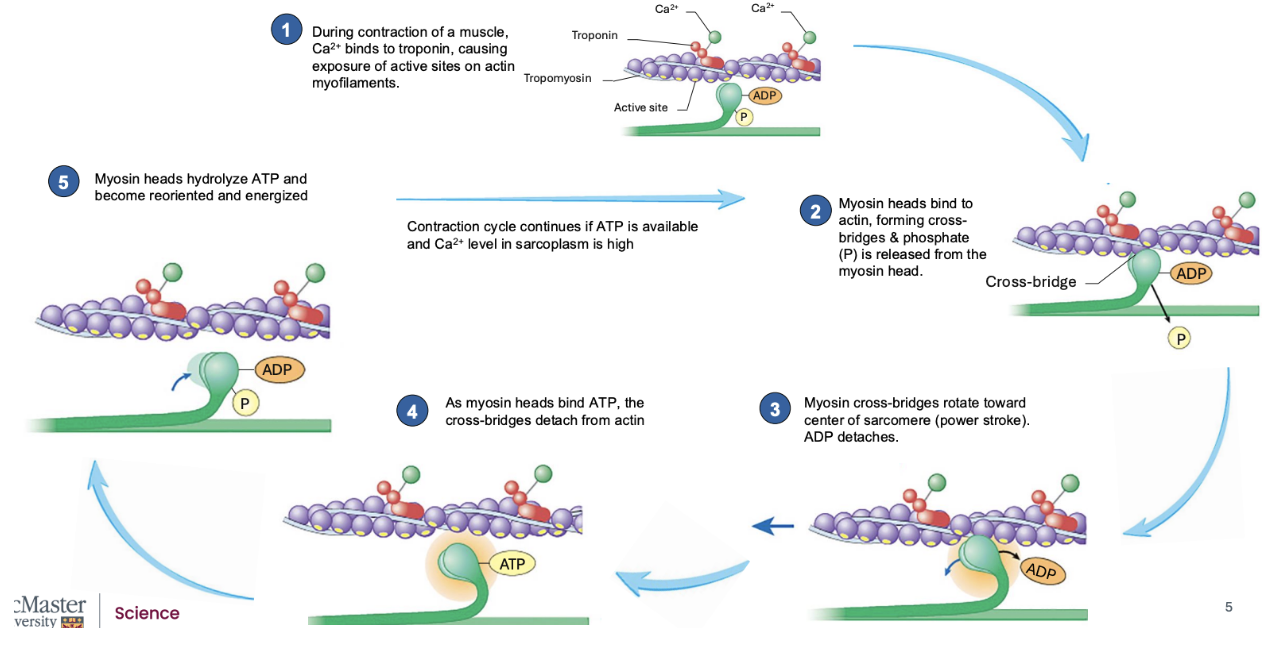
describe the cross-bridge cycle
Ca2+ released from SR ⇒ induces contractions of a muscle
Ca2+ binds to troponin → changes shape of troponin → moves tropomyosin off active site on actin → exposes active site to myosin → myosin attaches onto actin
Troponin is held over active sites on actin myofilaments
When Ca2+ binds to tropin
Active myosin heads (w stored energy (from breakdown of ATP) in upright position)
Has myosin ATPase: hydrolyzes ATP to form ADP + inorganic Ⓟ (gets bound to myosin heads)
Myosin heads bind to actin, forming cross-bridges and Ⓟ is released from the myosin head, but ADP is still bound to the head of myosin myofilament
After formation ⇒ energy in head is used to create “power stroke”: when myosin head moves towards M-line ⇒ ∴ pulls the actin myofilament too
∴ ADP is released
If NO ATP ⇒ rigor mortis: when myosin heads cant detach from actin
⤷ occurs in dead people bc their muslces will release a lot of Ca2+ ⇒ myosin binds to actin ⇒ but theyre dead so no ATP ⇒ ∴ no ATP to bind onto myosin head and cannot detect from actin
Myosin cross-bridges rotate towards center of sarcomere (power stroke) and ADP detaches
Results in myosin head still bound onto myosin myofilament ⇒ to release myosin (needs ATP)
As myosin heads bind ATP ⇒ the cross-bridge detacts from actin
The ATPase on myosin head hydrolyzes ATP to reform ADP + inorganic Ⓟ ⇒ reoriented and energized
Gets ready to bind again to actin, but now, will bind to region of actin thats closer to Z-disks bc we’ve pulled it towards M-line & we want to grab it even closer to the Z-disks
rigor mortis
when no ATP —> so myosin heads cannot detach from actin
⤷ occurs in dead people bc their muslces will release a lot of Ca2+ ⇒ myosin binds to actin ⇒ but theyre dead so no ATP ⇒ ∴ no ATP to bind onto myosin head and cannot detect from actin
what is necessary for the cross-bridge cycle to continue
ATP available and high levels of calcium in sarcoplasm
resting membrane potential in skeletal muscles
Resting membrane potential → higher in muscles (more # of K+ leak channels)
⤷ ∴ greater difference btw inside/outside of sacrolemma
↑ K+ inside cell, ↑ Na+ outside cell
Depolarization → will cause muscle action potential (MAP)
whats the neurotransmitter in synaptic vesicles in NMJ
ALWAYS acetylcholin (ACh)
motor end plate
portion of sarcolemma where we have ACh-ligand gated ion channels
steps of NMJ
|
|
|
|
⤷ where AP moves in many DIR (wants to go entire length of sarcolemma) ⤷ ∴ moves away from NMJ towards either end of muscle fibers and wraps around muscles fiber sarcolemma + move thru T-tubule |
|
|
difference between muscle and nervous system neurons
Always ACh released
Always excitatory (depolarization)
Has voltage-gated channels that are very close to ligand-gated change on sarcolemma
AP move in 𝓂 diff DIR (but always away from NMJ)
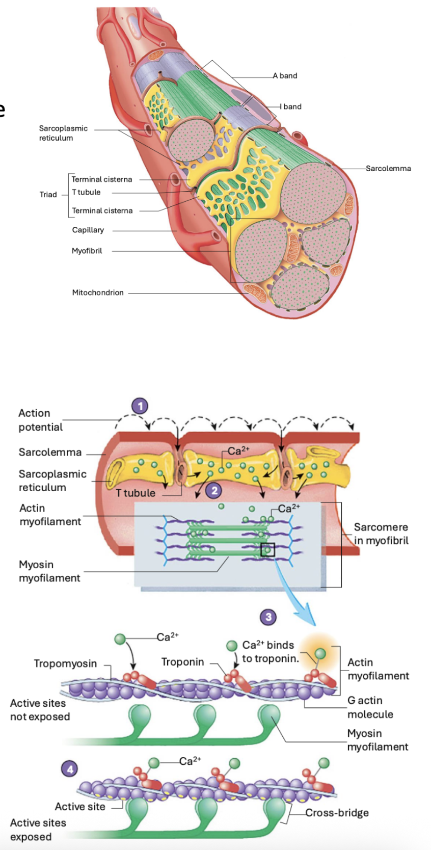
excitation-contraction coupling
Before the cross-bridge cycling process occurs
Triad:
Forms @ regular repeating intervals along length of muscle fibers
Occurs in regions where actin and myosin overlaps
2 triads per sarcomere (bc there's 2 regions where actin is able to overlap w myosin)
At RESTING:
Terminal cistern → has alot of Ca2+ stored
Ca2+ released channels are closed (and stored in SR)
⤷ ∴ troponin in resting state (holding tropomyosin over active site on actin)
At CONTRACTION:
Neuron signals MAP ⇒ moves along sarcolemma ⇒ when reached T-tubules ⇒ will drop down inside muscle fibers & move thru T-tubules
As membrane potential changes, detected by Ca2+ released channel ⇒ ∴ will open and spill into sarcoplasm ⇒ diffuse into regions where actin and myosin myofilaments
⤷ Ca2+ binds onto troponin → shape changes → moves tropomyosin out of way → expose active site → myosin is able to bind onto actin myofilaments
⤷ ∴ allow cross-bridge cycling to begin
summary of skeletal muscle contractions
An AP travels along an axon membrane to NMJ
Voltage-gated Ca2+ channels open adn Ca2+ enters the presynaptic terminal
Acetylcholine is released from synaptic vesicles
Acetylcholine stimulates ligand-gated Na+ channels on the motor end-plate to open
Na+ diffuses into muscle fibers, initiating an AP that travels along sarcolemma and T tubule membrane
AP in the T tubule causes opening of voltage-gated Ca2+ channels in the SR, releasing Ca2+
On the actin, Ca2+ binds to troponin, which moves tropomyosin and exposes myosin head binding sites
Start of cross (X)-bridge cycling
ATP molecules on myosin heads are broken down to ADP and Ⓟ, which release energy needed to move the myosin heads
The heads of the myosin myofilaments bend (power stroke), causing actin to slide past the myosin
To rid of Ca2+: (release of muscles)
Ca2+ must be taken back up into SR → via SERCA pumps (sacroendoplasm reticulum Ca2+ ATPase) ⇒ which stops muscles from contracting
Ca2+ moves away from tropin ⇒ and tropin moves tropomyosin back over the binding site & blocks myosin from further binding ⇒ muscle relax
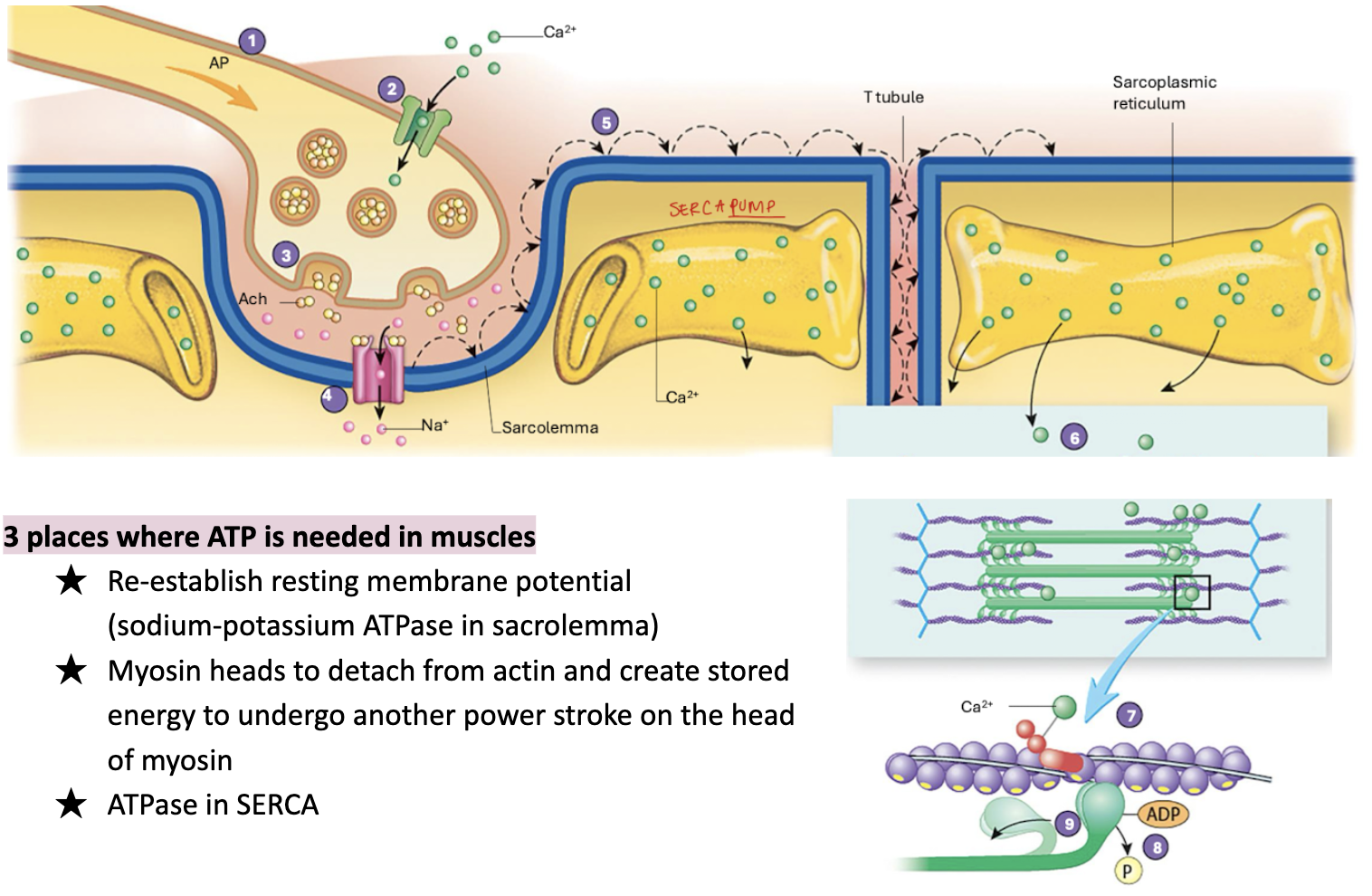
3 places ATP is needed in muscles
Re-establish resting membrane potential (sodium-potassium ATPase in sacrolemma)
Myosin heads to detach from actin and create stored energy to undergo another power stroke on the head of myosin
ATPase in SERCA (Sarcoplasmic/Endoplasmic Reticulum Ca2+ ATPase)